


INTRODUCTION
Aging is associated with cognitive decline, yet less is known about the neurobiological basis of this change (Salthouse, Reference Salthouse2011). Identifying risk factors and mechanisms of age-related cognitive decline is among the greatest challenges to improving the health of older adults. The brain relies on proper functioning of the vascular neural network for the maintenance of cognitive function. This network includes the neurovascular unit (endothelial cells, pericytes, glia, and neurons) and the upstream arteries and arterioles that feed into the microcirculation of the brain (Lo & Rosenberg, Reference Lo and Rosenberg2009; Zhang et al., Reference Zhang, Badaut, Tang, Obenaus, Hartman and Pearce2012). Research suggests that dysfunction within the vascular neural network can lead to neuronal injury and degeneration (Lo & Rosenberg, Reference Lo and Rosenberg2009; Zlokovic, Reference Zlokovic2010).
Cerebral blood flow (CBF), or the rate of delivery of arterial blood to the capillary bed of a particular mass of tissue, is a functional measure of the vascular neural network and has been implicated in both normal aging and Alzheimer’s disease (AD)-related cognitive decline (Bertsch et al., Reference Bertsch, Hagemann, Hermes, Walter, Khan and Naumann2009; Hays, Zlatar, & Wierenga, Reference Hays, Zlatar and Wierenga2016; Heo et al., Reference Heo, Prakash, Voss, Erickson, Ouyang, Sutton and Kramer2010). CBF has demonstrated reliable correlations with cognition across the lifespan in both normal and pathologic aging (Bangen et al., Reference Bangen, Restom, Liu, Wierenga, Jak, Salmon and Bondi2012; Bertsch et al., Reference Bertsch, Hagemann, Hermes, Walter, Khan and Naumann2009; Okonkwo et al., Reference Okonkwo, Xu, Oh, Dowling, Carlsson, Gallagher and Johnson2014; Wierenga et al., Reference Wierenga, Dev, Shin, Clark, Bangen, Jak and Bondi2012). Furthermore, CBF measurement has been shown to distinguish between normal controls and those with AD, identify those at risk for mild cognitive impairment (MCI) and AD, and predict conversion to MCI and AD, suggesting its usefulness as a preclinical marker of cognitive decline (Hays et al., Reference Hays, Zlatar and Wierenga2016; Wierenga, Hays, & Zlatar, Reference Wierenga, Hays and Zlatar2014).
Subjective cognitive decline (SCD), or self-reported cognitive decline despite normal neuropsychological test performance, has been identified as a risk factor for objective cognitive decline (Glodzik-Sobanska et al., Reference Glodzik-Sobanska, Reisberg, De Santi, Babb, Pirraglia, Rich and de Leon2007; Lista et al., Reference Lista, Molinuevo, Cavedo, Rami, Amouyel, Teipel and Hampel2015; Reisberg, Shulman, Torossian, Leng, & Zhu, Reference Reisberg, Shulman, Torossian, Leng and Zhu2010; Sun, Yang, Lin, & Han, Reference Sun, Yang, Lin and Han2015; Wang et al., Reference Wang, van Belle, Crane, Kukull, Bowen, McCormick and Larson2004). Although our understanding of SCD is in its infancy, some speculate that SCD represents an early stage of cognitive decline, with very subtle cognitive deterioration undetectable by current neuropsychological testing (Reisberg & Gauthier, Reference Reisberg and Gauthier2008), while others suggest that SCD results from early neuronal dysfunction together with compensatory mechanisms that preserve cognitive functions (Sun et al., Reference Sun, Yang, Lin and Han2015).
There is also evidence that SCD is strongly related to depression and other affective symptoms, highlighting the importance of controlling and/or adjusting for psychiatric symptoms when investigating SCD in the context of cognitive decline (Schmand, Jonker, Geerlings, & Lindeboom, Reference Schmand, Jonker, Geerlings and Lindeboom1997; Sun et al., Reference Sun, Yang, Lin and Han2015). Studies suggest that those with SCD are not only at a greater risk for future cognitive decline but that decline occurs at an accelerated rate, compared to those without SCD (Reisberg et al., Reference Reisberg, Shulman, Torossian, Leng and Zhu2010). Moreover, those with SCD are more likely to convert to MCI and AD (Glodzik-Sobanska et al., Reference Glodzik-Sobanska, Reisberg, De Santi, Babb, Pirraglia, Rich and de Leon2007; Lista et al., Reference Lista, Molinuevo, Cavedo, Rami, Amouyel, Teipel and Hampel2015; Sun et al., Reference Sun, Yang, Lin and Han2015), as demonstrated by findings from a recent meta-analysis indicating adults with SDC are twice as likely to develop dementia compared to those without SCD, with annual conversion rates of 2.3% in adults with SCD compared to 1% in those without SCD (Mitchell, Beaumont, Ferguson, Yadegarfar, & Stubbs, Reference Mitchell, Beaumont, Ferguson, Yadegarfar and Stubbs2014).
Individuals with SCD also exhibit pathologic markers of AD, such as amyloid-beta and tau protein deposition (Barnes, Schneider, Boyle, Bienias, & Bennett, Reference Barnes, Schneider, Boyle, Bienias and Bennett2006), regional brain atrophy (Cherbuin, Sargent-Cox, Easteal, Sachdev, & Anstey, Reference Cherbuin, Sargent-Cox, Easteal, Sachdev and Anstey2015; Stewart et al., Reference Stewart, Godin, Crivello, Maillard, Mazoyer, Tzourio and Dufouil2011), and altered brain function (Sun et al., Reference Sun, Yang, Lin and Han2015), with evidence of both higher and lower levels of regional cerebral perfusion and metabolism compared to those without SCD. For example, Hohman and colleagues found that high levels of cognitive complaints were associated with higher positron emission tomography (PET)-measured CBF in the insula, inferior parietal cortex, lingual gyrus, fusiform gyrus, and the cerebellum (Hohman, Beason-Held, Lamar, & Resnick, Reference Hohman, Beason-Held, Lamar and Resnick2011).
Similarly, another study showed that those with cognitive complaints demonstrated higher arterial spin labeling (ASL)-measured CBF along midline default mode network regions compared to those without cognitive complaints (Wang et al., Reference Wang, West, Risacher, McDonald, Tallman, Ghetti and Saykin2013). Conversely, Mosconi and colleagues found that those with subjective memory complaints showed lower 2-[18F]fluoro-2-deoxy-D-glucose (FDG)-PET-measured glucose metabolism in parietotemporal and parahippocampal regions compared to those without memory complaints while another study found higher FDG-PET-measured glucose metabolism in the medial temporal lobe and lower glucose metabolism in the precuneus among a similar group of older adults with memory complaints (Mosconi et al., Reference Mosconi, De Santi, Brys, Tsui, Pirraglia, Glodzik-Sobanska and de Leon2008; Scheef et al., Reference Scheef, Spottke, Daerr, Joe, Striepens, Kolsch and Jessen2012).
Although cerebral perfusion and glucose metabolism are thought to be tightly linked (Roy & Sherrington, Reference Roy and Sherrington1890; Verfaillie et al., Reference Verfaillie, Adriaanse, Binnewijzend, Benedictus, Ossenkoppele, Wattjes and Barkhof2015), it appears that SCD-related PET and ASL studies have produced conflicting results, likely due to differences in sample characteristics (e.g., operational definitions of SCD or normal control) or methodology (e.g., imaging modality limitations, statistical or experimental control of confounding variables). As such, it may be important to extend these prior studies using cutting-edge methodologies within well-characterized samples to further clarify the relationship between SCD and cerebral perfusion. Overall, current evidence suggests that those with SCD may exhibit altered CBF in regions typically associated with memory function, aging and AD-risk. This notion is consistent with the vascular theory of AD, which holds that vascular damage contributes to the development of AD. Notably, SCD has also been independently associated with vascular risk factors (Paradise, Glozier, Naismith, Davenport, & Hickie, Reference Paradise, Glozier, Naismith, Davenport and Hickie2011).
Taken together, evidence showing that SCD is associated with alterations in cerebral perfusion and future cognitive decline suggests that those with SCD may be experiencing vascular dysregulation, or a disruption in the normal relationship between CBF and cognition, yet no study to date has examined whether SCD modifies the relationship between CBF and cognition. The exploration of this moderating effect combined with the use of non-invasive neuroimaging techniques among a well-characterized sample of cognitively normal older adults might help elucidate the underlying mechanisms of SCD and enable early intervention strategies aimed at preventing cognitive decline and AD.
The current study used ASL magnetic resonance imaging (MRI) to determine if the relationship between CBF and memory function was modified by SCD. Based on previous reports, we hypothesized that SCD status would have a direct association with CBF in areas associated with aging and AD risk (hippocampus, parahippocampal gyrus, posterior cingulate, precuneus). We also hypothesized that the association of resting CBF and cognitive function (memory) would be moderated by SCD status (SCD+, SCD-), whereby those in the SCD+ group would display an inverse relationship, suggesting evidence of vascular dysregulation and supporting its role in cognitive decline and AD risk.
METHODS
Participants
See Table 1 for participant demographic and cognitive characteristics. Participants were community-dwelling older adult volunteers between the ages of 65 and 88 enrolled in a longitudinal study of aging and/or other ongoing research studies at the Veterans Administration (VA) San Diego Healthcare System and the University of California San Diego (UCSD). Of the 80 participants with available data who met inclusion/exclusion criteria, 35 reported SCD (SCD+) and were matched to 35 controls who reported no SCD (SCD-), based on age, sex, and Geriatric Depression Scale (GDS) score using the nearest neighbor matching method (Stuart, Reference Stuart2010).
Table 1 Participant demographic and cognitive characteristics

ApoE=apolipoprotein E; DRS=Mattis Dementia Rating Scale; rCBF=resting cerebral blood flow; GDS=Geriatric Depression Scale; SMRS=Subjective Memory Rating Scale; VM=Verbal Memory; WMS-R=Wechsler Memory Scale-Revised; LM=Logical Memory; CVLT-2=California Verbal Learning Test-2; SD=short delay; LD=long delay; df=degrees of freedom for independent samples T-test.
* Equal variances not assumed (df reported are adjusted for unequal variances).
Normal cognitive function was determined based on a comprehensive neuropsychological test battery. Participants were excluded if they met the empirically derived criteria for mild cognitive impairment developed by Jak and colleagues (Reference Jak, Bondi, Delano-Wood, Wierenga, Corey-Bloom, Salmon and Delis2009). Potential participants were also excluded if they had a history of dementia, severe head injury, uncontrolled hypertension, had a Diagnostic and Statistical Manual of Mental Disorders, 4th Edition, diagnosis of learning disability, attention deficit disorder, mood disorder, or substance abuse, or if they reported a significant level of depressive symptoms on the 30-item GDS (i.e., GDS≥10). Participants were also excluded if they had contraindications to MRI scanning, or if they were taking prescription psychoactive medications. All data were collected in accordance with UCSD and VA institutional research standards for human research and the Helinski Declaration.
Testing Sequence
All participants completed a comprehensive neuropsychological battery to determine normal cognitive functioning. Self-report measures, including the Subjective Memory Rating Scale (SMRS), were administered directly after the completion of cognitive testing. All participants were tested in one of three similar testing suites at UCSD and most were tested in the morning hours, though specific times varied. Participants also underwent an fMRI scan at the same location within 90 days of completing cognitive testing.
Verbal Memory Composite
A verbal memory composite score was created using trials 1–5, short delay free-recall, and long delay free-recall raw scores from the California Verbal Learning Test 2 (CVLT-2), measuring word list learning, and the Logical Memory immediate and delayed recall subtests of the Wechsler Memory Scale-Revised (WMS-R), measuring story recall. These tests were selected based on results from a principal component analysis previously reported by our group on a similar sample of older adults (Wierenga et al., Reference Wierenga, Dev, Shin, Clark, Bangen, Jak and Bondi2012). Verbal memory composite scores were derived by averaging the Z-scores for each of the tests within the composite for the entire sample.
SCD Assessment
The SMRS is a five-item questionnaire asking: “In the past year, do you think: (1) Your ability to remember the names of people you have just met has changed (2) Your ability to remember the faces of people you have just met has changed (3) Your ability to remember the names of close friends or relatives has changed (4) Your ability to remember appointments correctly has changed (5) Your ability to judge the passage of time and guessing the time of day without looking at a clock or the sun has changed.” Response categories for each item were: 1=definitely improved, 2=slightly improved, 3=no change, 4=slightly deteriorated, and 5=definitely deteriorated. SCD+ was operationally defined by normal cognitive function based on current neuropsychological performance and the presence of scores of “4=slightly deteriorated” and/or “5=definitely deteriorated” and the absence of any scores of “1=definitely improved” or “2=slightly improved”. SCD- was operationally defined by normal cognitive function and the presence of scores of “1=definitely improved”, “2=slightly improved”, and/or “3=no change” and the absence of scores of “4=slightly deteriorated” or “5=definitely deteriorated.”
Therefore, those in the SCD- group reported only improvements and stability in memory, while those in the SCD+ group reported deterioration and no improvements. For ease of interpretation, total scores on the SMRS were centered around “3=no change” so that positive total scores represented subjective improvements and negative total scores represented subjective declines. Although this measure is focused on memory changes, we use the term “subjective cognitive decline” rather than “subjective memory decline” as is recommended by the Subjective Cognitive Decline Initiative working group (Jessen et al., Reference Jessen, Amariglio, van Boxtel, Breteler, Ceccaldi, Chételat and Wagner2014). Among a sample of 1883 dementia-free older adults, the SMRS demonstrated a Cronbach alpha coefficient of 0.6. Exploratory factor analysis found one common factor with an Eigen value greater than 1 and factor loadings from 0.4 to 0.5 for the five items. Furthermore, the SMRS demonstrated adequate face validity among this same sample (Wang et al., Reference Wang, van Belle, Crane, Kukull, Bowen, McCormick and Larson2004). In the current sample, the SMRS demonstrated a Cronbach alpha coefficient of 0.68.
Apolipoprotein E Genotyping
Genotyping was performed by the ADCS Biomarker Core at UCSD using real-time polymerase chain reaction (PCR) restriction fragment length polymorphism analysis. Genomic DNA was collected from participants using buccal swab and extracted using Qiamp DNA blood mini kit (Qiagen) followed by PCR amplification (Wierenga et al., Reference Wierenga, Dev, Shin, Clark, Bangen, Jak and Bondi2012).
MRI Acquisition
Imaging data were acquired on a GE Discovery MR750 3 Tesla whole-body system with a body transmit coil and an eight-channel receive-only head coil at the UCSD Center for Functional MRI. The structural brain sequence consisted of a high-resolution T1-weighted fast spoiled gradient recall (three-diemnsional FSPGR) scan: 172 1 mm contiguous sagittal slices, field of view (FOV)=25 cm, TR=8 ms, TE=3.1 ms, flip angle=12, T1=600 ms, 256×192 matrix, bandwidth=31.25 kHz, frequency direction=S-I, NEX=1, scan time=8 min 13 s. Resting CBF was acquired with the multiphase pseudocontinuous ASL (MPPCASL) sequence, which is optimized for robust CBF quantification (Jung et al., Reference Jung, Wong and Liu2010): tagging duration=2 s, TI=3.6 s, TR=4.2 s, TE=minimum, reps=64, FOV=22×22 cm, twenty 5-mm axial slices with a single shot spiral acquisition, collecting eight cycles where each cycle consists of eight images acquired with unique phase offsets, acquisition time=4:46 min.
A spiral scan with a long TR (4000 ms) and short TE (3.4 ms) was also acquired to obtain an estimate of the equilibrium magnetization of cerebral spinal fluid, which is used to convert the perfusion signal into calibrated CBF units (mL blood/100 g tissue/min). Finally, a minimum contrast image was acquired to adjust for transmit and receive coil inhomogeneities. Two field map scans were also acquired and used for off-line field map correction to help correct for signal bunching and dropouts in the frontal/medial temporal lobes.
MRI Preprocessing
Image processing was performed with Analysis of Functional NeuroImages (AFNI, afni.nimh.nih.gov) (Cox, Reference Cox1996), FMRIB Software Library (FSL, Oxford, United Kingdom), and locally created Matlab scripts. Field map correction was applied to the ASL time series before co-registration to the middle time point to minimize the effects of participant motion. For each participant, a mean ASL image was formed from the average difference of the control and tag images using surround subtraction to create an uncorrected perfusion time series, and slice timing delays were accounted for, making the inversion time (TI2) slice specific (Liu & Wong, Reference Liu and Wong2005). This mean ASL image was then converted to absolute units of CBF (mL/100 g tissue/min) using an estimate of the equilibrium magnetization of CSF as a reference signal (Chalela et al., Reference Chalela, Alsop, Gonzalez-Atavales, Maldjian, Kasner and Detre2000).
This procedure resulted in a calibrated perfusion value for each voxel. Skull stripping of the high-resolution T1-weighted image was performed using AFNI’s 3dSkullStrip. Tissue segmentation was performed using FSL’s Automated Segmentation Tool algorithm to define CSF, gray matter (GM), and white matter (WM) regions. The high-resolution T1-weighted image and partial volume segmentations were registered to ASL space, and partial volume segmentations were down-sampled to the resolution of the ASL data. To correct the CBF measures for partial volume effects and ensure that CBF values were not influenced by known decreased perfusion in white matter or increased volume of CSF (Parkes et al., Reference Parkes, Rashid, Chard and Tofts2004), we used the method previously reported by Johnson and colleagues (Reference Johnson, Jahng, Weiner, Miller, Chui, Jagust and Schuff2005). These calculations assume that CSF has 0 CBF, and that CBF in GM is 2.5 times greater than that in WM.
The following formula was used to compute partial volume corrected CBF signal intensities: CBFcorr=CBFuncorr/(GM + 0.4*WM). CBFcorr and CBFuncorr are corrected and uncorrected CBF values, respectively. GM and WM are gray matter and white matter partial volume fractions, respectively. Information from the high resolution structural image and the FSL FAST was used to determine the tissue content of each perfusion voxel. A 4.0 mm full-width, half-maximum Gaussian filter was applied to the CBFcorr data. Voxels with negative intensities were replaced with zero (Brown et al., Reference Brown, Zorrilla, Georgy, Kindermann, Wong and Buxton2003), and GM voxels were thresholded at 0.9 probability. CBFcorr data were registered to the MNI-152 atlas using FMRIB’s non-linear image registration tool (FNIRT), part of FSL (http://fsl.fmrib.ox.ac.uk/fsl/), and resampled to a 3×3×3 mm resolution grid.
Statistical Analyses
The t tests were used to compare groups on age, years of education, GDS score, SMRS score, whole-brain resting CBF, and cognitive variables. Chi-square tests were used to compare groups on sex, apolipoprotein E (APOE) status, and family history of dementia. A voxel-wise linear mixed-effects (LME) regression model was conducted in R with resting CBF as the dependent variable and with independent variables: (1) verbal memory composite score, (2) SCD status (SCD+ vs. SCD-), and (3) the interaction term between verbal memory composite score and SCD status. In addition to matching groups on age, GDS score, and sex, statistical analyses also adjusted for these same variables to further decrease the chance of confounding (Pearce, Reference Pearce2016). The LME yielded statistical maps displaying the brain regions for which there were significant main effects of SCD status on CBF and the interactive effects of SCD status and verbal memory composite on CBF.
Significance was determined by applying cluster-size correction derived from Monte-Carlo simulations (via AFNI’s 3dClustSim) to guard against false positives on data initially thresholded at a value of p<.025 (uncorrected). Based on these simulations, it was determined that a cluster size of 28 contiguous voxels (756 mm3) ensured an overall p<.025 (corrected). To characterize the direction and magnitude of interactive and main effects, post hoc regression analyses were carried out using the mean CBF extracted from each significant cluster. Post hoc analyses were conducted in IBM SPSS Statistics, version 22 (bootstrapped with 1000 samples) and were conducted only to characterize the significant LME interaction terms.
RESULTS
Groups did not differ significantly on age, APOE status, family history of dementia, years of education, sex, DRS score, GDS score, whole-brain resting CBF, the verbal memory composite score, nor on any of the cognitive tests that comprised the verbal memory composite score (see Table 1).
Main Effect of SCD on CBF
Significant main effects of SCD on CBF were found in two clusters within the medial orbitofrontal cortex and the right putamen. Cluster locations with coordinates and corresponding Beta values by group are listed on Table 2. To characterize the direction and magnitude of the main effects of SCD on CBF, mean CBF was extracted from the two main effect clusters, with evidence of both positive and negative associations between SCD status and CBF. Compared to the SCD- group, those in the SCD+ group demonstrated lower CBF in the medial orbitofrontal cortex and higher CBF in the right putamen (see Figure 1).

Fig. 1 Main effects of SCD status on CBF. SCD=subjective cognitive decline; CBF=cerebral blood flow; L=left; R=right. * Significance at p<.025. Error bars represent standard error.
Table 2 Main effect of SCD status on CBF
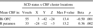
CBF=cerebral blood flow; OFC=orbital frontal cortex; Bi=bilateral; R=right. X, Y, and Z coordinates represent the peak F-value in MNI space. Beta values represent the standardized partial regression coefficients, with higher absolute values representing larger effect sizes.
Main Effect of Memory Function on CBF
A significant main effect of memory function on CBF was found in one cluster within the right inferior frontal gyrus. Cluster location with coordinates and corresponding Beta values by group are listed on Table 3. To characterize the direction and magnitude of the main effect of memory function on CBF, mean CBF was extracted from the main effect cluster, showing that memory function was positively associated with CBF in both groups (SCD+/-) within the right inferior frontal gyrus (see Figure 2).

Fig. 2 Main effect of memory function on CBF. SCD=subjective cognitive decline; CBF=cerebral blood flow; R=right. * Significance at p<.025.
Table 3 Main effect of verbal memory function on CBF
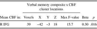
CBF=cerebral blood flow; R=right; X, Y, and Z coordinates represent the peak F-value in MNI space; IFG=inferior frontal gyrus. Beta value represents the standardized partial regression coefficient with higher absolute values representing larger effect sizes.
Interactive Effects of SCD Status and Verbal Memory on CBF
Significant interactions between SCD status and verbal memory composite scores on CBF were found in six clusters within the right hippocampus, inferior frontal gyrus, left middle temporal gyrus, left hippocampus and fusiform gyrus and inferior temporal gyrus, body of the corpus callosum, and bilateral posterior cingulate cortex and splenium of the corpus callosum. Cluster locations with coordinates and corresponding Beta values by group are listed on Table 4. To characterize the direction and magnitude of the interaction effects, mean CBF was extracted from the six significant clusters, demonstrating a consistent pattern of positive associations between CBF and verbal memory functions for those in the SCD- group, and negative associations between CBF and verbal memory functions for those in the SCD+ group (see Figure 3).

Fig. 3 Interactive effects of SCD status and verbal memory on CBF. SCD=subjective cognitive decline; CBF=cerebral blood flow; PCC=posterior cingulate cortex; CC=corpus callosum; MTG=middle temporal gyrus; FG=fusiform gyrus; ITG=inferior temporal gyrus; IFG=inferior frontal gyrus; L=left; R=right. * Significance at p<.025.
Table 4 Interactive effects of SCD status and verbal memory on CBF

CBF=cerebral blood flow; B=bilateral; L=left; R=right; X, Y, and Z coordinates represent the peak F-value in MNI space; PCC=posterior cingulate cortex; CC=corpus callosum; MTG=middle temporal gyrus; Hc=hippocampus; FG=fusiform gyrus; ITG=inferior temporal gyrus; IFG=inferior frontal gyrus. Beta values represent the standardized partial regression coefficients, with higher absolute values representing larger effect sizes.
Higher CBF was associated with better verbal memory function for those in the SCD- group within bilateral posterior cingulate cortices, left middle temporal gyrus, right inferior temporal gyrus, and the corpus callosum. In contrast, higher CBF was associated with worse verbal memory performance within the left middle temporal gyrus, bilateral hippocampi, bilateral posterior cingulate cortices, left fusiform gyrus, left inferior temporal gyrus, and right inferior frontal gyrus in the SCD+ group (see Figure 3).
DISCUSSION
Results showed that older adults with SCD demonstrated lower CBF in the orbitofrontal cortex and higher CBF in the putamen, compared to those without SCD. Furthermore, our sample of cognitively normal older adults demonstrated an overall positive association between memory function and CBF that was modified by SCD, such that those presenting without SCD showed positive associations between memory functions and CBF within frontal, temporal, and parietal regions, whereas those presenting with SCD showed negative associations between memory function and CBF within frontal, temporal, and parietal regions. Our results showing that those with SCD demonstrate both higher and lower regional CBF, compared to those without SCD, are consistent with other SCD-related perfusion studies, further supporting the notion that regionally specific perfusion differences exist between these groups in areas that have been implicated in normal aging and AD risk (Dai et al., Reference Dai, Lopez, Carmichael, Becker, Kuller and Gach2009; Hays et al., Reference Hays, Zlatar and Wierenga2016; Meltzer et al., Reference Meltzer, Cantwell, Greer, Ben-Eliezer, Smith, Frank and Price2000).
This is the first study, to our knowledge, to show that SCD modifies the relationship between voxel-wise CBF and memory function. These findings suggest that the normal beneficial effects of higher CBF on cognition may be disrupted among those with SCD, as higher CBF no longer appears to support better cognitive functioning in this group within regions associated with normal aging and AD-risk (Frederiksen, Reference Frederiksen2013; Grasby et al., Reference Grasby, Frith, Friston, Bench, Frackowiak and Dolan1993; Hays et al., Reference Hays, Zlatar and Wierenga2016; Wierenga et al., Reference Wierenga, Hays and Zlatar2014). Although the underlying mechanisms associated with the observed differences between our groups are still unknown, other SCD-related studies showing higher activation, perfusion, and nodal efficiency among those with SCD in similar regions, have suggested that compensatory mechanisms could explain these differences (Erk et al., Reference Erk, Spottke, Meisen, Wagner, Walter and Jessen2011; Sun, Reference Sun2015; Wang, Reference Wang2014).
Similarly, the observance of higher CBF among those at risk for cognitive decline or AD has been interpreted as recruitment of early compensatory mechanisms, thought to reflect efforts to maintain adequate brain oxygenation in the face of vascular aging or damage. The notion of CBF-related compensation among those at risk for AD supports the vascular theory of AD, which posits that vascular damage plays a role in the pathogenesis of AD (Wierenga et al., Reference Wierenga, Hays and Zlatar2014). This theory is further supported by longitudinal and cross-sectional studies showing that those in the preclinical phases of AD tend to show more areas of hyperperfusion early in the disease process, followed by more areas of hypoperfusion, thought to represent increasing heterogeneity of capillary flow patterns with disease progression (Ostergaard et al., Reference Ostergaard, Aamand, Gutierrez-Jimenez, Ho, Blicher, Madsen and West2013; Wierenga et al., Reference Wierenga, Hays and Zlatar2014). Taken together, this evidence suggests that the modifying effect of SCD on the relationship between CBF and cognition may reflect vascular dysregulation among those with SCD, and that the perfusion alterations observed in this group may reflect attempts to compensate for these vascular changes.
The results of this current study are similar to those of a recent study of AD-risk, which showed that APOE status modifies the relationship between CBF and cognition, such that those carrying the e4 allele, a known risk factor for AD, showed inverse relationships between CBF and memory function, while non-carriers displayed positive relationships (Zlatar et al., Reference Zlatar, Bischoff-Grethe, Hays, Liu, Meloy, Rissman and Wierenga2016), adding to an accumulation of evidence suggesting that SCD may reflect an early preclinical stage of AD (Glodzik-Sobanska et al., Reference Glodzik-Sobanska, Reisberg, De Santi, Babb, Pirraglia, Rich and de Leon2007; Sun, Reference Sun2015). We found no differences in APOE status or family history of dementia between those with and without SCD in our sample, indicating that SCD might represent an independent risk factor for cognitive decline.
The fact that group differences in the relationship between CBF and cognition were found despite normal cognitive function lends further support to the notion that SCD+ is distinct from normal aging (i.e., SCD-). It is also possible that current neuropsychological testing is not sensitive to the early subtle cognitive decline associated with SCD (Sun, Reference Sun2015). Results suggest that vascular dysregulation is occurring in those with SCD, even in the absence of clinical symptoms, further supporting its role as a preclinical marker of risk for cognitive decline.
The accurate assessment of SCD is a significant challenge facing the field. To address the lack of standardized and well-validated assessment tools for SCD, the Subjective Cognitive Decline Initiative (SCD-I) Working Group was established in 2014 with the goal of developing a conceptual framework and research criteria for SCD (Jessen et al., Reference Jessen, Amariglio, van Boxtel, Breteler, Ceccaldi, Chételat and Wagner2014). They recommended “well-constructed, easy-to-administer items with adequate reliability across diverse samples of older adults” (Rabin et al., Reference Rabin, Smart, Crane, Amariglio, Berman, Boada and Sikkes2015). Given that such a measure has yet to be developed or validated, the working group offered preliminary recommendations for instrument selection: (1) Select measures with appropriate demographic characterization. (2) Select measures with adequate content coverage for the target population. (3) Consider issues of psychometric adequacy (Rabin et al., Reference Rabin, Smart, Crane, Amariglio, Berman, Boada and Sikkes2015).
Despite the psychometric limitations of the SMRS, its use represents an improvement over much of the SCD literature, which consists of studies using only one or two questions to determine SCD. Furthermore, the five questions on the SMRS, which ask about changes in memory for names, faces, appointments, and the ability to judge time over the past year, provide better clinical insight to specific subjective experiences of cognitive decline over a specified period, compared to questions that ask about cognitive difficulties in general without reference to decline over time. Future SCD-related research would benefit from the development and widespread use of a more comprehensive consensus measure of SCD that has been shown to be valid and reliable.
In conclusion, this evidence supports the notion that those with SCD may be experiencing dysregulation within the vascular neural network. Elucidating the early vascular changes that accompany risk for cognitive decline and AD could lead to the identification of vasoprotective treatments with the potential to delay or prevent the onset of cognitive decline and AD. Although future research is needed to determine whether vascular dysregulation in those with SCD reflects normal or pathologic processes, the current results support SCD as a valid construct to detect those who might be at risk for cognitive decline and AD. Integrating SCD with other known markers of cognitive decline and AD could lead to earlier and more accurate identification of those who would likely benefit from treatments aimed at preventing cognitive decline.
Strengths and Limitations
As mentioned above, this study was limited by the challenge faced by all studies of SCD, namely that of accurate assessment of subjective cognitive decline. We chose to use the SMRS because it offers several advantages over other available measures. However, the SMRS consists of only five self-report questions, from which we wholly defined our groups. Moreover, most of the questions on the SMRS are memory-related, reducing the likelihood of capturing those with SCD in non-memory cognitive domains, and the Cronbach alpha coefficient of 0.68 is not particularly high, although it is still within the range of acceptability for reliability (Loewenthal, Reference Loewenthal2001). Nevertheless, the proportion of participants classified as having SCD was in line with prevalence rates in other studies (Jonker, Geerlings, & Schmand, Reference Jonker, Geerlings and Schmand2000; Mitchell, Reference Mitchell2008).
It is also possible that the timing of SCD assessment in this study may have influenced the way in which participants responded to this questionnaire, as we administered the SMRS directly after objective cognitive testing. However, it is important to note that the SMRS asks about changes in specific abilities (memory for names, faces, appointments, and the ability to judge time) that were not tested within the cognitive battery.
Furthermore, the SMRS asks participants to report changes in these specific abilities over the last year (rather than changes over weeks or months), further reducing the likelihood that the objective cognitive testing influenced responding on this questionnaire. Nonetheless, future studies should consider evaluating SCD and objective cognitive functioning on separate days. Overall, these limitations further highlight the need for a comprehensive consensus measure of SCD, together with standardized administration instructions, including recommendations for the sequencing of SCD assessment in relation to objective cognitive testing.
Despite a well-characterized and relatively large sample, the use of a cross-sectional design restricted our ability to draw causal conclusions and limited our ability to determine whether the effects of SCD on CBF and the relationship between CBF and cognition represent normal or pathologic processes. It is also important to note that our sample had relatively high levels of education, and although this demographic factor was not associated with SCD status, its limited range may reduce the generalizability of these findings.
Although groups (SCD+/SCD-) did not differ significantly in the mean time interval between cognitive testing and fMRI scanning (23 and 17 days, respectively), decreasing the time interval between cognitive testing and fMRI may improve accuracy of brain-behavior associations. Although only linear relationships were explored in the current study, future studies should consider exploring non-linear relationships between CBF and cognitive performance. Future investigations should also include longitudinal designs with larger, more diverse samples to replicate and extend the current findings. Furthermore, the inclusion of additional markers of AD, such as CSF biomarkers would help better characterize those with SCD.
The strengths of the current study include the use of non-invasive ASL MRI to measure CBF, the availability of several cognitive test performances to characterize cognitive status, and the use of voxel-wise LME models, which allowed us to examine the associations between SCD, verbal memory performance, and CBF across the entire brain. Despite these strengths, ASL methods are limited by low spatial resolution, transit time effects, and low signal-to-noise ratio in deep white matter. Lastly, the current study represents an improvement over other studies of SCD, in our inclusion of a well-characterized, well-controlled sample of older adults. Many other studies of SCD have failed to exclude or control for depressive symptoms and other psychiatric disorders, making it difficult to determine whether observed results were due to SCD. The current study matched participants on age, sex, and GDS, in addition to statistically correcting for these same variables within the analyses.
CONCLUSIONS
This study found that the relationship between CBF and cognition is disrupted in cognitively normal older adults with SCD, compared to those without SCD. Whereas higher CBF supports verbal memory functions in those without SCD, it appears that higher CBF is no longer supportive of verbal memory function among those with SCD. The current findings suggest that those with SCD may be experiencing vascular dysregulation and support SCD as a marker of risk for cognitive decline and AD. Future longitudinal studies should examine changes in perfusion and cognition over time among those with SCD to determine whether the moderating effect of SCD on the relationship between CBF and cognition represents normal age-related or pathologic processes and to further characterize the role of SCD in the trajectory from normal to pathologic aging.
ACKNOWLEDGMENTS
This work was supported by VA CSR&D Merit Award 5I01CX000565 (to C.E.W.); K23 AG049906 (to Z.Z.Z.); and National Science Foundation Graduate Research Fellowship Program 2015207525 (to C.C.H). There are no conflicts of interest to disclose.


