


INTRODUCTION
Although traditional models have long considered that medial temporal lobe (MTL) structures are crucial for the formation of long-term memories (Eichenbaum, Reference Eichenbaum1997), but not for working memory (WM) (Cave & Squire, Reference Cave and Squire1992; Gabrieli, Reference Gabrieli1998; Squire et al., Reference Squire, Stark and Clark2004), there is an emerging view supporting the essential contribution of MTL in WM processes (for a review, see Gazzaley et al., Reference Gazzaley, Rissman and Desposito2004; Ranganath & Blumenfeld, Reference Ranganath and Blumenfeld2005). Data from animal lesion and neurophysiological experiments (Davachi & Goldman-Rakic, Reference Davachi and Goldman-Rakic2001; Holscher & Rolls, Reference Holscher and Rolls2002; Suzuki et al., Reference Suzuki, Miller and Desimone1997; Young et al., Reference Young, Otto, Fox and Eichenbaum1997), as well as findings from human neuropsychological studies (Haettig et al., Reference Haettig, Schepler, Lehmann and Meencke2004; Lancelot et al., Reference Lancelot, Samson, Ahad and Baulac2003; Nichols et al., Reference Nichols, Kao, Verfaellie and Gabrieli2006; Olson et al., Reference Olson, Moore, Stark and Chatterjee2006; Owen et al., Reference Owen, Morris, Sahakian, Polkey and Robbins1996; Wagner et al., Reference Wagner, Sziklas, Garver and Jones-Gotman2008), provide evidence for the critical involvement of MTL in WM. Recent human neuroimaging studies provide further support for the relevant contribution of MTL activation during WM (Axmacher et al., Reference Axmacher, Mormann, Fernandez, Cohen, Elger and Fell2007, Reference Axmacher, Schmitz, Wagner, Elger and Fell2008; Cabeza et al., Reference Cabeza, Dolcos, Graham and Nyberg2002; Glabus et al., Reference Glabus, Horwitz, Holt, Kohn, Gerton, Callicott, Meyer-Lindenberg and Berman2003; Karlsgodt et al., Reference Karlsgodt, Shirinyan, van Erp, Cohen and Cannon2005; Lee et al., Reference Lee, Buckley, Gaffan, Emery, Hodges and Graham2006; Manoach et al., Reference Manoach, Greve, Lindgren and Dale2003; Monk et al., Reference Monk, Zhuang, Curtis, Ofenloch, Tottenham, Nelson and Hu2002; Petersson et al., Reference Petersson, Gisselgard, Gretzer and Ingvar2006; Ranganath & D’Esposito, Reference Ranganath and D’Esposito2001; Schon et al., Reference Schon, Hasselmo, Lopresti, Tricarico and Stern2004; Stern et al., Reference Stern, Sherman, Kirchhoff and Hasselmo2001; Tesche & Karhu, Reference Tesche and Karhu2000).
While most of the neuroscientific research has focused on the study of brain activity during WM maintenance, “the neural mechanisms for WM encoding have not been extensively investigated” (Ranganath et al., Reference Ranganath, DeGutis and D’Esposito2004). Several authors (Cowan, Reference Cowan1995, Reference Cowan, Miyake and Shah1999; Miyake & Shah, Reference Miyake and Shah1999) have considered an issue of central importance to understanding WM, the precise characterization of how the encoding of task-relevant information takes place in the context of WM tasks. In a previous study (Campo et al., 2005b), we used magnetoencephalography (MEG), a noninvasive technique with high temporal resolution and good spatial resolution, to investigate the encoding phase of a verbal task and a spatial WM task. The temporal pattern of neural activity provides valuable information to understand the cognitive functions supported by different cortical areas (Axmacher et al., Reference Axmacher, Mormann, Fernandez, Elger and Fell2006; Helenius et al., Reference Helenius, Salmelin, Service and Connolly1998). Thus, using MEG, we could specify not only which brain structures were involved but also a detailed time course of their activation. We observed a material-specific modulation of the activity of MTL, consisting of a greater activation in left MTL between 500 and 700 ms after stimulus presentation during the verbal task and a preponderance of right MTL activation between 400 and 800 ms during the spatial task. These findings, along with others (Karlsgodt et al., Reference Karlsgodt, Shirinyan, van Erp, Cohen and Cannon2005; Ranganath et al., Reference Ranganath, DeGutis and D’Esposito2004), provide support for the contribution of MTL activation in WM encoding operations (Martin, Reference Martin1999).
The aim of the current study was to further investigate our previous findings suggesting the involvement of left MTL during the encoding phase of verbal WM. As stated by some authors (Grippo et al., Reference Grippo, Pelosi, Mehta and Blumhardt1996; Sawrie et al., Reference Sawrie, Martin, Knowlton, Faught, Gilliam and Kuzniecky2001), epilepsy patients suffering from hippocampal sclerosis (HS), consisting of neural loss and gliosis restricted to hippocampus and surrounding tissue (Thom et al., Reference Thom, Zhou, Martinian and Sisodiya2005; Trenerry et al., Reference Trenerry, Jack, Ivnik, Sharbrough, Cascino, Hirschorn, Marsh, Kelly and Meyer1993), provide a useful lesional model for studying memory processes. While patients with HS typically exhibit episodic memory impairment (Jones-Gotman et al., Reference Jones-Gotman, Zatorre, Olivier, Andermann, Cendes, Staunton, McMackin, Siegel and Wieser1997; Rausch & Babb, Reference Rausch and Babb1993; Sass et al., Reference Sass, Buchanan, Kraemer, Westerveld, Kim and Spencer1995), some studies have shown that performance in WM tasks is also reduced (Abrahams et al., Reference Abrahams, Morris, Polkey, Jarosz, Cox, Graves and Pickering1999; Axmacher et al., Reference Axmacher, Mormann, Fernandez, Cohen, Elger and Fell2007; Grippo et al., Reference Grippo, Pelosi, Mehta and Blumhardt1996; Krauss et al., Reference Krauss, Summerfield, Brandt, Breiter and Ruchkin1997; Wagner et al., Reference Wagner, Sziklas, Garver and Jones-Gotman2008). The nature of this localized lesion allows furthering exploration of the essential contribution of MTL activation in WM. Of these mentioned studies, only two reported neurophysiological data comparing verbal WM functioning in epilepsy patients and matched healthy controls. Grippo et al. (Reference Grippo, Pelosi, Mehta and Blumhardt1996) used event-related potentials, a methodology inferior to MEG in terms of precise source localization. A study by Axmacher et al. (Reference Axmacher, Mormann, Fernandez, Cohen, Elger and Fell2007) used intracranial electroencephalogram (iEEG), a methodology with excellent temporal and spatial resolution but a highly invasive neurosurgical procedure.
In this study, we used MEG, a noninvasive procedure, to compare the neuromagnetic patterns of MTL activation evoked by a verbal–semantic WM task in a group of epilepsy patients with left HS, who were evaluated for surgical treatment, versus a group of matched healthy controls. The verbal WM task was designed to ensure that participants encoded words semantically (i.e., deep encoding) as prior neuroimaging investigations have demonstrated that the depth to which verbal stimuli are processed modulates the activity in the MTL (Kapur et al., Reference Kapur, Craik, Tulving, Wilson, Houle and Brown1994; Lepage et al., Reference Lepage, Habib, Cormier, Houle and McIntosh2000). The focus of the study was to determine whether MTL is essentially contributing to verbal WM encoding. If so, restricted left MTL damage should modify the dynamics of the activity of its constituent structures during the process of encoding. Based on previous neuroimaging studies investigating verbal episodic memory in patients with HS (Golby et al., Reference Golby, Poldrack, Illes, Chen, Desmond and Gabrieli2002; Henke et al., Reference Henke, Treyer, Weber, Nitsch, Hock, Wieser and Buck2003; Rabin et al., Reference Rabin, Narayan, Kimberg, Casasanto, Glosser, Tracy, French, Sperling and Detre2004; Richardson et al., Reference Richardson, Strange, Duncan and Dolan2003, Reference Richardson, Strange, Duncan and Dolan2006), we hypothesized that left MTL activation would be greater in the control group and that these differences would be time modulated. A reorganization of verbal WM to the undamaged MTL in patients with HS was also expected.
METHODS
Participants
Nine patients (five males) with medically refractory MTL epilepsy were consecutively recruited following evaluation at the “Hospital Ruber Internacional” and participated in the study. They ranged in age from 24 to 43 years (M = 33.00, SD = 7.08). Diagnosis was established according to clinical, electroencephalogram (EEG), and magnetic resonance imaging (MRI) data. All of them underwent neurological examination, continuous video-EEG monitoring, high-resolution 1.5-T brain MRI, and neuropsychological testing (Table 1). Patients were included in the study when clinical data and MRI and EEG findings were suggestive of unilateral mesial temporal lobe epilepsy related to left HS. All patients had seizures with typical temporal lobe semiology that were not controlled with antiepileptic drugs and moderate-to-severe decreased volume, and abnormally increased T2 and FLAIR signal, of the left hippocampus on brain MRI. No lesions were observed in other structures beyond left MTL. Bedside video-EEG monitoring showed interictal epileptiform activity ipsilateral to the side of HS and in five cases complex partial seizures with an ictal onset in left anterior temporal electrodes. No seizure occurred within 24 hr before the experiment.
Table 1. Neuropsychological memory performance of epilepsy patients and controls

Note
Verbal memory was assessed by two free recall tests: the HVLT (maximum score of 12) and the LM subtest from the Wechsler Memory Scale-Revised (maximum score of 50). The measures represent the percentage of words on immediate recall that are remembered on the delayed free recall trial. HVLT, Hopkins Verbal Learning Test; LM, Logical Memory.
The control group comprised 10 healthy volunteers (5 males) ranging in age from 24 to 37 years (M = 30.9, SD = 4.82). Participants were interviewed and entered in the study if they met the following inclusion criteria: (a) absence of a history of neuropathological conditions or psychopathological diseases and (b) no antecedent of drug or alcohol abuse. There was no significant difference between groups in terms of age (t = 0.76; p < .05).
Participants were right-handed according to the Edinburgh Handedness Inventory (Oldfield et al., Reference Oldfield1971), and Spanish was their primary language. We did not make any formal studies for hemispheric language dominance in the patient group. Only handedness was evaluated. However, a recent study (Fontoura et al., Reference Fontoura, Branco Dde, Anes, Costa and Portuguez2008) found that 92.3% of right-handed epilepsy patients with HS showed left-hemisphere language dominance (see also Springer et al., Reference Springer, Binder, Hammeke, Swanson, Frost, Bellgowan, Brewer, Perry, Morris and Mueller1999). Loring et al. (Reference Loring, Strauss, Hermann, Perrine, Trenerry, Barr, Westerveld, Chelune, Lee and Meador1999) observed that language transfer to the right hemisphere is more related to extensive lesions in areas beyond the temporal lobe. Chugani et al. (Reference Chugani, Muller and Chugani1996) reported that small neocortical lesions are associated with compensation of ipsilateral brain regions, whereas more extensive lesions induce compensatory modifications on the contralateral hemisphere. Based on these results, it can be considered that our patients have a left dominance for language, although it has not been systematically explored.
All participants signed a consent form detailing the procedures of the study in accordance with the Declaration of Helsinki (1991).
Stimuli and Tasks
We employed the same verbal WM task as in our previous studies (Campo et al., 2005b, Reference Campo, Maestu, Capilla, Morales, Fernandez, del Rio and Ortiz2008). In each trial, participants first saw a stimulus array comprising four words, located centrally in the display, which was projected for 3000 ms (encoding phase). Participants were informed that, later on, three consecutive probe displays comprising a semantic category name would be presented and that they would be asked to decide if one of the words in the stimulus display belonged to the semantic category represented by one of the words in the probe displays. Participants had 500 ms to respond to each probe (recognition phase). There was an interval between probe displays of 500–700 ms. A delay interval of 2500 ms during which participants viewed a black screen (maintenance phase) was presented between the offset of the stimulus display and the onset of the probe displays. Concrete words were used, four to seven letters in length (5.62 ± 1.57) and of moderate frequency (Algarabel, Reference Algarabel1996). A total of 120 trials were presented.
The stimuli were projected through an LCD video projector (SONY VPL-X600E), situated outside the shielded room, onto a series of in-room mirrors, the last of which was suspended approximately 50 cm above the subject’s face and subtended a visual angle of 1–3° horizontally and 0.5° vertically.
Data Collection and Analysis
All MEG recordings were done using a whole-head neuromagnetometer comprising an array of 148 magnetometers (4-D 2500®; 4-D Neuroimaging, San Diego, CA) housed in a magnetically shielded room. The magnetic flux measurements were band-pass filtered between 0.1 and 50 Hz and digitized at 678 Hz. MEG data were submitted to an interactive noise reduction procedure to reduce environmental noise (4-D 2500®). Single-trial event-related fields (ERFs) in response to 120 stimulus presentations were averaged separately for each sensor after excluding those containing eye movement (as indicated by peak-to-peak amplitudes in the electrooculogram channel in excess of 50 μV) or other myogenic or mechanical artifacts. Finally, the averaged epochs (3000-ms duration) were digitally filtered with a low-pass 20-Hz filter to improve signal quality and adjusted relative to the mean amplitude in the 150-ms prestimulus baseline. A minimum of 80 ERF epochs were collected to calculate each averaged waveform.
The sources of magnetic fields were modeled as equivalent current dipoles (ECDs). The location, orientation, and amplitude of the best-fitting single ECD were estimated using a spherical volume conductor model (Sarvas, Reference Sarvas1987). For a given point in time, the ECD-fitting algorithm (Levenberg–Marquardt; Moré, Reference Moré and Watson1977) was applied to the magnetic flux measurements obtained from a group of 34–38 magnetometers, always including both magnetic flux extremes. The ECD solutions were considered satisfactory after meeting the following criteria: (a) correlation coefficient of at least .9 between the observed and the “best” predicted magnetic field distribution, (b) a goodness of fit of at least 0.9 or higher, and (c) a confidence volume <1 cm3.
Brain response sources, represented as ECDs, were superimposed on the individual T1-weighted magnetic resonance images (1.5 T, relaxation time 13.6 ms, echo time 4.8 ms, recording matrix 256 × 256 pixels, one excitation, 240 mm field of view, and 1.4 mm slice thickness) to delineate the anatomical location. Anatomical landmarks (nasion, cz, inion, and the two auricular points corresponding to the external meati) were localized in each individual and used for the coregistration of the MRI and MEG scans. The degree of activation of a particular area was estimated by the total number of successive dipoles that accounted for the ERF components. The reason to elect this procedure for constructing brain activation is twofold: First, because it has demonstrated sufficient accuracy for lateralizing neurophysiological activity associated with language function (Breier et al., Reference Breier, Simos, Zouridakis and Papanicolaou2000; Maestu et al., Reference Maestu, Ortiz, Fernandez, Amo, Martin, Fernandez and Sola2002; Papanicolaou et al., Reference Papanicolaou, Simos, Castillo, Breier, Sarkari, Pataraia, Billingsley, Buchanan, Wheless, Maggio and Maggio2004) and for distinguishing healthy controls from patients with moderate-to-severe memory impairments (Maestu et al., Reference Maestu, Arrazola, Fernandez, Simos, Amo, Gil-Gregorio, Fernandez, Papanicolaou and Ortiz2003, Reference Maestu, Fernandez, Simos, Lopez-Ibor, Campo, Criado, Rodriguez-Palancas, Ferre, Amo and Ortiz2004, Reference Maestu, Garcia-Segura, Ortiz, Montoya, Fernandez, Gil-Gregorio, Campo, Fernandez, Viano and Portera2005). Second, its concurrent validity has been successfully tested in comparison with the results of invasive techniques considered as the “gold standards” in the field (Wada Test and electrocortical stimulation) in a large patient series (Papanicolaou et al., Reference Papanicolaou, Simos, Castillo, Breier, Sarkari, Pataraia, Billingsley, Buchanan, Wheless, Maggio and Maggio2004). The rationale for using the number of sequential activity sources as a dependent measure is based on the premise that processing of an incoming stimulus requires a transient increase in neural signaling in one or more neural populations. This in turn produces a time-limited increase in intracellular currents that, once integrated, can be represented as an electrical dipole.
While detection of activity generated in deep sources has been a major concern in the MEG literature, several studies have reported activation of MTL during encoding or retrieval phase of memory tasks, showing similar results to those commonly reported by studies using hemodynamic techniques, which also constitute a convergent validity (Castillo et al., Reference Castillo, Simos, Davis, Breier, Fitzgerald and Papanicolaou2001; Hanlon et al., Reference Hanlon, Weisend, Huang, Lee, Moses, Paulson, Thoma, Miller and Canive2003; Maestu et al., Reference Maestu, Campo, Gil-Gregorio, Fernandez, Fernandez and Ortiz2006; Papanicolaou et al., Reference Papanicolaou, Simos, Castillo, Breier, Katz and Wright2002; Riggs et al., Reference Riggs, Moses, Bardouille, Herdman, Ross and Ryan2009; Tendolkar et al., Reference Tendolkar, Rugg, Fell, Vogt, Scholz, Hinrichs and Heinze2000; Tesche & Karhu, Reference Tesche and Karhu2000; Ver Hoef et al., Reference Ver Hoef, Sawrie, Killen and Knowlton2008).
As the accuracy of MEG localization in deep structures is about 1 cm (Tarkiainen et al., Reference Tarkiainen, Liljestrom, Seppa and Salmelin2003; see also Gonsalves et al., Reference Gonsalves, Kahn, Curran, Norman and Wagner2005), we decided to collapse all the sources identified on different MTL structures into one anatomical variable referred as “MTL.” The identification of the cerebral areas where activity sources were observed was determined using a standard MRIcro software atlas of the human brain (Rorden & Brett, Reference Rorden and Brett2000) as guidance. Here, our interest focused on MTL, including hippocampal gyrus, parahippocampal gyrus, entorhinal cortex, and subiculum as described by Amaral and Insausti (Reference Amaral, Insausti and Paxinos1990).
For statistical purposes, activity sources observed were grouped into nonoverlapping successive latency windows of 100-ms duration from 200 to 800 ms. We only considered physiological effects occurring in a 200- to 800-ms interval since most previous studies have found effects in this time period (Aine et al., Reference Aine, Adair, Knoefel, Hudson, Qualls, Kovacevic, Woodruff, Cobb, Padilla, Lee and Stephen2005; Campo et al., Reference Campo, Maestu, Capilla, Fernandez, Fernandez and Ortiz2005a, Reference Campo, Maestu, Ortiz, Capilla, Fernandez and Fernandez2005b; Fernandez et al., Reference Fernandez, Effern, Grunwald, Pezer, Lehnertz, Dumpelmann, Van Roost and Elger1999, Reference Fernandez, Klaver, Fell, Grunwald and Elger2002; Paller & Wagner, Reference Paller and Wagner2002; Staresina et al., Reference Staresina, Bauer, Deecke and Walla2005).
RESULTS
Behavioral Performance
Performance in the verbal WM task was assessed in terms of corrected hits for each set of stimuli. We observed a mean accuracy level of 76.44% (SD = 8.29) in the control group and mean accuracy level of 56.23% (SD = 16.24) in the patient group. Control subjects performed significantly better than patients in terms of accuracy (t[17] = 3.47; p < .05).
Brain Imaging Data
The number of sequential activity sources in MTL regions was used as a dependent measure. Data were analyzed using a repeated measures analysis of variance (ANOVA) with one between-subject factor, group (patient and control), and two within-subject factors, hemisphere (left and right) and latency (200–300, 300–400, 400–500, 500–600, 600–700, and 700–800 ms). Due to the large number of comparisons, results were Bonferroni corrected. Effects were regarded as statistically significant when p remained <.05 after correction. Post hoc analyses were conducted using unpaired t tests.
Group Differences in Spatiotemporal Activation Profiles in MTL Activity
A significant Group × Hemisphere × Latency interaction was explained by a cubic effect (F = 12.55, p < .05, η 2 = .43). Post hoc comparisons indicated that left MTL showed a greater number of activity sources in the control group (M = 2.33, SD = 2.66) as compared to the patient group (M = 0.22, SD = 0.44) between 600 and 700 ms (t[17] = 2.31; p < .05) (Figure 1). In the same interval, patients showed a higher activation in right MTL (M = 4.44, SD = 5.07) than controls (M = 0.80, SD = 1.96), although the results of the post hoc comparisons yielded a marginal effect (t[17] = 2.11; p = .050). A Group × Hemisphere effect was also found (F = 4.56, p < .05, η 2 = .21). Further analyses revealed that left MTL showed more activity sources in the control group (M = 17.70, SD = 10.77) as compared to the patient group (M = 7.67, SD = 3.16) in the analyzed interval (between 200 and 800 ms) (t[17] = 2.68; p < .05) (Figure 1).

Fig. 1. Spatiotemporal profiles of brain magnetic activity for the control group and the patient group, as indicated by the mean number of activity sources found in MTL at the time period between 600 and 700 ms (upper panel) and between 200 and 800 ms (lower panel). Light-shaded vertical areas represent left MTL activity, and dark-shaded vertical areas represent right MTL activity. Activity sources for a representative control and patient projected on the participants’ MRI in the intervals during which activity was significantly different between groups are also depicted. In the upper panel, coronal slices through MTL show the activation between 600 and 700 ms. Lower panel shows coronal slices through MTL representing a composite map of activity sources observed between 200 and 800 ms. LH, left hemisphere; RH, right hemisphere.
Laterality Indexes of MTL Activity
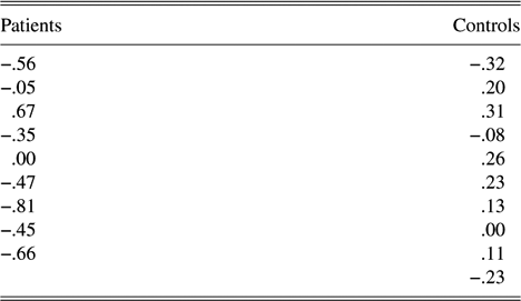
Previous language lateralization studies with English- and Spanish-speaking epilepsy patients adopted a cutoff laterality index (LI) value of .25, which was associated with the highest concordance with judgments of hemispheric dominance derived from the Wada Test (Maestu et al., Reference Maestu, Ortiz, Fernandez, Amo, Martin, Fernandez and Sola2002; Papanicolaou et al., Reference Papanicolaou, Simos, Breier, Zouridakis, Wilmore, Wheless, Constantinou, Gormley and Maggio1999, Reference Papanicolaou, Simos, Castillo, Breier, Sarkari, Pataraia, Billingsley, Buchanan, Wheless, Maggio and Maggio2004). Additionally, as pointed out by Binder et al. (Reference Binder, Bellgowan, Hammeke, Possing and Frost2005), “the capacity to detect a unilateral abnormality of hippocampal activation is greatly enhanced by computing an asymmetry index, which is feasible only when activation is bilateral.” Here, we apply this criterion, focusing on MTL activity and increasing the interval considered, in our case 200–800 ms. This modification was based on previous iEEG studies of verbal memory where relevant activity in MTL structures in very early latencies was observed (between 150 and 300 ms) (Fell et al., Reference Fell, Klaver, Elger and Fernandez2002, Reference Fell, Klaver, Elfadil, Schaller, Elger and Fernandez2003; Fernandez & Tendolkar, Reference Fernandez and Tendolkar2001; Fernandez et al., Reference Fernandez, Weyerts, Schrader-Bolsche, Tendolkar, Smid, Tempelmann, Hinrichs, Scheich, Elger, Mangun and Heinze1998). In the current study, the LI intends to reflect the relatively greater activation of one MTL as compared to the other. Accordingly, we calculated the LI by subtracting the number of activity sources localized in left MTL from the number of activity sources localized in the right MTL and dividing by the total number of observed sources [LI = (R −L)/(R + L)] (Ver Hoef et al., Reference Ver Hoef, Sawrie, Killen and Knowlton2008). According to this criterion, the proportions of patients who would be classified as right MTL dominant for memory were six (M = −0.55, SD = 0.16), two were bilateral (M = −0.02, SD = 0.03), and one was left MTL dominant (LI = .67). Considering controls, two were classified as left MTL dominant (M = 0.29, SD = 0.04), seven were bilateral (M = 0.05, SD = 0.16), and one was considered as right MTL dominant (LI = −.32). LI data for each participant are presented in Table 2.
Table 2. LIs of MTL activity for patients and controls
Note
LIs were calculated by subtracting the number of activity sources localized in left MTL from the number of activity sources localized in the right MTL and dividing by the total number of observed sources [LI = (R − L)/(R + L)].
In order to further explore the asymmetry of activation of MTL, we conducted a two-way ANOVA (Hemisphere × Latency) for each group. No significant effects were found in the control group. A main effect of hemisphere (F = 6.82, p < .05, η 2 = .46) and a Hemisphere × Latency (F = 2.63, p < .05, η 2 = .25) interaction were found in the patient group. Follow-up analyses indicated that right MTL (M = 19.44, SD = 11.10) was more activated than the left MTL (M = 7.66, SD = 3.16) (t[8] = 3.40; p < .05). Additionally, the right MTL (M = 4.44, SD = 5.07) was more activated than the left MTL (M = 0.22, SD = 0.44) between 600 and 700 ms after stimulus presentation (t[8] = 2.40; p < .05) (Figure 2). Inspection of brain activation profiles indicated that differences in the degree of activation between hemispheres were highly consistent across patients: Greater activation of right MTL was found in seven of nine patients.

Fig. 2. Spatiotemporal profiles of brain magnetic activity for the control group (upper panel) and the patient group (lower panel), as indicated by the mean number of activity sources found in MTL at successive 100-ms time windows (200–800 ms). Light-shaded vertical areas represent left MTL activity, and dark-shaded vertical areas represent right MTL activity.
Since previous studies have found differences in activation patterns between correct and incorrect trials (Fell et al., Reference Fell, Klaver, Elfadil, Schaller, Elger and Fernandez2003; Mormann et al., Reference Mormann, Fernandez, Klaver, Weber, Elger and Fell2007), it could be argued that the observed left MTL decreased activity in epilepsy patients could be due to a greater amount of incorrect trials in this group. Thus, with the aim of teasing apart the potential contribution of inclusion of greater number of incorrect trials in the patient group, patients were divided into two subgroups, according to level of performance. The threshold of group membership was the median value of performance. The high-performance group (n = 5; M = 68.99, SD = 5.75) showed a clear right LI (M = −0.47, SD = 0.27). Thus, we can conclude that the observed right MTL preponderance in patients is not due to a greater number of error trials included in the analyses.
DISCUSSION
In the current study, we investigated the contribution of MTL to encoding processes of verbal WM by comparing MEG profiles of a group of epilepsy patients with temporal lobe epilepsy related to HS with those of a group of healthy controls. Our findings corroborate our previous results with healthy controls (Campo et al., 2005b) and demonstrate behavioral verbal WM impairment and changes in the dynamics of MTL activity in epilepsy patients. Consistent with previous neuropsychological studies, we observed that patients with left MTL sclerosis demonstrated a reduced performance in verbal WM in comparison to healthy controls (Grippo et al., Reference Grippo, Pelosi, Mehta and Blumhardt1996; Owen et al., Reference Owen, Morris, Sahakian, Polkey and Robbins1996; Petrides & Milner, Reference Petrides and Milner1982; Wagner et al., Reference Wagner, Sziklas, Garver and Jones-Gotman2008).
Analysis of the spatiotemporal patterns of MTL activity showed that the dynamics of activation differed between patients and controls. MTL activation became evident at 200 ms in both groups and was sustained for the next 600–700 ms. Consistent with our predictions, controls showed greater number of activity sources in left MTL than patients. Differences between groups were significant for the whole time interval (200–800 ms). However, no differences were found in right MTL activity between groups (Figure 1). The absence of differences was explained by a high activation of right MTL structures in both groups. This finding suggests a bilateral recruitment of MTL structures in controls and a “normal” activation of the nonlesioned MTL in patients. Previous neuroimaging studies have also reported activation in right MTL during episodic verbal memory in healthy controls, that has been related to the use of elaborated (i.e., semantic) strategies (Davachi & Wagner, Reference Davachi and Wagner2002; Lepage et al., Reference Lepage, Habib, Cormier, Houle and McIntosh2000), as is the case in the current study. LIs and within-group analyses corroborated these data, with most patients showing right lateralization of MTL activity and most controls showing LIs indicative of bilateral activation (Table 2). When the temporal profiles of the activity on MTL were examined (Figure 2), a left preponderance of MTL activation in controls and a right preponderance of activation in patients became apparent from 400 ms after the stimulus presentation. These latencies have been shown to be crucially related to the formation of memories in previous electrophysiological studies (Castillo et al., Reference Castillo, Simos, Davis, Breier, Fitzgerald and Papanicolaou2003; Fell et al., Reference Fell, Klaver, Elger and Fernandez2002, Reference Fell, Klaver, Elfadil, Schaller, Elger and Fernandez2003; Fernandez et al., Reference Fernandez, Klaver, Fell, Grunwald and Elger2002). However, we only found time-modulated significant differences in MTL activation between groups in the 600- to 700-ms interval (Figure 1).
Specifically, controls exhibited greater activation than patients in left MTL, while patients showed an opposite pattern, greater right MTL activation than controls. Decreases in MTL activity ipsilateral to lesion and greater activation of the contralateral MTL have been previously reported in epilepsy patients with HS for episodic memory, either for verbal or for nonverbal material, with functional magnetic resonance imaging (Dupont et al., Reference Dupont, Van de Moortele, Samson, Hasboun, Poline, Adam, Lehericy, Le Bihan, Samson and Baulac2000; Golby et al., Reference Golby, Poldrack, Illes, Chen, Desmond and Gabrieli2002; Powell et al., Reference Powell, Richardson, Symms, Boulby, Thompson, Duncan and Koepp2007; Richardson et al., Reference Richardson, Strange, Duncan and Dolan2003) and iEEG/MEG (Mormann et al., Reference Mormann, Fernandez, Klaver, Weber, Elger and Fell2007; Ver Hoef et al., Reference Ver Hoef, Sawrie, Killen and Knowlton2008). This finding has been interpreted as a reorganization of memory function, although not sufficient to enable normal memory performance (Dupont et al., Reference Dupont, Van de Moortele, Samson, Hasboun, Poline, Adam, Lehericy, Le Bihan, Samson and Baulac2000; Powell et al., Reference Powell, Richardson, Symms, Boulby, Thompson, Duncan and Koepp2007). The double dissociation we observe between 600 and 700 ms after stimulus presentation is consistent with this explanation. As stated by Ver Hoef et al. (Reference Ver Hoef, Sawrie, Killen and Knowlton2008), the identical temporal character of left and right MTL activity supports its similar function. However, the fact that patients exhibited a decreased activity in the lesioned MTL and a similar activation in the nonlesioned MTL to that of controls in the interval between 200 and 800 ms, along with a worse performance, could be indicating that patients with left HS, rather than showing a reallocation, are using a residual component of a damaged memory structure subserving verbal WM. Further studies should be conducted to explore this alternative explanation. Nonetheless, current results suggest that MTL activation is contributing to verbal WM and that this activation is not signaling a parallel process not specifically related to this cognitive function (Wagner et al., Reference Wagner, Sziklas, Garver and Jones-Gotman2008).
Three concerns should be considered related to current results. First, as previous studies have found differences in activation patterns between correct and incorrect trials (Fell et al., Reference Fell, Klaver, Elfadil, Schaller, Elger and Fernandez2003; Mormann et al., Reference Mormann, Fernandez, Klaver, Weber, Elger and Fell2007), it could be argued that the decrease of activation on the pathological MTL could be due to a greater amount of incorrect trials made by epilepsy patients. To address this possible caveat, we demonstrate that a subgroup of patients with better performance (i.e., greater amount of correct trials) showed a clear right MTL preponderance of activation (Mormann et al., Reference Mormann, Fernandez, Klaver, Weber, Elger and Fell2007). Thus, the significant decrease in left MTL activity is due to underlying pathology. Second, MTL findings could be reflecting retrieval from semantic memory rather than specific WM activity. However, several lines of evidence suggest that while patients with damage to hippocampal structures sometimes exhibit impairments in semantic abilities, it appears that this deficit is less apparent in epilepsy patients than in patients with MTL damage resulting from other etiologies (Giovagnoli et al., Reference Giovagnoli, Erbetta, Villani and Avanzini2005; Ribbler & Rausch, Reference Ribbler and Rausch1990). Additionally, neuroimaging and neuropsychological studies suggest that semantic processes rely more on lateral aspects of the temporal lobe (Levy et al., Reference Levy, Bayley and Squire2004). Therefore, we suggest that MTL activation observed in this study is not signaling a semantic retrieval process but an encoding mechanism. Finally, some authors have claimed that MTL is recruited in WM only during processing of novel or complex stimuli (Ranganath & Blumenfeld, Reference Ranganath and Blumenfeld2005). This notion has led to an overwhelming number of studies investigating the role of MTL in WM using novel/complex visual stimuli (Haxby et al., Reference Haxby, Ungerleider, Horwitz, Rapoport and Grady1995; Piekema et al., Reference Piekema, Kessels, Mars, Petersson and Fernandez2006; Ranganath & D’Esposito, Reference Ranganath and D’Esposito2001). However, neuropsychological studies (Owen et al., Reference Owen, Morris, Sahakian, Polkey and Robbins1996; Petrides & Milner, Reference Petrides and Milner1982; Wagner et al., Reference Wagner, Sziklas, Garver and Jones-Gotman2008) and neuroimaging experiments of WM in young controls (Cabeza et al., Reference Cabeza, Dolcos, Graham and Nyberg2002; Campo et al., Reference Campo, Maestu, Capilla, Fernandez, Fernandez and Ortiz2005a, Reference Campo, Maestu, Ortiz, Capilla, Fernandez and Fernandez2005b; Karlsgodt et al., Reference Karlsgodt, Shirinyan, van Erp, Cohen and Cannon2005; Petersson et al., Reference Petersson, Gisselgard, Gretzer and Ingvar2006), older controls (Cabeza et al., Reference Cabeza, Daselaar, Dolcos, Prince, Budde and Nyberg2004), and patient groups (Grippo et al., Reference Grippo, Pelosi, Mehta and Blumhardt1996) demonstrate an additional involvement of MTL in WM for familiar verbal stimuli. The present study provides neurophysiological support for these findings, and highlights the potential application of MEG in exploring material-specific memory effects for WM in epilepsy patients (Wagner et al., Reference Wagner, Sziklas, Garver and Jones-Gotman2008), similar to that reported for episodic memory (Golby et al., Reference Golby, Poldrack, Illes, Chen, Desmond and Gabrieli2002; Jones-Gotman et al., Reference Jones-Gotman, Zatorre, Olivier, Andermann, Cendes, Staunton, McMackin, Siegel and Wieser1997; Powell et al., Reference Powell, Richardson, Symms, Boulby, Thompson, Duncan and Koepp2007).
Although it was not the main focus of our study, our results suggest the potential clinical utility of the current paradigm for the presurgical evaluation protocol assessing the suitability of epilepsy patients for surgery. However, larger samples should be used before this can be considered, and taking into account the material-specific lateralization of memory processing (Golby et al., Reference Golby, Poldrack, Illes, Chen, Desmond and Gabrieli2002; Powell et al., Reference Powell, Koepp, Richardson, Symms, Thompson and Duncan2004; Wagner et al., Reference Wagner, Sziklas, Garver and Jones-Gotman2008), nonverbal tasks should also be included.
In summary, the present study provides, for the first time, direct evidence for the alteration of the dynamics of MTL activity in epilepsy patients suffering from left HS in a verbal WM task using MEG. The observed changes in the timing and laterality of MTL activation, as well as a reduced behavioral performance, in patients with a restricted left MTL damage strongly support the crucial contribution of these structures to verbal WM encoding. Along with previous neuropsychological and neuroimaging studies, current results provide compelling evidence for the proposal that MTL contributes to both episodic memory and WM encoding.
ACKNOWLEDGMENTS
The manuscript is new and original, is not currently under review by any other publication, and has not been previously published either electronically or in print. The project did not receive any financial support, and there are no conflicts of interest affecting this manuscript.


