


INTRODUCTION
The use of primary, neoadjuvant, adjuvant and concurrent chemoradiotherapy has been proven to be efficacious in treating cancer.Reference Seiwert, Salama and Vokes1 Several mechanisms of action are responsible for the advantageous combination of chemotherapy and radiation therapy. These mechanisms are rooted in disrupting the cell cycle, and both chemotherapy and radiotherapy act on similar cytotoxic response pathways. In 1979, a theoretical framework was introduced by Steel and Peckham that described the chemoradiation paradigm.Reference Seiwert, Salama and Vokes1 The term ‘spatial cooperation’ was used to describe systemic and local disease control when chemotherapy and radiation are used, respectively.Reference Bentzen, Harari and Bernier2 Combined therapies could potentially improve overall disease-free survival in patients who have both locoregional and micrometastatic cancers.Reference Bentzen, Harari and Bernier2 In addition, local control can be reached if both modalities can target the tumour through its respective mechanisms of action while minimally impacting normal tissue.Reference Steel and Peckham3 The enhancement effect where one agent increases the effects of the other agent is referred to as additive or supradditive.Reference Bentzen, Harari and Bernier2, Reference Steel and Peckham3 An increasing number of treatments in radiation oncology are applying these frameworks to achieve better outcomes. The treatment of brain, rectum, cervical, breast, and head and neck cancer are examples of disease sites where radiotherapy is delivered in a timely sequence with chemotherapy. Understanding the underlying mechanisms and rationale for chemoradiotherapy is essential in optimising treatment efficacy.
Treatment efficacy is impacted by several biological and treatment factors. Biological factors include inefficient tumour vasculature and hypoxia that ensues within the tumour microenvironment. Oxygen deficiency increases radiation resistance and inefficient tumour vessels limit the delivery of chemotherapeutic agents into tumours.Reference Tran, El Kaffas, Al-Mahrouki, Gillies and Czarnota4 Treatment factors involve patient compliance to treatment and the timely administration of chemoradiation regimens. The purpose of this review is to examine the theoretical frameworks behind the chemoradiation paradigm and to describe current chemoradiation practices in radiation oncology. A review of the mechanisms of these cytotoxic agents is presented with respect to hypoxic microenvironments, pharmacokinetic frameworks and the potential for causing serious normal tissue side effects when escalating dose.
The chemoradiation paradigm mechanisms of chemoradiation action
Several rationales exist for combining chemotherapy and radiation therapy, with the first being the preservation of organ function and improved cosmesis when compared with surgery. Additional benefits of combinatory treatments include chemotherapy-driven cell cycle disruption and radiosensitization by reducing the hypoxic microenvironments within the tumour, and the capability of targeting both local and systemic disease when administered concurrently.Reference Seiwert, Salama and Vokes1 These rationales stem from the conceptual framework outlined by Steel and Peckham,Reference Steel and Peckham3 which analysed chemotherapy and radiation interactions. Within this context, a major benefit to combinatory treatments is the advantageous targeting of tumour cells in different phases of the cell cycle.Reference Nishimura5 Cells in the G2/M phase are more radiosensitive than cells in the radioresistant S phase.Reference Nishimura5 Therefore, treatment strategies have used chemotherapy to interrupt mitotic programming by arresting cells in the more radiosensitive G2/M phasesReference Nishimura5 (Figure 1).

Figure 1 Combination treatments and the cell cycle. Combinatory treatments capitalise on tumour cells in different phases of the cell cycle. Cells in the G2/M phase are more radiosensitive than cells in the radioresistant S phase. Therefore, treatment strategies have used chemotherapy to interrupt mitotic programming by arresting cells in the more radiosensitive G2/M phases.
The main clinical objective for combining chemotherapy and radiation therapy is to increase overall survival and quality of life in patients while minimising or reducing potentially life-threatening side effects.Reference Steel and Peckham3 In order to achieve this, combined treatment regimens need to be tailored according to the therapeutic ratio. The therapeutic ratio is commonly described as two sigmoid-shaped dose curves that define both the lethal dose and therapeutic dose. It can be described as the dose ratio that results in tumour and normal tissue damage.Reference Nishimura5 The amount of tumour control that can be expected from radiation administration depends on the dose tolerance of normal tissues.Reference Steel and Peckham3 The therapeutic ratio is an important consideration when choosing the appropriate anti-cancer treatments, where the intention to increase tumour control is influenced by the potential side effects caused by normal tissue damage. These side effects can be dose limiting, especially when critical anatomical structures are involved.
Additive effects from vascular normalisation
Tumour vasculature plays an important role in radiation sensitivity, drug delivery and tumour survival.Reference Tran, El Kaffas, Al-Mahrouki, Gillies and Czarnota4 Tumour blood vessels are immature, and structurally disorganised throughout the tumour.Reference Tran, El Kaffas, Al-Mahrouki, Gillies and Czarnota4 Tumour vasculature has weak endothelial cell connections, abnormal basement membranes and exhibits large variances in diameter and length.Reference Tran, El Kaffas, Al-Mahrouki, Gillies and Czarnota4, Reference Tozer, Kanthou and Baguley6, Reference Shannon, Bouchier-Hayes, Condron and Toomey7 The abnormal scaffolding of vessels creates hypoxic regions from insufficient blood flow into the tumour stroma.Reference Wachsberger, Burd and Dicker8 Hypoxia, as a result of inefficient tumour vasculature, plays a principal role in the biological effectiveness of radiation therapy. During radiation exposure to the cell, fast charged particles are produced.Reference Hall and Giaccia9 These particles are responsible for the production of ion pairs, which then lead to the production of reactive oxygen species, otherwise known as free radicals.Reference Hall and Giaccia9 Free radicals are known for their lethality to cellular DNA owing to the production of organic peroxide from molecular oxygen, which has been known to cause irreparable DNA damage.Reference Hall and Giaccia9 Molecular oxygen is a powerful radiosensitiser.Reference Rockwell, Dobrucki, Kim, Marrison and Vu10 The method of radiosensitization is the result of oxygen’s high electron affinity and its involvement in cascading reactions that lead to DNA damage.Reference Rockwell, Dobrucki, Kim, Marrison and Vu10
Similar to radiotherapy, chemotherapeutic treatment success is partly dependent on perfusion and oxygen saturation. This is commonly a net result of vascular disorganisation within tumours.Reference Harrison and Blackwell11 The unstable vascular network ultimately results in poor perfusion and thus, delivery of chemotherapy.Reference Harrison and Blackwell11 Harrison and BlackwellReference Harrison and Blackwell11 noted that hypoxic conditions cause cells to cycle slower than normal and get ‘stuck’ in the radioresistant S phase of the cell cycle. This hinders the efficacy of certain chemotherapeutic agents, such as alkylating agents and antimetabolites, whose main targets are cells in S phase.Reference Harrison and Blackwell11 In recent years, newly discovered classes of drugs known as vascular targeting agents have gained notoriety in their ability to normalise the erratic vascular architecture within tumours. Anti-angiogenic agents are responsible for targeting new tumour vessel growth, a process termed angiogenesis, while anti-vascular agents eradicate pre-existing tumour vessels.Reference Tozer, Kanthou and Baguley6, Reference Wachsberger, Burd and Dicker8, Reference Tozer and Bicknell12 Inefficient vessels in the network are eliminated, therefore improving blood flow, drug delivery and oxygen levels.Reference Wachsberger, Burd and Dicker8, Reference Tozer and Bicknell12 The increase in oxygen availability within the tumour microenvironment has been described to enhance sensitization to both radiationReference Wachsberger, Burd and Dicker8 and chemotherapy.Reference Shannon, Bouchier-Hayes, Condron and Toomey7
Additive effects from cell cycle disruption
Chemotherapeutic agents act specifically at different points in the cell cycle. Each agent has a unique mechanism of action that is targeted to various cellular components, ultimately resulting in cell death. The exact mechanism of action of the alkylating agents, antimetabolites and taxanes are described in the following section (Table 1).
Table 1 Summary of cytotoxic agents and their mechanisms of action
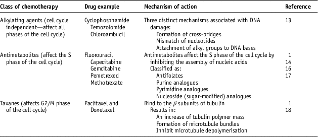
Alkylating agents modulate DNA, by way of cross-linking and DNA strand breaks. These effects lead to inhibition of cell division, abnormal base pairing and ultimately cell death.Reference Ralhan and Kaur13 These agents typically affect cells in all phases of the cell cycle, and are beneficial in the treatment of slow growing cancers such as leukaemia.Reference Ralhan and Kaur13 There are three distinct mechanisms associated with DNA damage: (1) formation of cross-bridges, which prevents DNA strands from being separated for synthesis or transcription,Reference Ralhan and Kaur13 (2) mismatch of nucleotides, thus leading to mutations,Reference Ralhan and Kaur13 and (3) attachment of alkyl groups to DNA bases. This activates DNA repair enzymes, which attempt to replace the alkylated bases, therefore causing the DNA to become fragmented.Reference Ralhan and Kaur13
Antimetabolites typically affect the S phase of the cell cycle by inhibiting the assembly of nucleic acids.Reference Scagliotti and Selvaggi14 Antimetabolites can be classified as: (1) antifolates, (2) purine analogues, (3) pyrimidine analogues and (4) nucleoside (sugar-modified) analogues.Reference Scagliotti and Selvaggi14 Antifolates such as Methotrexate interfere with cell activity by targeting folate-dependent enzymes.Reference Scagliotti and Selvaggi14 Pyrimidine analogues, such as Gemcitabine, are known to prevent DNA synthesis and repair by exhausting deoxynucleoside triphosphates. These are essential for maintaining DNA polymerase and ribonucleotide reductase.Reference Seiwert, Salama and Vokes1 Gemcitabine has been shown to induce radiosensitization in cells when administered 24 hours before radiation therapy, with lasting effects for up to 48 hours.Reference Seiwert, Salama and Vokes1 Nucleoside analogues such as 5-fluorouracil (5-FU) prevent nucleic acid production.Reference Scagliotti and Selvaggi14 This mechanism of action relies on cleavage into the DNA and RNA, resulting in disrupted DNA synthesis and transcription, thus affecting protein synthesis.Reference Scagliotti and Selvaggi14 5-FU is used widely with radiation in the treatment of rectal and stomach cancer, and mainly affects cells in the radioresistant S phase of the cell cycle.Reference Gez, Sulkes and Yablonsky-Peretz15 Typically, 5-FU is administered continuously owing to its short half-life in plasma, thus not necessitating radiotherapy treatment time frames for optimal results.Reference Seiwert, Salama and Vokes1, Reference Kvols16, Reference Lawerence, Blackstock and McGinn17 The suggested administration method for 5-FU is through intravenous administration.Reference Kvols16, Reference Lawerence, Blackstock and McGinn17 However, issues with infection and long-term venous access leading to thrombosis can complicate the course of treatment for patients, as well as the need for specialised medical equipment (i.e., pumps), which can be costly.Reference Lawerence, Blackstock and McGinn17 To counteract these issues, 5-FU is also available in pill form and is known as Capecitabine.Reference Lawerence, Blackstock and McGinn17 Hydroxyurea, another example of an antimetabolite, is a known radiosensitiser and is commonly used in the treatment of head and neck cancer.Reference Seiwert, Salama and Vokes1 It has been shown to also affect cells at the G1/S checkpoint in the cell cycle.Reference Seiwert, Salama and Vokes1
Taxanes are microtubule-stabilising agents that are widely used for the treatment of metastatic breast and head and neck cancers.Reference Frame18 Paclitaxel and Doxetaxel are common taxanes that are similar in function where both agents bind to the β subunits of tubulin, resulting in an increase of tubulin polymer mass, formation of microtubule bundles and inhibit microtubule depolymerisation.Reference Seiwert, Salama and Vokes1, Reference Frame18 As a consequence of this interaction, the cell cycle comes to a halt at the G2/M phase that leads to cell death and enhances radiation lethality.Reference Seiwert, Salama and Vokes1, Reference Frame18
Clinical applications of chemoradiation
Chemoradiation has been used in various tumour sites as standard therapy. These tumour sites include: brain, rectum, cervix, breast, and head and neck.
For brain lesions, such as glioblastoma, Temozolomide (TMZ) is typically administered daily at 75 mg/m2 of body surface area, 7 days a week for the entire course of radiation therapy.Reference Stupp, Mason and van den Bent19 Following this, six additional cycles are given at 150–200 mg/m2 for 5 days, every 28 days.Reference Stupp, Mason and van den Bent19 It has been noted that the optimal concentration of TMZ in a patients’ plasma is approximately within 1 hour of consumption, and is eliminated within ~1·8 hoursReference Brada, Judson and Beale20 (Figure 2). Pharmacokinetic drug effects are most optimal when patients take TMZ continuously and ~1 hour before radiation treatments.Reference Stupp, Mason and van den Bent19–Reference Portnow, Badie, Chen, Liu, Blanchard and Synold21 The continuous dose administration of TMZ enables an increase in the dose intensity by almost two-fold, without increasing toxicity to the patient, and enables the reduction of the amount of a particular enzyme that is responsible for the repair of DNA damage caused by alkylating agents such as TMZ.Reference Stupp, Mason and van den Bent19
Figure 2 Concentration of Temozolomide (TMZ) versus time for the treatment of glioblastoma multiforme (GBM). TMZ is used in the adjuvant treatment of GMB. The per cent optimal concentration of TMZ in a patients’ plasma is approximately within 1 hour of consumption, and is eliminated within ~1·8 hours.Reference Brada, Judson and Beale20
It has been shown that Capcetabine in combination with radiation therapy is effective in the treatment of advanced rectal cancer.Reference Yu, Kim and Kim22 It is typically absorbed by the body in the gastrointestinal tract and converted to 5-FU by a particular enzyme (thymidine or uridine phosphorylase).Reference Yu, Kim and Kim22 Typically, the time frame for administration of Capcetabine is orally 1 hour before radiation therapy treatments in order to maximise the radiosensitization of cells, therefore increasing the effectiveness of radiation therapy treatments.Reference Yu, Kim and Kim22
Other disease sites do not necessitate timing regimens in order to achieve optimal radiotherapy results, as drug concentrations are steadily maintained by continuous infusion. Cervical cancer has been shown to benefit from chemoradiation, and progression free survival is increased when concomitant treatments are delivered.Reference Rose, Bundy and Watkins23 A study by Rose et al.Reference Rose, Bundy and Watkins23 showed that concomitant radiation and cisplatin, fluorouracil, and hydroxyurea were beneficial in the treatment of locally advanced cervical cancer.
For head and neck cancers, Calais et al.Reference Calais, Alfonsi and Bardet24 showed that overall and disease-free survival increased for patients who received chemotherapy and radiation therapy as opposed to the cohort of patients who received radiation therapy alone for squamous cell carcinoma of the oropharynx. In a similar study conducted by Brizel et al.,Reference Brizel, Albers and Fisher25 it was also shown that the combination of chemotherapy and hyperfractionated radiation therapy was proven to be more beneficial versus the administration of radiation alone. The common aetiology of cervical and head and neck cancers involves the expression of human papilloma virus (HPV), suggesting that HPV-driven cancers may have a potential biological susceptibility to chemoradiation. However, there is limited data describing the biological interactions.
Emerging data is demonstrating novel treatment paradigms for disease sites that have been traditionally treated by mono-modalities. Lee et al.Reference Lee, Brackstone, Gandhi, Arce and Dinniwell26 demonstrated local disease control for triple-negative breast cancer patients undergoing salvage treatment for resistant disease. Cisplatin was delivered weekly, with a median dose of 30 mg/m2 concurrent with external beam radiotherapy (total dose 65 Gy).Reference Lee, Brackstone, Gandhi, Arce and Dinniwell26 A large percentage of patients in this observational study demonstrated complete clinical response.Reference Lee, Brackstone, Gandhi, Arce and Dinniwell26
Although the timing of chemotherapy and radiotherapy is not necessitated, or explicitly stated/determined in the cases of cervical, breast, and head and neck cancer, it is clear that the administration of both treatment modalities offers improved survival and disease-free progression because of the enhancement of normal tissue effects that can be observed with simultaneous drug and radiation therapy administration.Reference Fu27
CONCLUSION
Understanding the mechanistic activity of chemotherapy and radiation therapy, and the effects these treatments have on the cell cycle is crucial to finding a balance between increasing dose to patients while minding normal tissue side effects. The administration of chemotherapy in conjunction with radiation therapy is dependent on treatment timing and type of agents. There is great opportunity to explore the clinical application of combining radiation and chemotherapy, as novel therapies are emerging within the treatment landscape.
Acknowledgements
The authors would like to acknowledge the kind support from the department of Radiation Therapy, Simcoe Muskoka Regional Cancer Program, and in particular to Ms. Jennifer Montgomery.
Financial Support
This research received no specific grant from any funding agency, commercial or not-for-profit sectors.
Conflicts of Interest
None.


