


INTRODUCTION
The success of curative prostate radiotherapy depends on accurate delivery of high radiation doses to a defined tissue volume. Intensity-modulated radiotherapy (IMRT) can deliver increased dose to the tumour without increased toxicity but requires smaller delineated treatment margins. Hence, an accurate knowledge of the position of the prostate is mandatory.Reference Kupelian, Elshaikh, Reddy, Zippe and Klein1–Reference Cheung, Sixel, Morton, Loblaw, Tirona, Pang, Choo, Szumacher, Deboer and Pignol3 Departmental data from 100 patients treated with IMRT in 2004 observed a 14% Grade 2 and 2% Grade 3 bowel toxicity at 5 years.
The use of implanted marker seeds has proven to be more accurate than alignment of bony anatomical structures,Reference Nederveen, Dehnad, van der Heide, van Moorselaar, Hofman and Lagendijk4–Reference Van den Heuvel, Fugazzi, Seppi and Forman5 and by using online protocols where images are acquired, analysed and corrections made prior to treatment, greater accuracy can be achieved on a daily basis.Reference Chen, Lee, Handrahan and Sause6 Until recently, only off-line decision protocols have commonly been used, analysing images after a fraction has been delivered but prior to the next fraction. However, with the advent of image-guided radiotherapy (IGRT) capable linear accelerators, online correction strategies are now a possibility.
Planar kilovoltage (kV) imaging techniques produce images with improved contrast definition using smaller radiation doses than conventional megavoltage (MV) portal images. The Varian On-Board Imager (OBI) allows for daily imaging with automatic positional corrections prior to each treatment, directly from the treatment console. This therefore gives the possibility of daily imaging and online corrections within a shorter time frame during the routine working day. For this technique to be implemented, our department developed an online imaging schedule for a prostate tracking technique. The prostate can also move intrafractionally,Reference Ghilezan, Jaffray, Siewerdsen, Herk, Shetty, Sharpe, Zafar Jafri, Vicini, Matter, Brabbins and Martinez7 so we wanted to know the amount of intrafraction movement when deciding the action levels to use for the online correction schedule and to advise our clinicians which Planning Target Volume (PTV) margin to use.
For this analysis, we used data from a randomised study which was carried out to compare the accuracy of online compared with off-line correction schedules using implanted markers. These data were used to assist in the production of an online imaging schedule.
MATERIALS AND METHODS
Data from 60 patients were used. This included 1,636 image-guided session information.
Patient preparation
Three or four small (5 mm × 1 mm) gold markers were implanted using transrectal ultrasound guidance and antibiotic cover approximately 2 weeks prior to the Computed Tomography (CT) planning scan, so that any initial swelling of the prostate gland caused by marker insertion had subsided and the marker seeds were fixed in position. Before CT scanning and daily treatments, patients followed a protocol for rectal and bladder preparation, using a microenema (RelaxitTM) to empty the rectum and drinking 500 ml of water 20 minutes prior to CT/treatment. During the planning scan and treatment, patients were positioned supine on a flat couch top using in-house indexed head, knee and feet supports.
Patient treatment
Patients were treated daily following either the National Cancer Research Institute conventional or hypofractionated high-dose intensity-modulated-radiotherapy for prostate cancer (CHHIP) protocol, using either 74Gy/37#, 60Gy/20# or 57Gy/19# dose/fractionation schedules or an in-house IMRT schedule using 72Gy/32#. Treatment plans were produced using the Pinnacle treatment planning system using 10 MV photon beams and either five IMRT fields or three field brick with three concurrent boost fields for treatment delivery.
Patient positioning and online position correction
Patients were aligned for treatment using the standard laser alignment technique and a table-isocentre height, superior/inferior and lateral shifts according to the individual patient’s treatment plan. A correction was made to the table-isocentre height position to account for couch deflection prior to first treatment on an individual patient basis. The position of the marker seeds on the digitally reconstructed radiographers (DRRs) obtained during treatment planning was used as the reference for the positional verification. Daily kV planar images, using a 5 cm × 5 cm field size, were acquired from the anterior and lateral directions prior to treatment using the OBI equipment. To determine the position of the prostate, the seed reference template was overlaid on the acquired kV image and manually aligned. If a set-up correction greater than or equal to 2 mm was observed in any direction (right-left RL, superoinferior SI or anteroposterior AP), all corrections were made prior to treatment. The couch was moved remotely into position from outside the treatment room. Treatment field images were also acquired which were subsequently analysed off-line to assess any intrafraction motion.
The 2 mm action level rounds up a combination of a 0.5 mm interobserver variation (shown in an unpublished study from our institution) using marker seeds and a 1 mm measured accuracy limit of the treatment couch.
Weekly cone beam CT images (CBCT) were acquired to verify that the markers had not been displaced since the planning stage. Other studies have shown that there is no significant marker migration within the prostate.Reference Pouliot, Aubin, Langen, Liu, Pickett, Shinohara and Roach8–Reference Kupelian, Willoughby, Meeks, Forbes, Wagner, Maach and Langen9 This is in agreement with our previous departmental experience of using prostate marker seeds. However, our previous data were for 45 patients, and we felt that marker migration needed to be checked on an increased number of patients to give increased confidence in our department.
Calculation of positional errors and margins
The image analysis results were used to calculate the individual and population systematic (Σ), and random (σ) errors (1 standard deviation) as described by van Herk and othersReference van Herk, Remeijer, Rasch and Lebesque10–Reference de Boer and Heijmen12 for a number of imaging strategies. For each imaging correction strategy, the appropriate PTV margins were calculated from the systematic and random errors using the van HerkReference van Herk, Remeijer, Rasch and Lebesque10 formula 2.5 Σ + 0.7 σ, which optimises the margin needed to deliver a 95% dose (D95) to the clinical target volume (V95) for 90% of the patients.
Imaging schedules
The imaging schedules included no corrections, two different no-action level (NAL) imaging approaches and daily imaging with online corrections. These schedules are summarised in Table 1.
Table 1. Summary of imaging schedules

RL, right-left direction; SI, superoinferior direction; AP, anteroposterior direction.
Effect of intrafraction motion
The images acquired prior to a treatment fraction were compared with those acquired at the end of the fraction in order to measure the intrafraction motion. Either anterior and lateral or posterior and lateral end of fraction images were acquired to establish the three-dimensional prostate position. This was carried out on an individual patient basis and the range, mean and standard deviation of values in each direction RL, SI and AP calculated. Each imaging schedule was rechecked to calculate their residual errors taking into account the intrafraction motion.
Resource implications
Daily in-room timings were made for 10 patients to quantify the resource implications of using this online technique, this gave a total of 280 individual timings. The timing began when the patient entered the linac room and stopped as they exited. Therefore, the timing was representative of the image acquisition, analysis and treatment delivery processes. This was compared to the 12-minute in-room time allocated to radical prostate patients in the department.
Statistical analysis
Data were analysed using Statistical Package for the Social Sciences (SPSS) statistical analysis software using paired Wilcoxon signed rank, Kolmogorov-Smirnov test of normality and F- tests.
RESULTS
Positioning accuracy
The maximum prostate displacements observed in each direction using the pretreatment imaging data as if no correction schedule had been applied were 11 mm, 12 mm and 11 mm in the RL, SI and AP directions, respectively. The percentage of displacements greater than standard 3 mm and 5 mm action levels was greatest in the AP direction with 48% > 3 mm and 26% > 5 mm. These data are summarised in Figure 1.

Figure 1. % shifts above particular levels. Abbreviations: RL = right-left direction, SI = superoinferior direction, AP = anteroposterior direction. +ve = right, inferior, posterior, −ve = left, superior, anterior isocentre correction.
Figure 2 describes the positional accuracy results of the four imaging schedules analysed. Mean, systematic and random errors were calculated for each direction. Making no corrections, the systematic errors Σ and the random errors σ in the various directions ranged from 2.4 to 3.3 mm and from 1.8 to 2.5 mm, respectively. The overall mean error M in the AP direction of −2.2 mm is larger than the calculated values in the RL and SI directions (0.7 mm and −0.01 mm).

Figure 2. (a) Mean errors for each imaging schedule. (b) Systematic and random errors for each imaging schedule. Abbreviations: M = overall population mean, ∑ = systematic error, σ = random error, RL = right-left direction, SI = superoinferior direction, AP = anteroposterior direction, intra = intrafraction motion.
The pretreatment image analysis results were used to simulate the effect of the NAL imaging schedules in order to calculate their systematic and random errors. In comparison with the no-correction schedule, the off-line correction schedules NAL3 and NAL5 reduce the systematic errors to approximately 1 mm in the LR and SI directions, 1.5 mm in the AP direction, but the random errors remain approximately the same, 2 mm in the LR and SI directions, 3 mm in the AP direction.
If daily imaging is performed together with online corrections prior to treatment, using the image-guided technique, both the systematic and random errors are significantly reduced to negligible values with a range of 0.04–0.2 mm and 0.09–0.4 mm, respectively. The overall mean deviation is reduced to negligible values less than 0.5 mm. Figure 2 shows the improvement in the systematic and random errors, in all directions, that can be achieved when this imaging schedule is used.
Intrafraction motion
Positional differences between the prefraction image analysis results and the last two treatment field image analysis results were measured. The prostate location observed on the last two treatment field images was used to represent the post-treatment fraction position. The intrafraction motion observed had mean values of -0.1, 0.2 and 0.6 mm with standard deviation of 0.7, 3.5 and 1.9 mm in the RL, SI and AP directions, respectively (Table 3). The maximum intrafraction motion observed was 20 mm in the AP direction, indicating a posterior displacement of the prostate. However, a similar displacement of 11 mm was indicated in the opposite direction, indicating an anterior prostate displacement. The standard deviation is higher in the SI direction indicating a larger spread. The intrafraction motion was less than 3 mm for 55% of patients in the AP direction, 70% in the SI direction and 76% in the LR direction.
We tested the distribution of errors calculated on data obtained at the start of each treatment fraction compared to the distribution of errors at the end of the fraction. For the IGRT schedule, set-up corrections were made prior to treatment delivery if the observed correction was 2 mm or above in any direction, and the distribution of errors at the end of the fractions was significantly wider than the distribution of the residual errors prior to each fraction, p-values are displayed in Table 2. A Kolmogorov-Smirnov test of normality of the distributions of the intrafraction motion are non-rejectable in each direction except for the lateral displacement. However, the displacements in the lateral direction are significantly lower than the other directions.
Table 2. P value significance of intrafraction change
Individualised imaging schedules
The four imaging schedules were compared using a paired Wilcoxon signed rank test to assess the advantages of each. This test showed that the IGRT schedule gave a very significant improvement (p = 0.000) in the systematic and random errors and that the NAL5 schedule did not show a significant advantage when compared to the NAL3 schedule.
The variance of the mean systematic prostate position during the first week of treatment to the last week of treatment was also compared to quantify any time trends that might exist. The results showed that there was significantly greater variation in the AP position late in the treatment compared to early (p = 0). In the SI direction, the variation was greater early in the treatment compared to late. This again was statistically significant with a p-value of 0.009. In the LR direction, there was no significant difference (p = 0.218).
Resource requirements
In-room timings on the first 10 patients in this group ranged from 10.12 to 22.15 minutes. The mean time was 14.36 minutes (SD 1.95).
The in-room timing for CBCT acquisition and treatment has been measured in a separate in-house study to be 13.5 minutes. Analysis of the CBCT images and set-up corrections will add a further 2 minutes and consequently may increase the amount of intrafraction motion. This is another contraindication for the use of routine daily CBCT imaging in comparison with the planar kV imaging method described in this article.
A further contraindication to the use of CBCT for prostate tracking is that the dose implication from daily planar kV imaging is minimal, especially compared to daily CBCT. For a pair of kV images, the dose is approximately 0.1 mSv, while a CBCT pelvis scan is approximately 4 mSv. The daily planar kV imaging technique leads to a small radiation dose of 3.7 mSv for a 37# course.
DISCUSSION
Positioning accuracy
In this study, similar maximum prostate displacements of approximately 11 mm were found in all three directions. Other groups have found maximum displacements in the AP direction,Reference Chung, Haycocks, Brown, Cambridge, Kelly, Alasti, Jaffray and Catton13–Reference van der Heide, Kotte, Dehnad, Hofman, Lagenijk and van Vulpen16 our results potentially differ due to the correction of table-isocentre position made prior to the first treatment to account for individual couch deflection. As the couch deflection has been accounted for, our pretreatment image assessment results will represent ‘real’ prostate movement. Van der Vight’s group showed that when this deflection is accounted for, the pretreatment mean AP deviation was reduced from −4.1 to −2.1 mm,Reference van der Vight, van Lin, Post, Visser and Louwe15 concurring with the mean value of -2.17 mm found in our study. Another factor may be the use of bladder and rectal preparation. All of the patients in our study used a rectal enema and drank a fixed amount of water throughout the planning and treatment process, whereas none of the other studies used any rectal preparation in enema form. McNair et al. asked the patients to empty their rectum prior to treatment;Reference McNair, Hansen, Parker, Evans, Norman, Miles, Harris, Acroix, Smith, Keane, Khoo, Thompson and Dearnaley14 van der VightReference van der Vight, van Lin, Post, Visser and Louwe15 used an endorectal balloon for 65 of the 102 patients in the study. This may explain why our mean AP displacement was lower than other groups. From the data, we calculated the percentage of displacements which were greater than standard action levels of 3 mm and 5 mm (Figure 1). This was considerable in the AP direction where 50% of the pretreatment image data showed prostate displacement of > 3 mm and 25% > 5 mm. These results include data from more patients than reported by McNairReference McNair, Hansen, Parker, Evans, Norman, Miles, Harris, Acroix, Smith, Keane, Khoo, Thompson and Dearnaley14 and ChungReference Chung, Haycocks, Brown, Cambridge, Kelly, Alasti, Jaffray and Catton13 who found 16% > 5 mm and 11% > 5 mm, respectively. In our study, it was interesting to note that of the AP displacements greater than 3 mm, the majority (83%) were in the negative direction, that is, the prostate had moved anterior. This may be due to rectal gas or changes in bladder filling. This is difficult to evaluate as the gas is difficult to evaluate using planar images as used for this study. Even though a strict bladder filling protocol is used throughout, patients often have difficulties holding this volume due to treatment-related toxicities towards the end of their treatment course. Also, it has been observed on CBCT images that the bladder volume reduces towards the end of treatment. This may help to explain the anterior displacement and will be a subject of future analysis using the CBCT image data acquired for the patients within this study.
The systematic and random positioning errors for each of the imaging schedules are in agreement with other studies.Reference McNair, Hansen, Parker, Evans, Norman, Miles, Harris, Acroix, Smith, Keane, Khoo, Thompson and Dearnaley14–Reference van der Heide, Kotte, Dehnad, Hofman, Lagenijk and van Vulpen16 Systematic displacements can be halved if an NAL protocol is used and are slightly less in each direction for the NAL5 protocol compared to the NAL3. Our NAL3 imaging schedule reduced systematic errors by 50%, 40% and 47% in the RL, SI and AP directions, respectively, while the NAL5 schedule reduced the systematic errors by 57%, 50% and 53%. McNair et al.Reference McNair, Hansen, Parker, Evans, Norman, Miles, Harris, Acroix, Smith, Keane, Khoo, Thompson and Dearnaley14 found similar results, with reductions of 39%, 54% and 62%. Random errors can only be reduced by using an IGRT daily online imaging schedule, a conclusion also reached by van der VightReference van der Vight, van Lin, Post, Visser and Louwe15 who showed that the random AP error using an off-line imaging schedule was 2.6 mm and this could be reduced to 1 mm if an online schedule was used. Our study showed a reduction in the AP random error from 2.7 to 0.1 mm.
Image-guided protocols have the potential to reduce (prefraction) systematic and random errors to small residual values,Reference de Neve, Van den Heuvel, de Beukeleer, Coghe, Thon, de Roover, van Lancker and Storme17–Reference Stroom, Quint and Seven18 our results showing residual errors of approximately 1 mm for systematic and random errors in each direction.
Intrafraction motion
Rank testing of our data (Table 2) showed that the intrafraction motion significantly adds to the prefraction small residual positioning errors achieved using the IGRT correction protocol and is random in nature. AP intrafraction motion was the most notable, with only 55% of displacements within 3 mm. This intrafraction motion may be caused by either patient movement or changes in bladder or rectal volumes causing the prostate to become displaced.
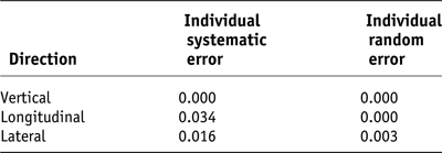
Table 3 shows a comparison of our results with other studies which have measured intrafraction motion in the same way, either comparing pre- and post-treatment images or comparing first two beams to last two beams. We found the SD was greater in the SI and AP directions. The largest observed intrafraction motion occurred in the AP direction which is in agreement with CheungReference Cheung, Sixel, Morton, Loblaw, Tirona, Pang, Choo, Szumacher, Deboer and Pignol3 and HuangReference Huang, Dong, Chandra, Kuban, Rosen, Evans and Pollack19. The Huang study used the b-mode, acquisition and targeting ultrasound system (BAT), while the CheungReference Cheung, Sixel, Morton, Loblaw, Tirona, Pang, Choo, Szumacher, Deboer and Pignol3, MadsenReference Madsen, Hsi, Pham, Presser, Esagui, Corman, Myers and Jones20 and McNairReference McNair, Hansen, Parker, Evans, Norman, Miles, Harris, Acroix, Smith, Keane, Khoo, Thompson and Dearnaley14 studies used imaging of markers as in our study. The KitamuraReference Kitamura, Shirato, Seppenwoolde, Onimaru, Oda, Fujita, Shimizu, Shinohara, Harabayashi and Miyasaka21 study reported lower mean and standard deviation values than most of the other studies, for 10 patients. They used a fluoroscopic real time tumour tracking system to image the markers during treatment. This technique may provide a more accurate estimation than the method used in the other studies, including ours.
Table 3. Intrafraction motion

RL, right-left direction; SI, superoinferior direction; AP, anteroposterior direction.
PTV margin
For the daily online correction schedule, it is important to include the intrafraction motion effect since the position error is corrected before the treatment fraction but the prostate location has moved by the end of the treatment fraction.
Figure 3 shows calculated PTV margins required for each of the imaging schedules and indicates a reduction in margin size in each direction (RL, SI and AP) obtained using an IGRT imaging schedule. However, this gain is limited by the intrafraction motion, which requires the margin to be increased from 0.7 to 1.4 mm, 0.2 to 2.3 mm and 0.2 to 3.9 mm in the RL, SI and AP directions. These results correspond well to the results of both Cheung and McNairReference Chung, Haycocks, Brown, Cambridge, Kelly, Alasti, Jaffray and Catton13–Reference McNair, Hansen, Parker, Evans, Norman, Miles, Harris, Acroix, Smith, Keane, Khoo, Thompson and Dearnaley14 who found the margins required were 3 mm/3.2 mm RL, 3 mm/3.4 mm SI and 4 mm/3.5 mm AP. Both our departmental IMRT protocol and the CHHIP protocol use a concurrent boost technique. For the high-dose PTV, a 2 mm posterior margin and a 5 mm margin in all other directions is applied, while for the lower dose PTV, a margin of 1 cm in each direction defines the PTV. Our results suggest that these margins can be reduced if online tracking is used, although the margins calculated in our study do not take into account some potential sources of error, such as target delineation accuracy on CT and/or magnetic resonance imaging (MRI)Reference Rasch, Barillot, Remeijer, Touw, van Herk and Lebesque22, prostate deformationReference Nichol, Brock, Lockwood, Moseley, Rosewall, Warde, Catton and Jaffray23 and rotationReference Dehnad, Nederveen, van der Heide, van Moorselaar, Hofman and Lagendijk24. However, perhaps due to our use of daily rectal preparation, we have not seen any rotation or deformation, and rotation has been shown to be negligible in a number of studies.Reference Dehnad, Nederveen, van der Heide, van Moorselaar, Hofman and Lagendijk24–Reference van Herk, Bruce, Kroes, Shouman, Touw and Lebesque26 De Crevoisier et al.Reference de Crevoisier, Tucker, Dong, Mohan, Cheung, Cox and Kuban27 showed that rectal distension at the planning stage is associated with a decreased probability of biochemical control, concluding that this was due to geographical miss and suggesting that daily image-guided technique should be used. The set-up margin should not be less than 1.4 mm, 2.3 mm and 3.9 mm in the RL, SI and AP directions for the online tracking technique. For any patients treated without markers and/or using an off-line schedule, this margin should be greater.

Figure 3. Margin required for set-up error using Van Herk et al formula 2.5∑ + 0.7 σ
Individualised imaging schedules
Rank testing of the imaging schedules confirmed the obvious advantages of using an online prostate tracking schedule. The time-trend analysis between the first and last weeks of treatment showed that the variance is significant systematically in the AP and SI directions and is different for each patient. The greater variation in the AP direction would be explained by changes in bladder and rectal volumes in the latter part of the treatment course when toxicities become an issue. This may also effect the SI displacement. Van der Heide’s groupReference van der Heide, Kotte, Dehnad, Hofman, Lagenijk and van Vulpen16 also showed that time trends occur frequently in the AP and SI directions and used a large database of 453 patients to identify this. They also found that individual patients show a substantial variation from the common group behaviour. The trend in the AP direction is partly explained by the effect of couch deflection which was not corrected for. The AP and SI trends are probably due to the variation in rectal filling as the patients were not given any rectal preparation or advice throughout the treatment.
Daily imaging was found to be necessary for the majority of patients due to the analysis of the systematic and random errors for each technique and the margins produced compared with the current margins used in our institution. A method to identify these patients was suggested to be necessary by Ghilezan et al.Reference Ghilezan, Yan, Liang, Jaffray, Wong and Martinez28 This study looked at the dose increments that can be achieved if an image-guided schedule is used based on the equivalent uniform dose to the rectal wall. However, they found that although all patients would benefit from a prostate tracking schedule in terms of facilitating an increase in dose, only for a third of the patients would this be significant (15–41%). They suggested that an off-line decision-making protocol to select which patients would benefit from online imaging schedules can be useful. Our data will be further analysed to assess whether a similar protocol can be developed to identify the group of patients who would benefit most from an online prostate tracking schedule.
An action level of 1.5 mm was suggested by van der Vight et al.Reference van der Vight, van Lin, Post, Visser and Louwe15 to take into account the accuracy limit of the treatment couch. As the OBI system measures in whole numbers, 2 mm is the recommended action level for this tracking technique, and corrections should be made in each direction simultaneously using OBI equipment if any one direction is ≤ 2 mm.
Resource requirements
The in-room time, taking into account the patient entering and exiting the treatment room, for a standard IMRT prostate treatment is currently 12.5 minutes (mean). The in-room timings of the patients in this study showed that online imaging can be performed with a 2-minute (mean) increase. Van der Vight et al.Reference van der Vight, van Lin, Post, Visser and Louwe15 estimated that the total time for repositioning in the AP direction only and treatment was 13.5 minutes and 10.5 minutes for IMRT and 3-field box technique, respectively, although they did not have the capability of remote couch repositioning as we have. This is comparable with our timings which take into account corrections in all directions. It is important to acknowledge that the range of in-room timings was 10.12 to 22.15 minutes, and with experience of using the technique, it is likely that the lower range value would be more representative of the required in-room time for this technique.
The department has recently implemented RapidArc for prostate IMRT patients. This gives a 3-minute reduction in beam-on time compared to the standard IMRT beam-on time. Due to this, the in-room time for the prostate tracking schedule together with the beam-on time is now less than the 12-minute time currently allocated for this group of patients.
Stroom et al. showed the advantage of online correction strategies but stated that the process was too time consuming for routine use. We are fortunate in our department to now have the equipment available to realise the potential of this technique. The National Radiotherapy Advisory Group report29, 2007 recommends that all new and replacement linacs should be capable of image guidance—hopefully most departments will eventually have the capacity to offer this technique.
CONCLUSION
Use of an online prostate tracking schedule, with daily isocentre correction before treatment and an action level of 2 mm, significantly reduces (prefraction) prostate positioning inaccuracy down to approximately 1 mm. By using this image-guided technique, the PTV set-up margin can be reduced to 4 mm (including the effects of intrafraction movement), which should help to reduce treatment-related toxicities without compromising local control rates. If other imaging schedules are used, these can reduce margins by a lesser amount. Online daily imaging schedule gives the smallest margin and there is a small difference between the margin of the NAL3 and NAL5 schedules. The use of other possible schedules will be explored in a further study. The online daily image-guided technique using marker seeds adds 2.5 minutes to the in-room time; this will be improved with experience of the use of the image-guided technique.


