


Background
During radiotherapy fractions, the positioning of the patient usually deviates to some degree from the treatment planning position. Indeed, the most important errors introduced during radiotherapy are setup errors and errors due to organ motion. These systematic errors should be reduced through clear protocols, multimodality imaging, computed tomography scan procedures and in-room imaging, such as fluorography or electric portal imaging devices, with decision rules.Reference Van Herk 1 These implementation procedures were reviewed by Langen et al.,Reference Langen and Jones 2 who reported that a potential risk of conformal radiotherapy is that organ motion may compromise clinical outcomes. Although movement during radiotherapy can be limited to a few millimetres using immobilisation devices such as noninvasive masks and vacuum bags,Reference Murphy, Chang and Gibbs 3 , Reference Hoogeman, Nuyttens, Levendag and Heijmen 4 monitoring of the target site’s position is required to maintain the margin of error during treatment fraction,Reference Murphy, Chang and Gibbs 3 and treatment time can contribute to the margin of error.Reference Hoogeman, Nuyttens, Levendag and Heijmen 4 With the advancement of radiotherapy qequipment and irradiation technology, the time that daily treatment takes has largely increased.Reference Lievens, Van den Bogaert and Kesteloot 5 – Reference Zabel-du Bois, Milker-Zabel and Bruns 7 In high-precision radiotherapy, the daily position of the target needs to be confirmed before irradiation by using a reliable imaging modality. Approaches to setup correction using cone beam computed tomography (CBCT) have been reported. Thilimann et al.Reference Thilmann, Nill and Tucking 8 concluded that CBCT target position correction may avoid the need for complex patient fixation. Meyer et al. and Guckenberger et al. reported that intrafractional patient motion significantly reduced the accuracy of patient position correction without the application of adequate immobilisation using CBCT and a robotic couch with 6 degrees of freedom.Reference Meyer, Wilbert and Baier 9 , Reference Guckenberger, Meyer, Wilbert, Baier, Sauer and Flentje 10 Infrared (IR) markers can also be used to achieve the required geometrical accuracy for patient alignment during radiotherapy, either aloneReference Baroni, Ferrigno, Orecchia and Pedotti 11 , Reference Soete, Van de Steene and Verellen 12 or in combination with other equipment such as a stereoscopic X-ray imaging device,Reference Verellen, Soete and Linthout 13 , Reference Meeks, Tome and Willoghby 14 ultrasound imaging device,Reference Meeks, Tome and Willoghby 14 motion tracking device,Reference Wilbert, Meyer and Baier 15 robotic couch with 6 degree of freedomReference Wilbert, Meyer and Baier 15 – Reference D’Souza and McAvoy 17 or three-dimensional surface imaging device.Reference Lyatskaya 18 , Reference Portalés, Gimeno, Vera and Fernández 19 These reports demonstrated the accuracy of patient alignment. To maintain the patient’s position according to the treatment planning position, it is necessary to determine the precise positions for placing the IR markers and rapid patient alignment associated with real-time computation.
The aim of this study was to apply already reported systems to a patient positioning guidance system developed in our institution that exploits optical-guided techniques to improve patient alignment accuracy and reduce the time needed for patient alignment for radiotherapy. The results of experimental laser centring procedures monitored by the guidance system were compared with a quantitative assessment of patient alignment accuracy and the required alignment time.
Methods
The patient positioning guidance system
The patient positioning guidance system used six IR cameras (Motion Analysis Corp., Santa Rosa, CA, USA), three on each side of the treatment bed as left side of Figure 1, to acquire the positions of IR markers attached to the patient. The system uses the positions of the markers acquired by the IR cameras to calculate the displacement of each marker from its reference position. Placement of the IR markers introduced uncertainty based upon needs to be paste in every treatment. A function was added to the patient positioning guidance system to paste on accurately patient skin position.

Figure 1 The left side of figure shows the placement of infrared camera. Black squares indicate the treatment couch, which is also the region of acquire for the displacement of each marker from its reference position. (a), (b) and (c) are positioning process. The function of each arrow is to change the size by quantity of displacement with the reference position.
The system’s guidance display images during patient alignment are shown in Figure 1, where (a) is an image before positioning and (b) an image during positioning. Each top depicts the current IR marker position, and displacement from the reference position is displayed quantitatively below it. The displayed angle was calculated from the change in shape of a triangle linking three tops from the reference position. Each rotation direction (rolling, pitching and yawing) is also indicated. The function of each arrow is to change the size by quantity of displacement with the reference position and separate it by colour on the basis of the tolerance level and allow this to be understood visually to operator. Figure 1c shows an image after the positioning was complete.
Patient alignment errors were calculated as follows. The IR marker position displacement caused by a rigid motion at the i n treatment delivery was denoted using a vector of three translational parameters and three rotational parameters, such that
 $$\vec {u}_{t} \left( v \right){\equals}\vec {\Delta }_{t} \left( p \right){\plus}R_{t} \left( p \right) \cdot \left[ {\vec {x}_{r} \left( v \right){\minus}\vec {x}_{r} \left( p \right)} \right].$$
 $\vec {\Delta }_{t} \left( p \right)$
is the translational vector with displacement
$\vec {\Delta }_{t} \left( p \right)$
is the translational vector with displacement
 $\delta _{t}^{{j^{n} }} $
along the j
n
axis, determined with respect to a reference point p predefined on the reference IR marker position, such that
$\delta _{t}^{{j^{n} }} $
along the j
n
axis, determined with respect to a reference point p predefined on the reference IR marker position, such that
 $$\vec {\Delta }_{t} \left( p \right){\equals}\left[ {\matrix{ {\delta _{t}^{1} } \cr {\delta _{t}^{2} } \cr {\delta _{t}^{2} } \cr } } \right].$$
 $R_{t} \left( p \right){\equals}R_{t}^{1} \cdot R_{t}^{2} \cdot R_{t}^{3} $
is the rotation matrix with respect to the same reference point and rotation around and individual axis, X, Y or Z, such that
$R_{t} \left( p \right){\equals}R_{t}^{1} \cdot R_{t}^{2} \cdot R_{t}^{3} $
is the rotation matrix with respect to the same reference point and rotation around and individual axis, X, Y or Z, such that
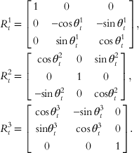 $$\eqalignno{ & R_{t}^{1} {\equals}\left[ {\matrix{ {{\rm }1} & {{\rm }0} & 0 \cr {{\rm }0} & {{\rm }{\minus}\!\!\cos \theta _{t}^{1} } & {{\minus}\!\!\sin \theta _{t}^{1} } \cr {{\rm }0} & {{\rm }\sin \theta _{t}^{1} } & {{\rm }\cos \theta _{t}^{1} } \cr } } \right], \cr & R_{t}^{2} {\equals}\left[ {\matrix{ {{\rm }\cos \theta _{t}^{2} } & {{\rm }0} & {{\rm }\sin \theta _{t}^{2} } \cr 0 & {{\rm }1} & {{\rm }0} \cr {{\minus}\!\!\sin \theta _{t}^{2} } & {{\rm }0} & {{\rm cos}\theta _{t}^{2} } \cr } } \right], \cr & R_{t}^{3} {\equals}\left[ {\matrix{ {{\rm }\cos \theta _{t}^{3} } & {{\rm }{\minus}\!\!\sin \theta _{t}^{3} } & {{\rm }0{\rm }} \cr {{\rm sin}\theta _{t}^{3} } & {{\rm }\cos \theta _{t}^{3} } & {{\rm }0{\rm }} \cr 0 & 0 & {{\rm }1{\rm }} \cr } } \right]. $$
Evaluation procedure
In total, ten operators with special knowledge of radiotherapy techniques and 1–15 years of experience were enrolled in the evaluation of the patient positioning guidance system. Each operator processed ten subjects chosen randomly. Each subject gave their informed written consent for participation, and this study was approved by the institutional review board at our hospital. The ten subjects for each operator were processed in three parts, targeting the craniocervical system, the torso and the pelvis. In the examination of the craniocervical system, the IR markers were attached to the skin directly within staved small holes in a noninvasive fixation mask. The positioning of the holes was determined according to the three places of minimum displacement on the thin skin: the temple, glabella and lower jawbone. Fixation of the subject’s torso and pelvis was accomplished with a suction bag as commonly used. IR markers were attached to the skin on the right and left elbows, the xiphoid process, the right and left of the abdomen, subcostal plane and iliac crests.
IR marker positioning time and accuracy
The patient positioning guidance system determined the final IR marker attachment positions. Although these positions cannot easily be found, few variable marker as sticking the treatment room wall was intended to facilitate positioning the IR marker correctly. The IR marker positioning time was the elapsed time from starting the attachment process until all nine IR markers were attached to the torso and pelvis. IR marker positioning accuracy was assessed by the mean of nine IR marker displacements from their reference positions.
Alignment accuracy
The examination of patient alignment accuracy was performed within a fixed measurement time of 5 minutes. This examination used experimental laser centring procedures, and was carried out by the same ten operators for the same ten subjects as the patient positioning guidance system. Comparisons were made between the two datasets to determine whether positioning differed between the two systems.
Alignment time
The time to complete the alignment of the subject was measured to evaluate the patient alignment time. The end condition for this measurement was defined such that the subject’s position was aligned within the results of the alignment accuracy examination. This examination and analysis was carried out similar to the previously mentioned examination.
Ethics statement
The study protocol was approved by the ethics committee of Ibaraki prefectural university of health sciences. All study participants provided written informed consent.
Results
The mean IR marker attachment time was 110·5 seconds and the mean accuracy was 0·76 mm (Figures 2 and 3). The maximum time taken for the attachments was 160 seconds; the markers were attached in up to 17·8 seconds/marker. The greatest inaccuracy in attachment was a displacement of up to 1·5 mm/marker.

Figure 2 The mean infrared (IR) marker attachment time. Notes: The plots are result of positioning by ten operators. Solid line indicates the mean IR marker attachment time.

Figure 3 The mean infrared (IR) marker attachment accuracy. Notes: The plots are results of positioning by ten operators. Solid line indicates the mean IR marker attachment accuracy.
Comparisons of alignment accuracy between the experimental laser centring procedures and the patient positioning guidance system for the craniocervical system, torso and pelvis are shown in Figures 4–6. Alignment accuracy with both methods was within 3·0 mm, and there was no significant difference between them. These results were independent of operator and subject. The intrafractional errors for the chest and abdomen are shown in Figures 6 and 7. The maximum displacement was observed in the value containing the respiratory motion. Displacement due to the respiratory motion varied according to the abdominal position. For the pelvis, the intrafractional error showed displacements of up to 10·0 mm in the right iliac crest. There was also a significant difference between the degree of pitching and yawing in the pelvis and degree of rolling, which was significantly greater.

Figure 4 A comparison between the two datasets to determine whether positioning differed in the craniocervical system between experimental laser centring procedures and the patient positioning guidance system. Notes: The plots are results of positioning by ten operators. Solid line indicates the mean patient alignment accuracy. Abbreviation: IR, infrared.

Figure 5 A comparison between the two datasets to determine whether positioning differed in the torso between experimental laser centring procedures and the patient positioning guidance system. Notes: The plots are results of positioning by ten operators. Solid line indicates the mean patient alignment accuracy. Abbreviation: IR, infrared.

Figure 6 A comparison between the two datasets to determine whether positioning differed in the pelvice between experimental laser centring procedures and the patient positioning guidance system. Notes: The plots are results of positioning by ten operators. Solid line indicates the mean patient alignment accuracy. Abbreviation: IR, infrared.

Figure 7 A comparison between the two datasets to determine whether positioning time differed in the craniocervical system between experimental laser centring procedures and the patient positioning guidance system. Notes: The plots are results of positioning by ten operators. Solid line indicates the mean patient alignment time. Abbreviation: IR, infrared.
A comparison of the positioning time for the craniocervical system between the experimental laser centring procedures and the patient positioning guidance system is shown in Figure 7. The experimental laser centring procedures took up to 138 seconds, with a minimum time of 70 seconds, whereas the patient positioning guidance system took up to 73 seconds, with a minimum time of 39 seconds. A comparison of alignment times for the torso and pelvis are shown in Figures 8 and 9, respectively. For the torso, the experimental laser centring procedures took up to 338 seconds, with a minimum time of 185 seconds, whereas the patient positioning guidance system took up to 152 seconds, with a minimum time of 98 seconds. The alignment time with the patient positioning guidance system was significantly shorter than with the experimental laser centring procedures in both cases (p<0·001). The positioning time was reduced for all operators.

Figure 8 A comparison between the two datasets to determine whether positioning time differed in the torso between experimental laser centring procedures and the patient positioning guidance system. Notes: The plots are results of positioning by ten operators. Solid line indicates the mean patient alignment time. Abbreviation: IR, infrared.

Figure 9 A comparison between the two datasets to determine whether positioning time differed in the pelvice between experimental laser centring procedures and the patient positioning guidance system. Notes: The plots are results of positioning by ten operators. Solid line indicates the mean patient alignment time. Abbreviation: IR, infrared.
Discussion
The patient positioning guidance system was added with the function to paste on accurately patient skin position. This function was shown to be sufficiently applicable for clinical usage, taking up to 17·8 seconds to attach one IR marker. This system was improved by using the irremovable IR marker on the wall of the treatment room to reduce the uncertainty when attaching the IR markers.
When applying radiotherapy for the head, it is necessary to guarantee precision within a mean displacement of within 3·0–5·0 mm with noninvasive fixation using a removable mask.Reference Rabinowitz, Broomberg, Goitein, McCarthy and Leong 20 ,21 The results of this study satisfied those requirements. However, the mean displacement on the torso and pelvis was >2 and 3 mm, respectively. These results indicated the importance of image-guided radiotherapy and monitoring during a fraction. We found that various motions in the torso and pelvis can become factors that disturb a patient’s alignment.
Using this system with the IR marker shortens the positioning time compared with experimental laser centring procedures. For operator, it is thought that this takes time in positioning, because it is difficult to understand the three axes and the rotation of the body by imagination alone. Rotatory displacement was particularly difficult to understand. In contrast, the patient positioning guidance system indicated a direction and the distance to move, with the length and numerical value of the arrow on the guidance monitor to guide positioning to the reference position. This reduced positioning time regardless of the operator’s number of years of experience. Previously, the attachment position of IR marker included uncertainty for attaching the IR marker on the body surface in every radiotherapy procedure.Reference Soete, Van de Steene and Verellen 12 – Reference Meeks, Tome and Willoghby 14
IR markers on the chest and abdomen are thought to be useful for monitoring respiratory motion. Respiratory-gated radiotherapy has become a robust technique by capturing the breathing state at multiple points such that the respiratory motion of tumour can be inferred from the complex movement of the abdominal surface. However, in the present study, IR marker position detection except the respiratory motion was difficult because detection of the chest and abdominal intrafractional error depended greatly on the respiratory motion. Thus, this system should be improved to measure intrafractional error except the respiratory motion.
Conclusion
The IR interactive patient position guidance and acquisition control system was shown to be a highly precise system for monitoring a patient’s position, posture and respiratory movements during radiotherapy.
Acknowledgements
None.
Authors’ Contributions: Conception and design of study: H. F., K. K., H. A.; acquisition of data: H. F., K. K., H.A.; analysis and/or interpretation of data: H. F., K. K., H. A., T. S., T.F.; drafting and approval of the manuscript: H. F., T. S., T.F.
Ethical Standards
This study was approved by the Ibaraki Prefectural University of Health Sciences Ethics Committee. Informed consent was obtained from all participants in the study.
Conflicts of Interest
The authors declare no conflicts of interest associated with this manuscript.
Financial support
This study supported by a grant of special power supply location prefecture technology promotion work from the Ministry of Education, Culture, Sports, Science and Technology (JP).


