Introduction
A spontaneous fistula between the temporomandibular joint and the external ear canal is a very rare entity. Otorrhoea is the most common symptom. An additional problem in diagnosing this condition is infection, which can appear to be the cause of the otorrhoea. Otorrhoea without middle or external ear pathology is a very rare condition, and in such cases a salivary or temporomandibular joint fistula should be considered. Iatrogenic fistulae following surgery on the parotid gland or temporomandibular joint, or following aural atresia reconstruction, have been described.Reference Miller, Jahrsdoerfer, Hashisaki and Kesser 1 – Reference Yavuzer, Unal, Ozmen, Latifoglu and Celebi 3 Spontaneous salivary fistulae with otorrhoea during mastication have also been reported.Reference Rushton and Pemberton 4
Defects of the anteroinferior part of the tympanic bone (termed foramen of Huschke) are considered to be one of the main factors responsible for the development of spontaneous fistulae.Reference Wang, Bingham, Hawke, Kwok and Li 5 – Reference Anson and Donaldson 7 The foramen of Huschke develops due to an ossification defect of the temporal bone in the first five years of life.Reference Anson and Donaldson 7 It has been described in cases of temporomandibular joint herniation into the external ear canal and salivary discharge into the external ear canal during mastication.Reference Hawke, Kwok, Shankar and Wang 8 , Reference Toyama, da Silva, Fugita and Scapini 9
We report a case with a foramen of Huschke and a spontaneous fistula between the external ear canal and the temporomandibular joint, which presented with otorrhoea and external ear infection.
Case report
A 53-year-old woman complained of occasional clear fluid otorrhoea from her right ear. Episodes of otorrhoea were followed by external ear infection, which was treated topically with drops containing dexamethasone and neomycin. The otorrhoea had first appeared three months earlier, and had since recurred five times. It usually commenced during chewing of food. The patient had no history of ear infection, trauma or surgery which could lead to the problem, and had no problems with temporomandibular joint pain or function.
Otoscopic examination showed a normal tympanic membrane and normal skin in the external ear canal. Otomicroscopy revealed a small bulging of the skin, similar to a small punctum, on the anteroinferior part of the external ear canal. Serous fluid was noticed at this site, but the amount was too small to be collected for analysis.
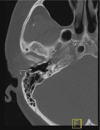
Computed tomography (CT) and magnetic resonance imaging (MRI) scans of the temporomandibular joint, salivary gland and temporal bone were performed. The CT scan revealed an anatomical variation of the tympanic portion of the temporal bone, also known as the foramen of Huschke or foramen tympanicum.Reference Wang, Bingham, Hawke, Kwok and Li 5 – Reference Anson and Donaldson 7 The foramen was characterised by loss of bone on the anteroinferior part of the external ear canal. The CT did not reveal any communication between the parotid gland or the temporomandibular joint and the meatus, but a small skin bulge was noticed, as well as proximity of the temporomandibular joint to the skin of the external ear canal (Figure 1). The MRI did not show any abnormalities of the parotid gland or other soft tissue structures, but a bone defect of the anteroinferior part of the external ear canal was seen. No communication was found between the temporomandibular joint or the parotid gland and the external ear.

Fig. 1 Axial computed tomography scan of the right temporal bone. A small bulge of skin and soft tissue into the external auditory canal is visible (arrow).
The patient had a history of allergic reactions to a contrast agent and various drugs. Therefore, we could not perform CT sialography.
Since the otorrhoea had recurred five times, a decision was made to pursue surgical treatment. A preauricular incision was made and the soft tissues were elevated off the anteroinferior aspect of the tympanic bone. A foramen was exposed. A small communication between the external ear canal and the temporomandibular joint was identified. The fistula was ligated and partially resected. In order to reconstruct the auditory canal, a tragal cartilage graft with perichondrium was harvested. The cartilage graft was placed between the defect and the temporomandibular joint, with the perichondrium facing the latter.
A small portion of the resected tissue was taken and sent for histopathological analysis; this showed connective tissue and inflammatory cells.
Post-operatively, the auditory canal was packed with antibiotic-impregnated gauze for 7 days.
The patient was followed for a year and a half. Post-operatively, she had neither otorrhoea nor external ear inflammation. At first, she could feel a mild pain when opening her mouth, but this stopped after three weeks.
Discussion
To our knowledge, this is only the second reported case of otorrhoea caused by a spontaneous fistula between the temporomandibular joint and the external ear canal.Reference Hawke, Kwok, Shankar and Wang 8
Otorrhoea is usually caused by middle or external ear pathology (e.g. chronic otitis media, cholesteatoma, tympanic membrane perforation or otitis externa). Cases of otorrhoea without middle or external ear pathology are very rare. Infection in the external ear canal may appear to be the cause of otorrhoea actually caused by more serious pathology. In our case, infection of the external ear canal occurred a few days after otorrhoea commenced.
The most commonly reported causes of otorrhoea without ear pathology are temporomandibular joint herniation, arthritis of the joint, meniscus herniation and salivary fistula.Reference Rushton and Pemberton 4 – Reference Hawke, Kwok, Shankar and Wang 8 Our patient had no temporomandibular joint problems or previous ear infections. However, her CT scan revealed a foramen of Huschke defect of the external ear canal, which is one of the factors that can contribute to fistula formation. However, the incidence of this defect is around 7 per cent, and complications are extremely rare.Reference Miller, Jahrsdoerfer, Hashisaki and Kesser 1 Only a few cases with CT findings of foramen of Huschke, temporomandibular joint herniation and salivary fistulae have been previously described.Reference Hawke, Kwok, Shankar and Wang 8 – Reference Fusconi, Benfari, Franco, Deriu, Dambrosio and Antonio 12 A salivary fistula usually occurs after parotid or atresia surgery.Reference Miller, Jahrsdoerfer, Hashisaki and Kesser 1 – Reference Yavuzer, Unal, Ozmen, Latifoglu and Celebi 3
-
• Spontaneous fistula between the temporomandibular joint and the external ear canal is very rare
-
• The presence of Huschke's foramen is one of the main causative factors
-
• In otorrhoea without middle or external ear pathology, a salivary or temporomandibular joint fistula should be considered
-
• Computed tomography and magnetic resonance imaging are very useful for diagnosis
-
• Surgery should be considered in cases with repeated infection or other complications
Since our patient's otorrhoea occurred while chewing food, a spontaneous salivary fistula was suspected. However, the amount of fluid observed was too small to be tested for amylase, and the patient was allergic to contrast medium, so we could not perform sialography. The CT and MRI scans did not show a connection between the parotid gland and the external ear canal; however, they did show a foramen of Huschke together with proximity of the temporomandibular joint to the skin of the external auditory canal.
The decision to pursue surgical treatment was made in the light of our patient's repeated episodes of otorrhoea and external ear infection. Computed tomography and/or MRI scanning is very useful in the diagnosis of congenital defects in this area; however, in some cases surgery is necessary to make the final diagnosis. Surgical treatment of the foramen of Huschke has been described in cases of temporomandibular joint herniation. The risk of complications is greater in such cases than in our case, since our patient had no herniation of the joint. If conservative therapy fails to provide any benefit, surgery is the next reasonable step. To our knowledge, our patient represents the first reported case of successful surgery of a spontaneous fistula between the temporomandibular joint and the external ear canal, described in detail.
Conclusion
This report presents a rare manifestation of a fistula between the temporomandibular joint and the external ear canal, associated with Huschke's foramen. Such a congenital defect may be difficult to detect. It should be suspected in cases of chronic otorrhoea resistant to medical therapy, in which neither middle nor external ear disease is apparent. Surgery is usually successful, and is advised in cases with repeated infections or other complications.