


Introduction
Sinonasal papillomas are benign neoplasms arising from the mucosal lining of the sinonasal cavity. Sinonasal papillomas account for 0.5–4 per cent of all nasal tumours.Reference Bawa, Allen and Ramadan1 The incidence of malignant change has been reported to range from 2 to 53 per cent, but is more likely approximately 3.6–7 per cent.Reference von Buchwald and Bradley2 Squamous cell carcinoma is the most commonly associated malignant tumour. Sinonasal papilloma classically originates from the lateral nasal wall and invades the paranasal sinuses, with maxillary and ethmoid sinuses most commonly involved.Reference Woodworth, Bhargave, Palmer, Chiu, Cohen and Lanza3, Reference Liu, Yu, Minovi, Wei, Wang and Zheng4
Traditionally, these lesions were treated via external approaches, with recurrence rates ranging between 0 and 60 per cent.Reference Lund, Stammberger, Nicolai, Castelnuovo, Beal and Beham5 The development of endoscopic sinus techniques has led to a progressive reduction in the utilisation of external approaches, with associated low morbidity and recurrence rates.Reference Busquets and Hwang6 Initially, frontal sinus involvement was thought to preclude endoscopic management, given the narrow, angulated and variable frontal recess. Development and refinement of endonasal endoscopic frontal sinus surgery, which facilitates good access to the frontal sinus, has led to a technically achievable procedure. Correspondingly, the requirement for accurate pre-operative diagnosis of frontal sinus disease has become particularly important for case-specific surgical planning.
The European position paper on intranasal tumours indicates that non-contrast computed tomography (CT) is the standard imaging modality for the evaluation of patients with inverted papilloma prior to surgery.Reference Lund, Stammberger, Nicolai, Castelnuovo, Beal and Beham5 Magnetic resonance imaging (MRI) is an equally accepted alternative modality. Computed tomography characteristically demonstrates a lesion within the middle meatus, associated with heterogeneous opacification and sclerosis in adjacent bone.Reference Lund7 It demonstrates areas of focal hyperostosis, which have been shown to correspond to the tumour origin in 89.1 per cent of cases.Reference Dammann, Pereira, Laniado, Plinkert, Lowenheim and Claussen8 The major diagnostic disadvantage of CT is its limited sensitivity for differentiating tumour from associated inflammatory disease.Reference Gomaa, Hammad, Abdelmoghny, Elsherif and Tawfik9 Conversely, MRI has excellent capacity to delineate soft tissues (Figure 1).
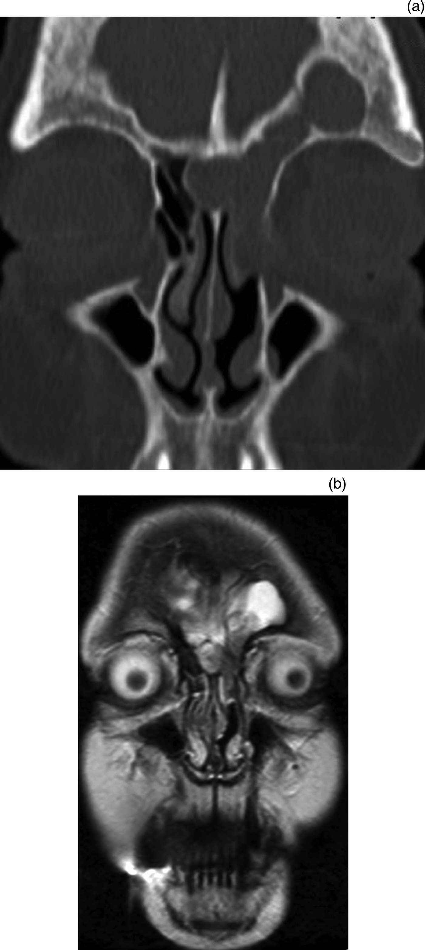
Fig. 1. (a) Coronal computed tomography scan demonstrating a left-sided sinonasal papilloma with possible extension into the frontal sinus. (b) Coronal T2-weighted magnetic resonance imaging (MRI) scan with a hyper-intense left frontal sinus depicting inflammatory fluid rather than sinonasal papilloma. The addition of MRI helped in the localisation of the tumour and in surgical planning.
We undertook a retrospective comparison study of a cohort of patients who underwent surgical management of sinonasal papilloma. This study aimed to determine the factors that influence the decision to request MRI, in order to elucidate a pre-operative strategy with accurate radiological diagnosis for the surgical management of sinonasal papilloma. In addition, we aimed to establish whether MRI is advantageous when used in addition to CT.
Materials and methods
Study cohort
A retrospective, single-blinded study methodology was employed. Patients who presented with sinonasal papilloma at the institution of the senior author (ACS) between 2007 and 2013 were identified from a database kept by the senior author. A retrospective chart review was performed on all patients. The senior author performed all surgery. We included patients with prior surgery conducted at other institutions. At the time of performing this study, all patients who had surgery for sinonasal papilloma underwent both CT and MRI as part of a standardised hospital protocol (Appendix 1). Approval was obtained from the local institutional audit department prior to initiation of the study.
Three clinicians (an ENT consultant (ACS), a head and neck radiology consultant (TK), and an ENT senior specialist registrar (AVK)) were asked to interpret CT imaging, to determine the location and extent of sinonasal involvement in cases with sinonasal papilloma. Clinicians were blinded to the surgical findings. The ENT specialist registrar and radiologist were unaware of the patients’ histories and operation details, and as such were blinded. The names and dates of scans were obscured from the ENT consultant. For the purposes of this article, a specialist registrar is defined as a trainee; a consultant is defined as an attending or surgeon who has finished their training and acts as an independent practitioner.
Computed tomography scans, and subsequently MRI scans, were analysed by the clinicians to evaluate the extent of sinonasal involvement and record where tumour was present. After reviewing the images, clinicians were asked if they thought an MRI in addition to CT was necessary to plan surgery and identify the extent of sinonasal involvement. Appendix 2 illustrates the proforma that the three raters were asked to complete.
An independent clinician correlated the clinicians’ responses with the operative findings to define accuracy of response following CT alone versus CT and MRI combined.
Reliability measures
The clinicians were asked to score sinus opacification using the Lund–Mackay staging system (0 = no abnormalities, 1 = partial opacification and 2 = total opacification). Inter-rater agreement of Lund–Mackay score was calculated with the intraclass coefficient.
Statistical analysis
Logistic regression was used to investigate the accuracy of predicting tumour location via CT. The clinician was used as the main effect. Values are reported as odds ratio ± 95 per cent confidence interval (CI). Logistic regression analysis was used to investigate the clinicians’ decision to conduct an MRI scan. The following outcomes were compared: radiologist versus ENT registrar; ENT consultant versus registrar; and ENT consultant versus radiologist. Finally, the model was adjusted to investigate which variables affected the decision to conduct an MRI scan. The factors investigated were: previous surgery, frontal sinus involvement and maxillary sinus involvement on CT.
All analyses were conducted using Stata statistical software version 13.0 (Stata, College Station, Texas, USA).
Results
Demographics and tumour characteristics
Between 2007 and 2013, 19 patients were identified as having undergone surgery for sinonasal papilloma (Table I). No patients were excluded. The most common presenting symptom was nasal obstruction, occurring in 58 per cent of patients. Patients presented with hyposmia, epistaxis and facial discomfort in 37 per cent, 26 per cent and 21 per cent of cases, respectively. Sixteen per cent of patients (n = 3) were referred after initial biopsy from other centres. No cases represented recurrence.
Table I Summary of 19 patients with sinonasal papilloma, according to tumour origin

M = males; F = females
Sixteen per cent of samples (n = 3) showed moderate dysplastic changes. Inverted papilloma was the most common pathological finding, occurring in 84 per cent of patients (n = 16). Exophytic and oncocytic papillomas occurred in one and two patients, respectively. All patients (100 per cent) underwent endoscopic tumour resection.
Reliability measures
The Lund–Mackay score had excellent inter-rater reliability when averaged over the three clinicians (intraclass coefficient = 0.92), and strong reliability for individual patients (intraclass coefficient = 0.79). There was no difference in the accuracy with which the three clinicians predicted the surgical findings based on the CT (p = 0.478) and MRI (p = 0.213) scans.
Computed tomography findings
A frontal sinus Lund–Mackay score of 1 or 2 on CT was significantly associated with intra-operative findings of frontal sinus tumour involvement (n = 11; odds ratio = 26.8, CI = 2.93–44.9; p = 0.004). However, this association was non-significant for maxillary sinus involvement (n = 7; odds ratio = 0.10, CI = 0.01–1.18; p = 0.068).
Addition of magnetic resonance imaging
The clinician responses regarding the extent of tumour involvement following CT alone and CT and MRI together were independently correlated with operative findings (accurate response coded as 1, mostly inaccurate response coded as 2 and largely inaccurate response coded as 3). Accuracy of prediction of the tumour extent was significantly improved with the addition of MRI (p = 0.0204; Table II). More specifically, the addition of MRI improved the accuracy of imaging interpretation in cases with frontal sinus involvement on CT (p = 0.017), and where the tumour originated in the ethmoid sinus (p = 0.04) or intranasally (p = 0.013). However, the small sample size for cases where the tumour originated intranasally (n = 3) should be noted.
Table II Accuracy of imaging interpretation following CT alone versus CT and MRI

* Coding: 1 = accurate, 2 = mostly inaccurate and 3 = inaccurate.
† Indicates statistical significance.
CT = computed tomography; MRI = magnetic resonance imaging
Value of magnetic resonance imaging
After viewing both the MRI and CT, the panel were asked whether MRI in addition to CT was necessary for surgical planning. Overall, the ENT clinicians (both registrar and consultant) were in agreement regarding the necessity for MRI (Figure 2). More specifically, in comparison to the radiologist, the ENT consultant and registrar were approximately nine and six times, respectively, more likely to think MRI was necessary.

Fig. 2. Odds ratios for decision on whether to conduct a magnetic resonance imaging (MRI) scan, comparing the ENT specialist registrar (‘Reg’), ENT consultant (‘ENT’) and radiology consultant (‘Rad’). The ENT clinicians were more likely to want an MRI scan in addition to computed tomography. Values are reported on a logarithmic scale as odds ratio ± 95 per cent confidence interval. *Indicates statistical significance.
Decision to conduct magnetic resonance imaging
Analysis was conducted to investigate whether any variables affected the decision to conduct an MRI (Figure 3). Frontal sinus opacification on CT (Lund–Mackay score 1 or 2) was significantly associated with the clinicians’ decision to conduct an MRI (n = 8; p = 0.002; 22 times more likely to request a scan). This finding was not true for maxillary disease or previous surgery. There was only one patient with sphenoid sinus involvement on CT; therefore, it was not possible to determine if this was an independent factor in the decision for an MRI request.

Fig. 3. Variables that affect the decision to conduct a magnetic resonance imaging scan: maxillary (n = 7) or frontal sinus disease on computed tomography (n = 8), and previous surgery (n = 7). Values are reported on a logarithmic scale as odds ratio ± 95 per cent confidence interval. *Indicates statistical significance.
Discussion
Sinonasal papilloma is a rare, benign tumour with unique characteristics, which make it difficult to treat. It has a tendency to recur following excision, it may erode bone and there is an appreciable risk of malignant transformation.Reference von Buchwald and Bradley2 Pre-operative surgical planning and imaging are crucial to ensure the correct approach is employed during the primary surgery, in order to enable complete extirpation of disease.
Several important observations were made in the present study. The majority of sinonasal papillomas occur in the fourth to sixth decades of life. The inverted papilloma subtype comprised 84 per cent of the current study population and is the most common subtype reported in the literature.Reference Barnes and Bedetti10 All patients presented with benign disease and were treated endoscopically.
Current surgical management options for sinonasal papilloma may be divided into external or endoscopic resections. Surgery for sinonasal papilloma was originally described in 1902 (PandeyReference Pandey11) using an external approach by lateral rhinotomy. Various external approaches remained the ‘gold standard’ for many years, providing the surgeon with good exposure and favourable recurrence rates. However, traditional external approaches are associated with undesirable morbidities and consequences, including facial incision, scarring, longer in-patient stay and prolonged recovery. With the improvement in minimally invasive approaches, there is a growing trend towards endoscopic techniques in cases where outcomes are comparable.Reference Karkos, Fyrmpas, Carrie and Swift12 It is this trend that prompted the present study.
In the past, the excellent exposure afforded by external approaches meant that pre-operative tumour localisation was less crucial. However, accurate tumour delineation is critical in planning the appropriate endoscopic surgical approach for sinonasal papilloma. Endoscopic surgery may not be suitable when the tumour is detected in endoscopically inaccessible regions, which are commonly considered to be the periorbital region, lacrimal sac, supraorbital ethmoidal air cells or frontal sinus.Reference Karkos, Khoo, Leong, Lewis-Jones and Swift13 Despite technological advances, frontal sinus sinonasal papilloma still poses an endoscopic, yet achievable, challenge, given the narrow anatomical boundaries of the frontal recess, limited working room and proximity to critical structures.Reference Walgama, Ahn and Batra14, Reference Kamel, Abdel Fattah and Awad15 The desirability of MRI in this study in cases with CT-proven frontal sinus disease would also attest to the surgical challenge of frontal sinus sinonasal papilloma. In practice, most rhinologists will tailor their approach based on the location and extent of tumour, using a variety of techniques for frontal sinus disease, including an osteoplastic flap, endoscopic frontal trephination, endoscopic frontal sinusotomy, endoscopic modified Lothrop, or, ultimately, a combination of open and endoscopic techniques.
In the resource-limited setting of the National Health Service, it is important that surgeons have the necessary tools to confidently predict the events of the surgical procedure and request appropriate instruments in advance of the operating day. This helps to accurately plan operative timing, thus informing surgery schedules, to prevent financial penalties associated with under- or over-utilised operating sessions. Accurate delineation of a tumour allows informed consent to be obtained regarding the surgical risks and complications associated with the planned surgical approach. For example, detection of involvement of the lateral aspects of the frontal or maxillary sinus would allow counselling regarding the additional morbidity transferred when drilling the sinus walls to reach the lateral margins of the tumour. The question therein is how can we aid the surgeon to best predict the surgical procedure?
Pre-operative radiographic assessment of sinonasal papilloma is important; hence, choosing the appropriate investigation is paramount. As reflected in this study, patients with sinonasal papilloma present with a variety of non-specific symptoms. Computed tomography and MRI are commonly requested. There is a clear preference in the literature for MRI; however, a systematic review published in 2009 indicated that there is no evidence to prove that MRI is better than CT in identifying inverted papilloma pre-operatively.Reference Karkos, Khoo, Leong, Lewis-Jones and Swift13 Three articles were found which compared both CT and MRI imaging; all studies were retrospective. Two studies concluded that MRI held advantages over CT in terms of improved differentiation from inflammatory material, and was more efficient than CT for detecting recurrence.Reference Savy, Lloyd, Lund and Howard16, Reference Petit, Vivarrat-Perrin, Champsaur, Juhan, Chagnaud and Vidal17 In contrast, one study concluded that both CT and MRI defined the extent of histologically proven recurrent disease; whereas, CT was more helpful for evaluating primary, non-recurrent inverted nasal papilloma.Reference Head, Sercarz, Luu, Collins and Blackwell18
A CT scan is usually the first-line method of evaluating for underlying disease, and it tends to illustrate non-specific sinus opacification. If the clinician feels it is warranted, they may request an MRI; however, there are no specific guidelines for this. The advantage of CT lies in its ability to identify invasion of bony structures or sclerosis.Reference Karkos, Khoo, Leong, Lewis-Jones and Swift13 However, CT will not usually differentiate sinonasal papilloma from inflammatory sinus content. This diagnostic difficulty is well reflected in the literature.Reference Dammann, Pereira, Laniado, Plinkert, Lowenheim and Claussen8 Sinonasal papilloma may block the ostium, which leads to mucus accumulation in the maxillary sinus and is difficult to differentiate from solid tumour on CT. In this study, the CT findings of maxillary sinus opacification were not always associated with intra-operative maxillary sinus disease.
There is a trend towards using MRI for investigating sinonasal papilloma because of its unique capabilities compared to CT. T2-weighted and T1-weighted gadolinium-enhanced images are capable of assessing tumour involvement. Typically, T2-weighted images illustrate areas of increased signal in tissue with high water content, for example inflammatory material. Sinonasal papilloma is highly cellular, which results in an intermediate signal on T2-weighted scans. In 80 per cent of cases, a convoluted cerebriform pattern on T2 may be present; where present, this pattern has a positive predictive factor of 95.8 per cent for inverted papilloma.Reference Ojiri, Ujita, Tada and Fukuda19–Reference Jeon, Kim, Chung, Dhong, Kim and Yim21 The disadvantages of MRI lie in the added expense and comparative scarcity of the modality in comparison to CT.
Diagnostic imaging is repeatedly targeted as a source of excess healthcare expenditure, particularly MRI, given its relative scarcity and expense in comparison to CT. Increased utilisation of diagnostic imaging significantly affects hospital funds and slows healthcare system operations. Radiologists prevent unnecessary investigations on patients and act as economic gatekeepers to avoid wastage of limited resources.Reference Knechtges and Carlos22 A radiologist's priority in interpreting images can be different to that of the surgeon. This subtle difference may partially explain the MRI requesting behaviour between the ENT clinicians and the radiologist. Radiologists aim to diagnose a disease and attempt to localise the lesion. The experienced head and neck radiologist who participated in this study was able to confidently diagnose sinonasal papilloma on CT. By contrast, the surgeon will also be planning surgical excision, and the best route to achieve safe and complete endoscopic resection. This study indicates that pre-operative CT imaging may be restricted in its ability to delineate the precise areas of tumour origin, including involvement of the frontal sinus, thus providing a role for MRI in certain cases.
The question ‘how can we aid the surgeon to best predict the surgical procedure’ is not equivalent to ‘which imaging modality best evaluates sinonasal papilloma’. The answer to the latter is MRI. The answer to the former is MRI in certain circumstances. Our results suggest that these circumstances are when CT illustrates involvement of the frontal sinus. Our results also indicated that the addition of MRI helped to predict the origin of sinonasal papilloma if located within the ethmoid sinus (n = 11) or intranasally (n = 3). Despite this, it is accepted by experienced surgeons that it is largely inconsequential whether CT opacification in the ethmoid sinuses or medial maxilla is mucus or tumour, as this area is easy to access surgically; this is equally true of opacification limited to the frontal recess. However, surgical access is more problematic if there is papilloma within the frontal sinus or lateral maxillary wall; therefore, opacification in such areas may require MRI to distinguish between mucus and tumour. This distinction for the frontal sinus is validated by our study. Interestingly, maxillary sinus disease was not significantly correlated with MRI desirability. This is likely a result of the study's consideration of the entire maxillary sinus, rather than differentiating between medial and lateral aspects. It was not feasible to adjust the study to reflect this given the small sample size of cases with maxillary involvement (n = 7).
The comparison between an ENT consultant and registrar is interesting. In the UK, a senior specialist registrar may act as an autonomous practitioner, including requesting further imaging without consultant approval. It is encouraging that the findings were not significantly different between the two clinicians, thereby indicating that resources were not being wasted unnecessarily.
Important limitations of this analysis must be considered. The sample size is relatively small (n = 19), which reflects the rarity of the disease. Our department is a tertiary referral centre, with a wide catchment area of around seven million, and the study took place over a six-year period. The small sample size leads to wide CIs, which reflect a degree of uncertainty within the estimates. The clinicians were blinded to the operative results at the time of performing the study, but as the study was retrospective and the senior author performed all surgery, there is a possibility that recall bias was introduced.
Note should be made of the use of the Lund–Mackay score. This is widely used in the assessment of chronic rhinosinusitis and provides an objective measure of sinus opacification. It does not make the diagnosis of sinonasal papilloma. The Lund–Mackay score was used to inform the study that the three independent observers were interpreting the CT scans similarly and, thus, providing a baseline competence.
Future studies should be conducted prospectively. Open and endoscopic cases should be compared. This comparison was not possible in our series as all cases were performed endoscopically.
• Sinonasal papillomas are benign neoplasms arising from mucosal lining of the sinonasal cavity
• There is variation in pre-operative imaging (computed tomography (CT), magnetic resonance imaging (MRI) or both)
• A shift from external approaches towards endoscopic techniques has led to the requirement for accurate pre-operative delineation of disease origin and extent
• Using MRI in addition to CT improves accuracy of pre-operative interpretation when correlated to operative findings
• Surgeons are more likely than radiologists to request MRI, specifically when CT indicates frontal sinus involvement
Conclusion
Successful management of sinonasal papilloma can be achieved with careful pre-operative planning and meticulous surgical technique. Magnetic resonance imaging is unquestionably valuable for the management of sinonasal papilloma; it provides accurate differentiation between inflammatory disease and sinonasal papilloma, and is sensitive for the identification of disease. However, its use should be weighed against performing scans that may not change the surgical plan or outcome. This paper suggests that the addition of MRI helps predict disease in the frontal sinus better than CT and is more useful for planning the surgical procedure. A close working relationship between the ENT and radiology departments is important to facilitate accurate localisation of the tumour and to ensure that the radiologist understands the surgeon's requirements.
Acknowledgements
The authors would like to acknowledge the help of the statistics department at the University of Liverpool, particularly James Dodd, Eftychia-Eirini Psarelli and Rebecca Asher.
Competing interests
None declared.
Appendix 1. Standardised hospital computed tomography and magnetic resonance imaging sequences.
CT sinus
Scanner: Toshiba
Bone 0.5 mm volume axial
Bone 2.0 axial
Bone 2.0 coronal
Bone 2.0 sagittal
Soft tissue 0.5 mm volume
Soft tissue 2.0 axial
Soft tissue 2.0 coronal
Soft tissue 2.0 sagittal
MRI sequences
Scanner: Siemens 1.5 Tesla
1. Localiser
2. T2 blade transverse
3. T2 TSE coronal cover frontal to sphenoid sinuses
4. T1 Se_Tra320
5. T1 post-gadolinium
Appendix 2. Proforma completed by the three raters
Patient hospital number: Pre-op CT date:
Operation date: Assessor:
Lund–Mackay staging system

Please state areas/sinuses involved with IP/tumour. Any other features on scan? ______________________________________________________________ ____________________________________
Pre-op MRI date: Please tick one box for each site
MRI left side

MRI right side

Please describe your thoughts on the likely extent of the sinonasal papilloma at specific sites. ______________________________________________________________ ______________________________________________________________ ______________________________________________________________ ____________________________
Do you think MRI in addition to CT was necessary for surgery/planning of surgery? ____________


