


Introduction
Extra-nasopharyngeal angiofibroma is a rare and distinct clinical entity, entirely different from juvenile nasopharyngeal angiofibroma (JNA).Reference Szymanska, Scymanska, Morshed, Czekajska-Chehab and Szczerbo-Trojanowska 1 , Reference Windfuhr and Remmert 2 Moreover, nasal septal angiofibroma is a rarity and is regarded by some as a subclass of extra-nasopharyngeal angiofibroma.Reference Garcia-Rodriquez, Rudman, Cogbill, Loehri and Poetker 3 Furthermore, the said lesion is extremely rare in the paediatric population. An extensive search of PubMed and Medline databases revealed only 18 cases of septal extra-nasopharyngeal angiofibroma in the English-language medical literature,Reference Garcia-Rodriquez, Rudman, Cogbill, Loehri and Poetker 3 – Reference Castillo, Timmons and McClay 9 with only 4 paediatric cases,Reference Hiraide and Matsubara 6 – Reference Castillo, Timmons and McClay 9 and all 4 children were male.
This report documents an extremely rare case of extra-nasopharyngeal angiofibroma arising from the septum in a female child, with a review of the contemporary literature. To the best of our knowledge, such a case has not been reported previously.
Case report
A nine-year-old female child presented in the ENT out-patient department of our institution with a three-month history of nasal obstruction and epistaxis. The frequency of epistaxis episodes had increased over the previous three months to three times per week. These episodes lasted for a short duration of 3–5 minutes, and ceased on pinching the nose. The patient did not require hospitalisation for any of these episodes.
Anterior rhinoscopy revealed a small reddish mass arising from the anterior part of the septum on the left side (Figure 1). On probing, the mass was painful and bleeding was observed. These findings were confirmed on nasal endoscopy. There were no palpable lymph nodes in the neck. The patient underwent routine blood and urine tests, the findings of which were normal.
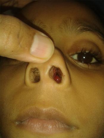
Fig. 1 Clinical photograph showing lesion in the left nasal cavity. Published with patient's permission.
Contrast-enhanced computed tomography (CT) revealed a soft-tissue lesion in the left anterior nasal cavity, with a broad base towards the nasal septum and no areas of intense enhancement (Figure 2). There was no radiological evidence of bony erosion or extension into the paranasal sinuses.

Fig. 2 Coronal computed tomography scan, showing a broad-based, non-enhancing lesion arising from the nasal septum on the left side, with no signs of sinus invasion. R = right
A presumptive diagnosis of a vascular tumour (probably haemangioma) was made. The lesion was endoscopically excised via electrosurgical cauterisation of the lesion base. No intra-operative complications were recorded, although there was minimal bleeding.
Post-operatively, patient was administered antibiotics and analgesics. The packing was removed after 24 hours, and the patient was subsequently discharged with no untoward incident to report.
Histopathology of the lesion revealed collagenised vascular stroma, with numerous irregularly shaped stag horn blood vessels, consistent with angiofibroma (Figure 3). Further, immunohistochemistry of the excised specimen was positive for markers of smooth muscle actin, cluster of differentiation 34 and vimentin (Figure 4). Hence, a final diagnosis of septal extra-nasopharyngeal angiofibroma was made.

Fig. 3 Histopathological examination showed interstitial collagen and characteristic stellate cells, with thin-walled blood vessels (H&E; ×400).

Fig. 4 Immunohistochemistry findings, showing positivity for markers: (a) cluster of differentiation 34 (×400), (b) smooth muscle actin (×400) and (c) vimentin (×400).
The patient has been followed up regularly for the past year, with no residual lesion or recurrence.
Discussion
Extra-nasopharyngeal angiofibroma has some salient clinical features that distinguish it from JNA. It is more common in females as compared to JNA, with 30 per cent of cases occurring in females.Reference Windfuhr and Remmert 2 , Reference Handa, Kumar, Singh and Chhabra 8 , Reference Singh, Aggarwal, Arora, Doloi and Kumar 10 In addition, it occurs across a wide age group of 6–78 years (mean age of 22–28 years), in contrast to 6–26 years for JNA (mean age of 15 years).Reference Szymanska, Scymanska, Morshed, Czekajska-Chehab and Szczerbo-Trojanowska 1 , Reference Windfuhr and Remmert 2 Moreover, it is less vascular and thus the chances of massive haemorrhage are lower too: only 16 per cent of previous cases required blood transfusion.Reference Windfuhr and Remmert 2 , Reference Garcia-Rodriquez, Rudman, Cogbill, Loehri and Poetker 3 , Reference Singh, Aggarwal, Arora, Doloi and Kumar 10 Lastly, recurrence has not been reported for extra-nasopharyngeal angiofibroma.Reference Szymanska, Scymanska, Morshed, Czekajska-Chehab and Szczerbo-Trojanowska 1 , Reference Windfuhr and Remmert 2 , Reference Akbas and Anadolu 11 In this context, it would also be pertinent to note that a CT scan is sufficient for diagnosis and surgery is the treatment of choice.Reference Szymanska, Scymanska, Morshed, Czekajska-Chehab and Szczerbo-Trojanowska 1 – Reference Garcia-Rodriquez, Rudman, Cogbill, Loehri and Poetker 3 , Reference Singh, Aggarwal, Arora, Doloi and Kumar 10 , Reference Akbas and Anadolu 11
The exact aetiology for extra-nasopharyngeal angiofibroma is unclear. Developmental, genetic and hormonal theories have been proposed.Reference Peric, Sotirovic, Cerovic and Zivic 12 , Reference Yokoi, Arakawa, Kuribayashi, Inoshita, Haruyama and Ikeda 13 However, the occurrence of extra-nasopharyngeal angiofibroma in a young female aged nine years leads us to refute the hormonal theory of angiofibroma, which states that it is a testosterone-dependent tumour and high oestrogen levels protect females against this tumour.Reference Akbas and Anadolu 11 These tumours arise from fascia basalis.Reference Brunner 14 Usually fascia basalis extends to the posterior part of the vomer and ethmoid bone, but as a developmental anomaly it can extend anteriorly and form small islands of ectopic tissue.Reference Peric, Sotirovic, Cerovic and Zivic 12 These islands of ectopic tissue may give rise to septal angiofibromas.
-
• This paper describes the first reported case of extra-nasopharyngeal angiofibroma in a female child
-
• The case refutes the hormonal theory of angiofibroma aetiopathogenesis
-
• The report highlights the importance of immunohistochemistry in the diagnosis of angiofibroma
Table I gives us a brief synopsis of the previous four cases of nasal septum extra-nasopharyngeal angiofibroma in the paediatric age group.Reference Hiraide and Matsubara 6 – Reference Castillo, Timmons and McClay 9 All the cases (including our patient) presented with chief complaints of nasal obstruction and epistaxis, and were misdiagnosed initially. Two of these cases had massive bleeding intra-operatively, which led to abandonment of the transnasal excision initially.Reference Sarpa and Novelly 7 , Reference Handa, Kumar, Singh and Chhabra 8 One case required lateral rhinotomy later on for excision.Reference Handa, Kumar, Singh and Chhabra 8 Spontaneous auto-amputation of the said lesion was also reported in one case.Reference Castillo, Timmons and McClay 9 It would be prudent to note that the lesion in our patient was non-enhancing. We removed it transnasally, with no post-operative complications. No massive haemorrhage was encountered, given the decreased vascularity of the tumour.Reference Windfuhr and Remmert 2 , Reference Garcia-Rodriquez, Rudman, Cogbill, Loehri and Poetker 3 , Reference Singh, Aggarwal, Arora, Doloi and Kumar 10 Interestingly, immunohistochemistry was not conducted in the previously reported cases to confirm the diagnosis of extra-nasopharyngeal angiofibroma.
Table I Brief synopsis of paediatric septal extra-nasopharyngeal angiofibroma cases

*Prior to presentation. Y = years; IHC = immunohistochemistry; M = male; N/A = not available; F = female
It would be imperative to note that, in accordance with evidence-based medicine, immunohistochemistry clinches the diagnosis.Reference Peric, Sotirovic, Cerovic and Zivic 12 Though a haemangioma can be distinguished from extra-nasopharyngeal angiofibroma by marked lobulation and decreased stromal fibrosis, it is difficult to differentiate from haemangiopericytoma. An immunohistochemical analysis of this lesion shows positive staining for vimentin, and negative staining for cluster of differentiation 34 and alpha smooth muscle actin.Reference Yokoi, Arakawa, Kuribayashi, Inoshita, Haruyama and Ikeda 13 Angiofibromas have positive staining for vimentin, cluster of differentiation 34 and alpha smooth muscle actin.Reference Peric, Sotirovic, Cerovic and Zivic 12 Moreover, a significantly high cluster of differentiation 34 expression in angiofibroma distinguishes it from cavernous haemangioma as well. In our patient, immunohistochemistry confirmed the diagnosis of extra-nasopharyngeal angiofibroma.
In summary, this report describes an exceptional atypical cause of epistaxis in a female child: angiofibroma arising from the septum. This is also the only case of immunohistochemistry-validated septal angiofibroma reported in a female child. The case also merits mention on account of the rarity of the lesion and its underreporting in medical literature, which limits the conclusions that can be drawn regarding its clinical course and management.


