Introduction
Acute mastoiditis is an otological emergency, and is usually successfully managed with intravenous antimicrobial therapy, with or without surgical intervention. Occasionally, infection can spread from the temporal bone to other head and neck sites; this complicates management, and often necessitates cross-sectional imaging for diagnosis and surgical planning.Reference Psarommatis, Voudouris, Douros, Giannakopoulos, Bairamis and Carabinos1
With the increasing sophistication of diagnostic radiology, the ability to identify complications of mastoiditis has greatly increased. However, by the same token, the interpretation of head and neck imaging is also becoming increasingly complex.
Using a portfolio of selected cases, we reviewed the appearances of common and rare complications of acute mastoiditis on computed tomography (CT) and magnetic resonance imaging (MRI). We propose a simple 16-point checklist, which can be used to guide interpretation for acute mastoiditis and its complications for time-critical out-of-hours assessment when a specialised radiologist opinion may be difficult to obtain.
Normal temporal bone anatomy
With a complex three-dimensional shape and a multitude of critical neighbouring structures, the temporal bone is a notoriously complex part of human anatomy. It articulates with five other bones, namely the zygoma, mandible, sphenoid, parietal and occipital bones, via a number of sutures, including the zygomaticotemporal, sphenosquamous, squamoparietal and occipitomastoid sutures. The temporal bone itself consists of five distinct parts: squamous, zygomatic, mastoid, petrous and tympanic parts.
The tympanic cavity is a six-sided space containing three ossicles: the malleus, incus and stapes. These are associated with the tendons of the tensor tympani and stapedius muscles, and ossicular ligaments. The tegmen tympani is a thin plate of bone that forms the roof of the tympanic cavity and separates it from the middle cranial fossa. The floor is composed of the fundus tympani, below which is the jugular fossa. The lateral wall is formed mostly by the tympanic membrane and scutum. The medial wall is the lateral wall of the inner ear, which itself comprises the vestibule, three semi-circular canals and cochlea. The tympanic portion of the facial nerve traverses the medial wall in the fallopian canal. The posterior wall is the mastoid bone populated with mastoid air cells, which communicate with the middle ear via the aditus ad antrum. The anterior wall of the middle ear is related to the carotid canal.Reference Lemmerling, Smet, De Foer, Lemmerling and De Foer2 Axial and coronal sections of normal temporal bone CT images are shown in Figures 1 and 2.
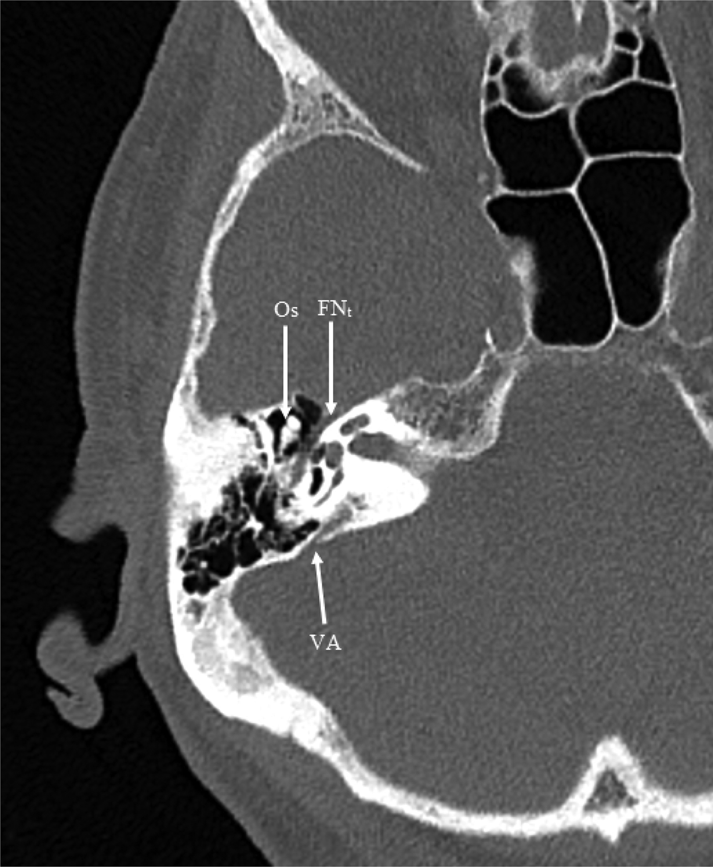
Fig. 1. Axial computed tomography scan of normal anatomy, showing ‘ice-cream cone’ appearance of ossicles (Os), tympanic facial nerve (FNt) and vestibular aqueduct (VA).
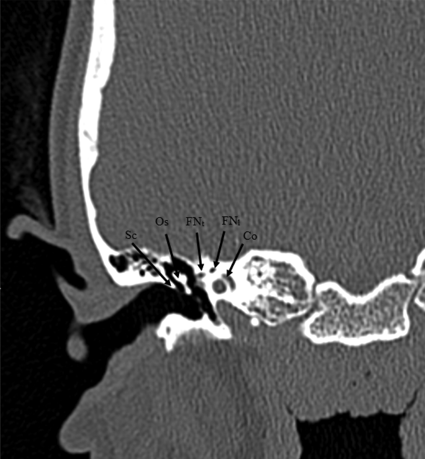
Fig. 2. Coronal computed tomography scan of normal anatomy, showing ‘snake eyes’ appearance of facial nerve (labyrinthine and tympanic parts, FNl and FNt), scutum (Sc), ossicles (Os) and cochlea (Co).
The appearance of the ossicles in axial images is often described as an ‘ice-cream cone’ shape, with the circular ‘ice-cream’ component representing the head of the malleus, and the ‘cone’ representing the body and short process of the incus. The ‘cone’ funnel points in the direction of the aditus ad antrum.
At its first genu, the facial nerve undergoes a sharp change in direction. This results in both the labyrinthine and tympanic parts being visible on cross-sectional imaging, a sign often referred to as ‘snake eyes’.Reference Juliano, Ginat and Moonis3 The apical turn of the cochlea can be seen medially at this level.
Radiology checklist
In order to assist the ENT surgeon with out-of-hours temporal bone imaging interpretation, when the opinion of radiologists with an interest in head and neck imaging may be hard to obtain, we have provided a systematic, radiologist-derived 16-point checklist (the ‘mastoid 16’), to identify common complications of acute mastoiditis.
Factors to specifically examine on imaging include: (1) intratemporal factors, such as middle-ear opacification, the state of the ossicles, tegmen tympani and scutum, any labyrinthine fistula, mastoid opacification or erosion of the cortex and septae, a subperiosteal abscess, petrous apex involvement, and the facial nerve and canal; (2) intracranial factors, including subdural empyema and extra-axial collection, pneumocephalus, a focal intracerebral or cerebellar lesion, and diffuse cerebritis or meningeal enhancement; and (3) other head and neck factors, such as any neck abscess, temporomandibular joint (TMJ) involvement, vascular status (thrombosis of the internal jugular vein (IJV), or the sigmoid or transverse sinuses; internal carotid artery (ICA) aneurysm; high jugular bulb or aberrant ICA, if considering surgical intervention), muscular involvement (sternomastoid, digastric, temporalis), and the mastoid tip.
Examples of common manifestations of the above pathologies are demonstrated below, with special reference to the interpretation of cross-sectional CT and MRI scans.
Intratemporal imaging
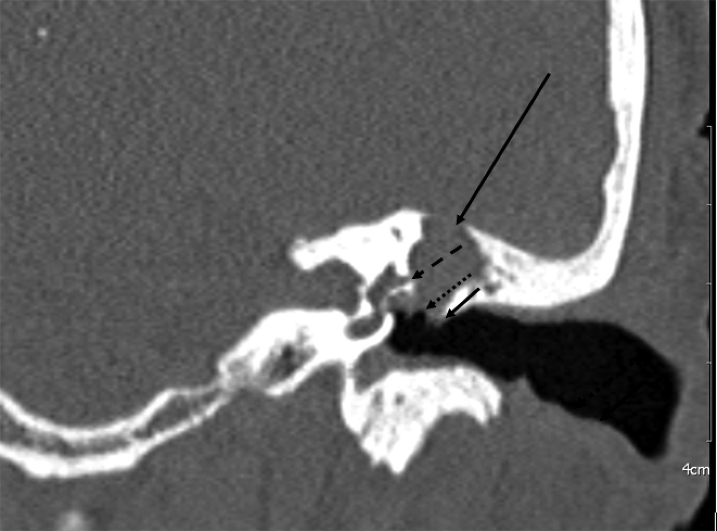
Initially, it is important to differentiate between incidental findings and pathology. For example, the opacification of mastoid air cells in an asymptomatic patient is not necessarily indicative of acute infection. Erosion of the septae, by contrast, would be a concerning feature of a destructive infection. Erosion of the scutum, accompanied by opacification of Prussak's space (the epitympanic space between the ossicles and the scutum), is a classical sign of pars flaccida cholesteatoma. Although often slowly destructive, such a cholesteatoma can become acutely infected and associated with mastoiditis.Reference Mirza, Varadarajan, Youshani and Willatt4 An example is shown in Figure 3.

Fig. 3. Coronal computed tomography scan of cholesteatoma in middle-ear cavity, showing scutum erosion (short arrow), eroded ossicles (dotted arrow), eroded tegmen tympani (long arrow) and labyrinthine fistula (dashed arrow).
Both cholesteatoma and middle-ear infection can lead to destruction of the ossicles, making this a non-specific sign. Medial erosion into the bony labyrinth can lead to a labyrinthine fistula (Figure 3). Similarly, the tympanic portion of the facial nerve may be dehiscent in up to 50 per cent of patients. These are both important to consider prior to mastoidectomy, given the risks of sensorineural hearing loss or facial nerve injury.
The tegmen tympani, or roof of the middle-ear cavity, must be closely inspected, to identify whether bony erosion has made the middle-ear space continuous with the middle cranial fossa, thereby permitting direct access for spreading infection (as shown in Figure 3). It is important, however, to note that meningitis can still occur even in the absence of tegmen erosion.
Tracking of infection to the petrous apex can cause cranial nerve involvement, resulting in Gradenigo's syndrome. The latter is a triad of suppurative otitis media, pain in the distribution of the trigeminal nerve, and an abducens nerve palsy.Reference Gradenigo5 It indicates extradural inflammation adjacent to the trigeminal ganglion within Meckel's cave and the abducens nerve. This spread can occur through air cells, vascular routes or direct fascial spread.Reference Allam and Schuknecht6 A combination of CT and MRI have been recommended for their respective abilities to delineate bony anatomy and identify central nervous system (CNS) involvement.Reference Jensen, Hansen, Møller and Saunte7,Reference Zammit-Maempel8
The outer or inner mastoid cortex can be eroded in acute mastoiditis, and the ‘bone window’ on CT can facilitate identification of this (Figure 4). Outer mastoid cortex erosion is often associated with a subperiosteal abscess (Figure 5), while inner mastoid cortex erosion may be seen with extra-axial collections. Subperiosteal abscesses can be seen particularly well on contrast-enhanced CT imaging and on MRI scans.Reference Platzek, Kitzler, Gudziol, Laniado and Hahn9
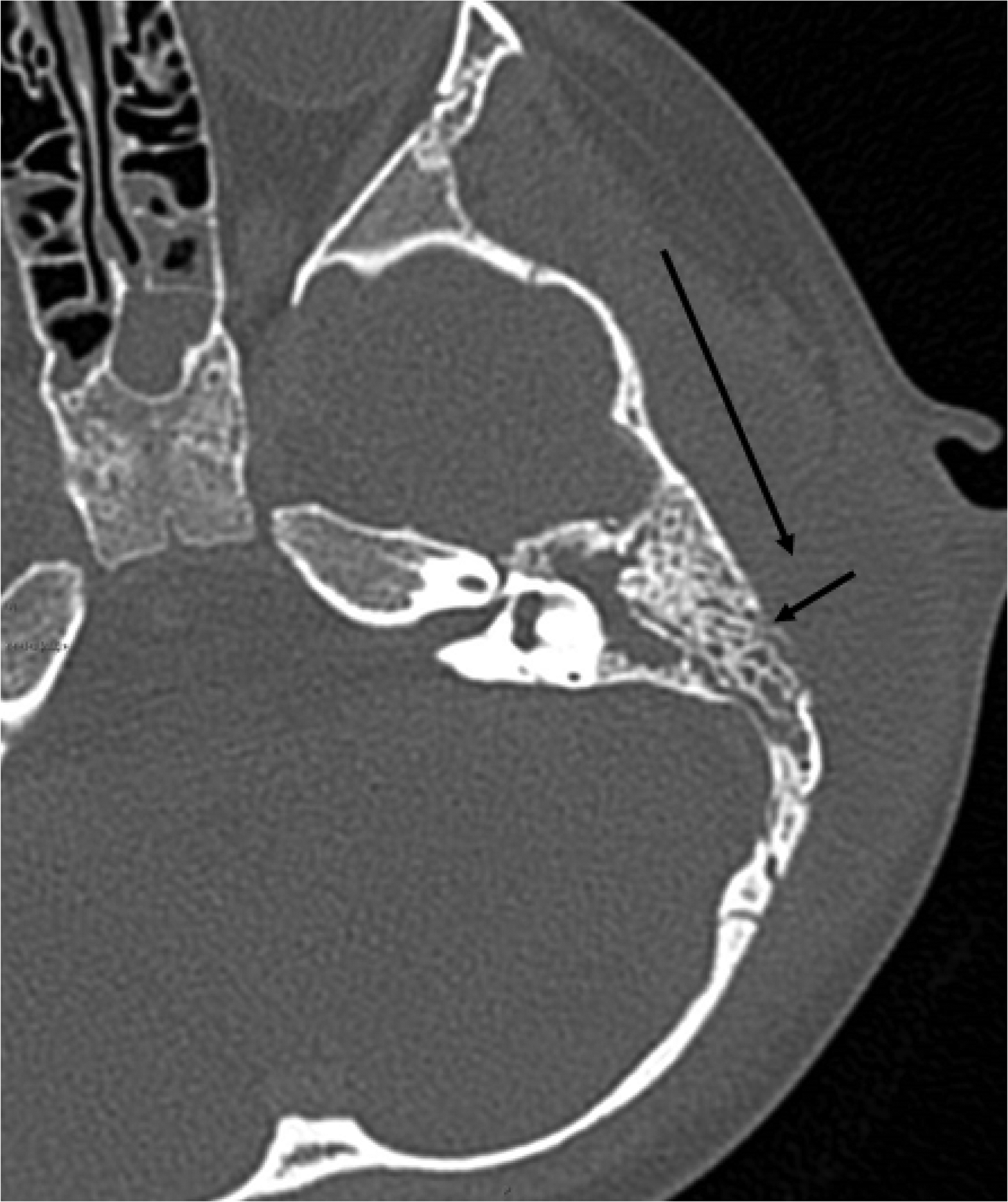
Fig. 4. Axial computed tomography scan in the ‘bone window’, showing erosion of the outer mastoid wall (short arrow) with associated soft tissue thickening secondary to subperiosteal abscess (long arrow).
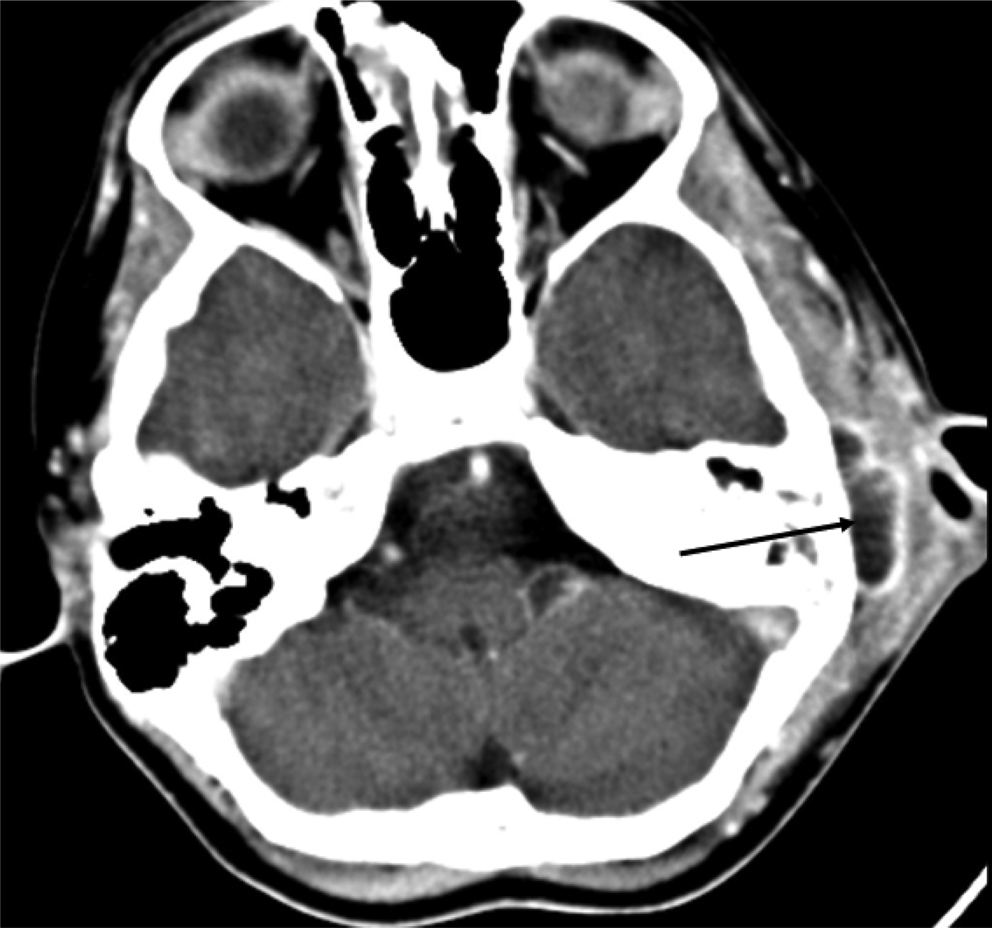
Fig. 5. Axial, contrast-enhanced computed tomography scan showing left subperiosteal abscess (black arrow).
Intracranial imaging
The CT and MRI scans both demonstrate high sensitivity for detecting intracranial complications of acute mastoiditis.Reference Platzek, Kitzler, Gudziol, Laniado and Hahn9,Reference Migirov, Duvdevani and Kronenberg10 Meningitis is the most common of these; radiologically, meningeal enhancement can often be seen, although the absence of this does not exclude the diagnosis. Focal abscesses may develop below the dural layer, which also enhance strongly on contrast-enhanced CT scans. The brain parenchyma itself may be involved too, identified either as diffuse ill-defined areas of low attenuation in cerebritis, or with focal lesions showing peripheral enhancement (such as a cerebral abscess, most commonly in the region of the temporal lobe),Reference Duarte, Kozin, Barshak, Reinshagen, Knoll and Abdullah11 as seen in Figure 6. The presence of air within the cranial cavity (pneumocephalus) can be seen as a post-operative change following mastoidectomy, but can also indicate middle-ear infection with an eroded tegmen and dural tear.Reference Abbati and Torino12
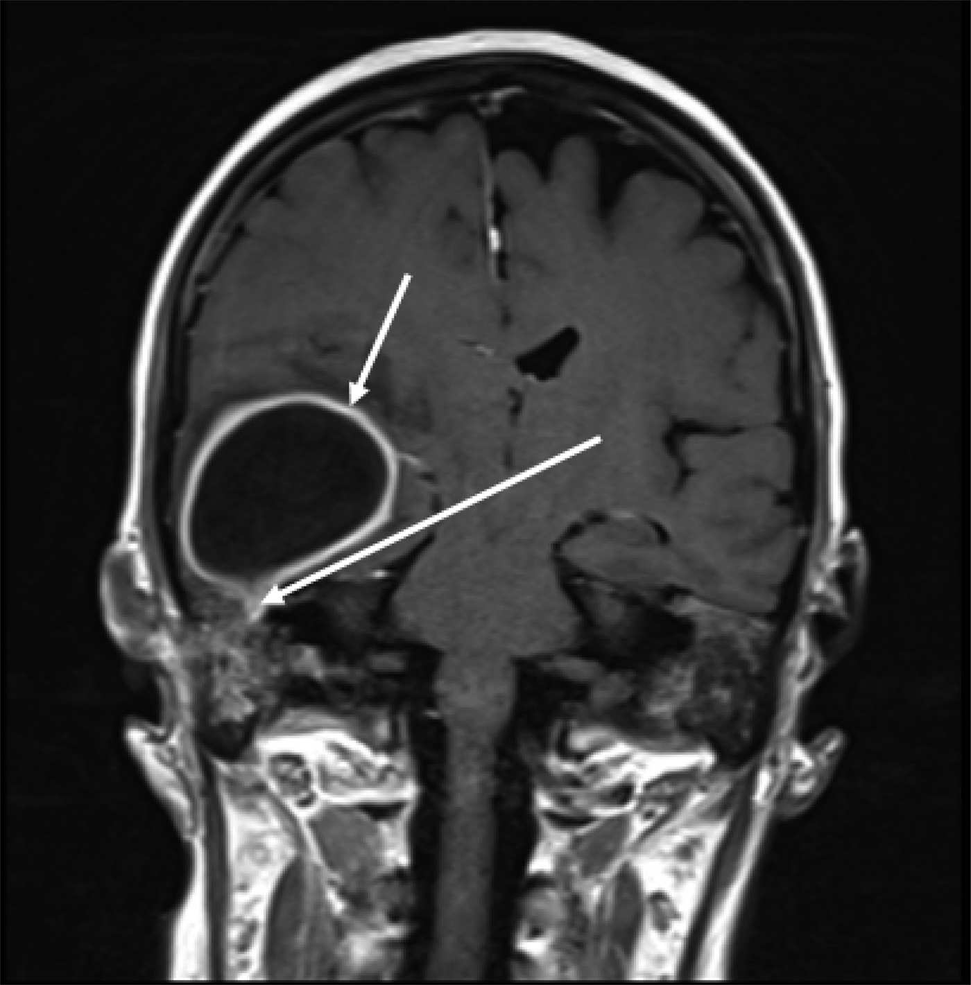
Fig. 6. Coronal, contrast-enhanced, T1-weighted magnetic resonance imaging scan, showing right temporal lobe abscess (short arrow), with ‘beak enhancement’ extending into the epitympanum suggesting tegmen dehiscence (long arrow).
Inferiorly, the cerebellum may also be affected. The MRI scans demonstrate well the presence of focal abscesses, and can potentially reveal meningeal or vascular involvement, as shown in Figure 7.
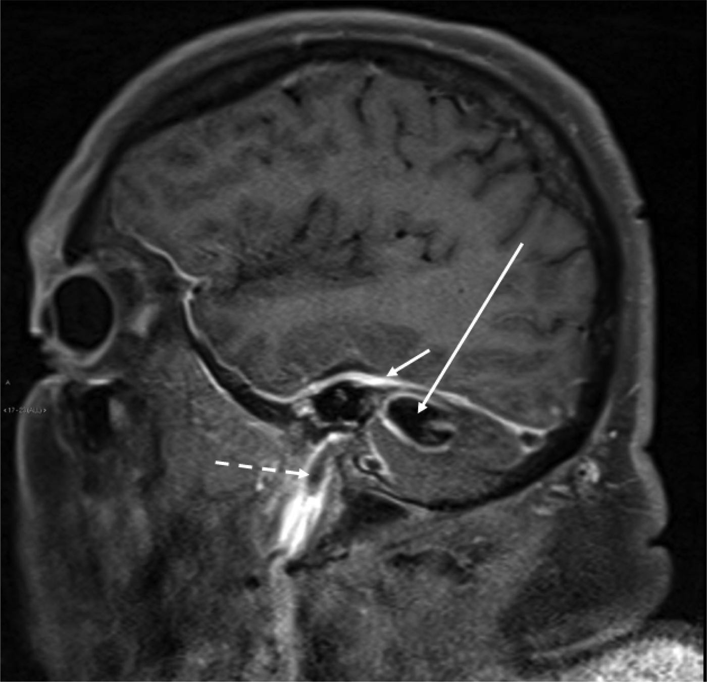
Fig. 7. Sagittal, fat-suppressed, T1-weighted, post-gadolinium magnetic resonance imaging scan, showing cerebellar abscess (long arrow) with associated meningeal tentorial enhancement (short arrow) and internal jugular vein thrombus seen as a filling defect (dashed arrow).
Other head and neck imaging
In addition to the intratemporal and intracranial extension of infection from the mastoid, other head and neck sites are at risk. In particular, septic arthritis or localised abscesses adjacent to the TMJ have been well-described previously. Indeed, one series evaluating consecutive patients admitted with mastoiditis identified associated TMJ abscesses in up to 13 per cent of cases.Reference Luscan, Belhous, Simon, Boddaert, Couloigner and Picard13 An example is shown in Figure 8. Such cases can also occur as a result of odontogenic infection; therefore, clinical examination of the oral cavity is also warranted.

Fig. 8. Axial, contrast-enhanced computed tomography scan showing multiloculated right-sided masticator space abscess containing a small locule of air and extending onto the temporomandibular joint (white arrow).
In addition to intracranial extension, infection from mastoiditis can spread to the neck, leading to a number of potentially serious soft tissue complications. One example is a Bezold's abscess (Figure 9), which represents suppuration within the sternomastoid and digastric muscle due to pus tracking to the mastoid tip.Reference Gaffney, O'Dwyer and Maguire14 This can spread along digastric muscle to the chin and fill the retromaxillary fossa, or into the deep neck spaces and descend into the thorax.

Fig. 9. Axial, contrast-enhanced computed tomography scan showing left-sided Bezold's abscess (white arrow).
Luc's abscess represents the development of a subperiosteal collection beneath temporalis muscle. The original descriptions of this attributed it to the spread of infection from the middle ear in acute otitis media to the temporalis muscle, via the notch of Rivinus and branches of the deep auricular artery.Reference Luc15 However, it has subsequently been described in association with acute mastoiditis. Temporal bone CT has demonstrated efficacy in surgical planning.Reference Scrafton, Qureishi, Nogueira and Mortimore16
Important vascular structures can be affected by acute mastoid infection. For example, sigmoid sinus thrombosis is seen in both adults and children with middle-ear and mastoid infections. Diagnosis can be made using contrast-enhanced CT or MRI, with a filling defect or no enhancement indicating thrombus (Figures 10 and 11). Management, particularly with regard to anticoagulation to prevent clot propagation, remains controversial.Reference Mather, Musgrave and Dawe17

Fig. 10. Coronal, contrast-enhanced computed tomography scan showing completely thrombosed left sigmoid and transverse sinuses (black arrow).
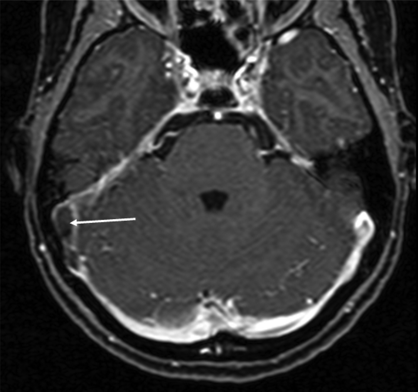
Fig. 11. Axial, contrast-enhanced, fat-suppressed, T1-weighted magnetic resonance imaging scan, showing right sigmoid sinus thrombus (white arrow).
As with sigmoid sinus thrombosis, IJV thromboses can be seen in the context of otogenic infection (Figure 12). They can be identified by the observation of filling defects of the jugular bulb on contrast-enhanced CT scanning or MRI and magnetic resonance venography.Reference Redaelli de Zinis, Gasparotti, Campovecchi, Annibale and Barezzani18 In addition to screening for thrombus, identifying the position of the IJV is also important in surgical planning, as a high jugular bulb may pose a risk of haemorrhage if myringotomy is to be performed to decompress the middle ear.
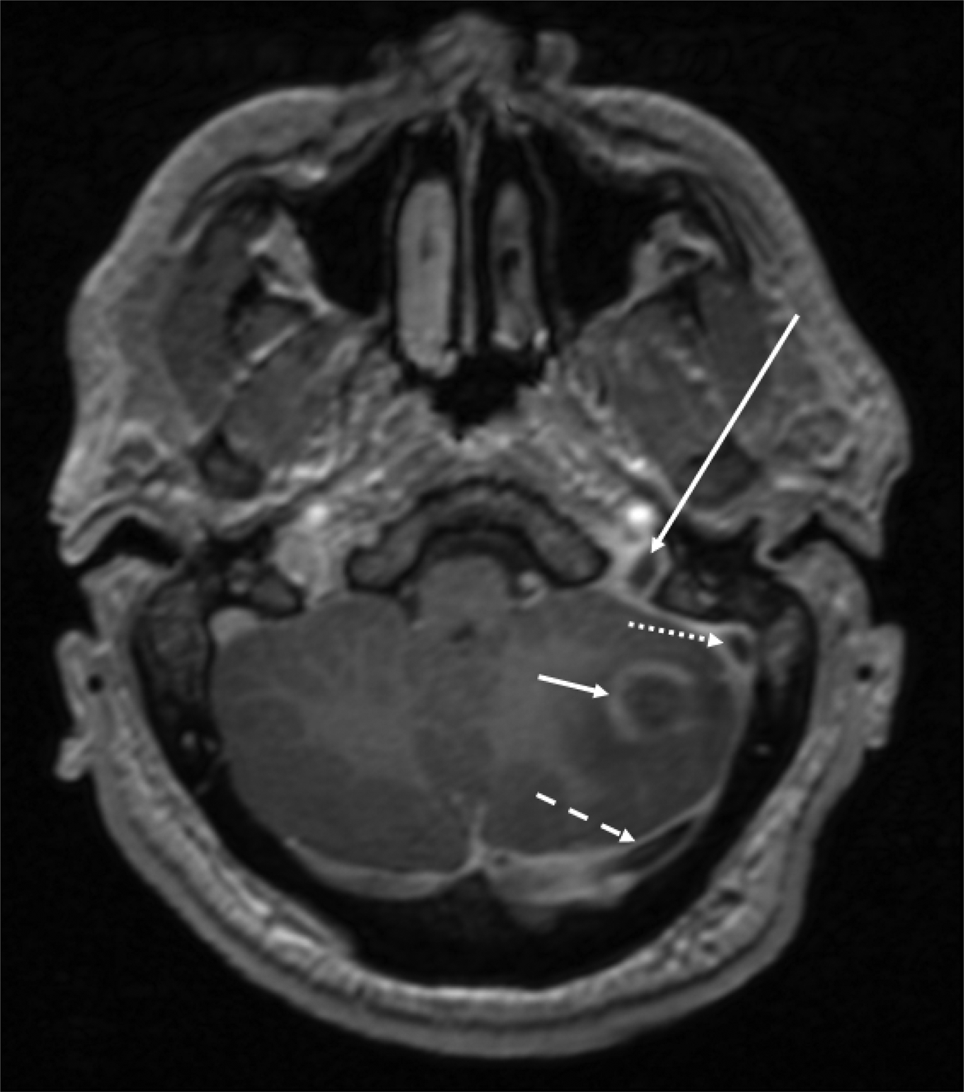
Fig. 12. Axial, contrast-enhanced, fat-suppressed T1-weighted magnetic resonance imaging scan, showing left cerebellar abscess (short arrow), as well as internal jugular vein, sigmoid sinus and transverse sinus thrombus (long, dotted, and dashed arrows respectively).
Although venous thromboses have been most commonly described and are frequently evaluated on imaging, it is also important to evaluate the arterial system, as internal carotid artery aneurysms secondary to acute mastoiditis have been described.Reference Shon and Berenson19 Systemically, it is important to highlight that infection from acute mastoiditis can spread outside the head and neck, resulting in medical emergencies, such as sepsis or rare distant complications such as bacterial endocarditis.Reference Smith, Jackson and Stewart20
Conclusion
Acute mastoiditis has a range of dangerous complications; modern cross-sectional imaging has a key role to play in their diagnosis and in surgical planning. The choice of imaging depends on the strengths and limitations of each modality, and the specific clinical scenario. Computed tomography is particularly suitable for defining bony anatomy, and MRI has a clear role in identifying CNS involvement. These approaches may be used in combination, to good effect. We have provided a 16-step checklist (the ‘mastoid 16’) and case-led examples to assist the ENT surgeon with interpreting CT and MRI scans in acute mastoiditis cases, when the advice of a head and neck radiologist may be difficult to obtain.
Competing interests
None declared