


Introduction
It can be difficult to manipulate a cautery stick when cauterising a visible bleeding point within the posterior nasal cavity. Surrounding healthy nasal mucosa may be cauterised given the small size of the nasal cavity and difficulties navigating the cautery stick to the required location. Complications of silver nitrate cauterisation include infection, necrosis and increased bleeding.Reference Morgan and Kellerman1 A technique is required that facilitates precise cautery of the posterior nasal cavity. Techniques using suction tubing and nasogastric tubes have previously been reported, however never using a plastic straw.Reference Eng, Hilmi and Ram2–Reference Judd4
Technique
The proposed technique requires a 4 mm, zero-degree endoscope, an ear suction handle, a silver nitrate cautery stick, crocodile forceps, cotton wool, Co-Phenylcaine™ spray and a plastic straw. The straw should be of 125 mm length and 3 mm diameter, allowing free movement of the silver nitrate stick within the straw without significantly changing the diameter.
Step one
Simply place the cautery stick into the straw, ensuring the end containing the silver nitrate is covered. Within the UK, nasal cautery sticks are non-sterile. Nasal cautery is a surface application that is single-use only. Therefore, non-sterile, single-use straws are required for the proposed technique.
Step two
Slowly advance the cautery stick forwards within the straw, ensuring the cautery stick moves forward but the straw remains static. Then use the thumb and second finger to pull the cautery back towards the straw.
Step three
First, the nasal cavity is cleared using microsuction, and the bleeding point is located with a 4 mm, zero-degree endoscope. Co-Phenylcaine-soaked cotton wool is then applied to the bleeding point using crocodile forceps. This allows direct pressure to cease the bleeding and vasoconstriction of surrounding blood vessels, and anaesthetises the local mucosa prior to cautery. Using the technique practised within the previous steps, insert the covered cautery stick into the nasal cavity (Figures 1 and 2). Under endoscopic control, navigate the equipment towards the recognised bleeding point within the posterior cavity.
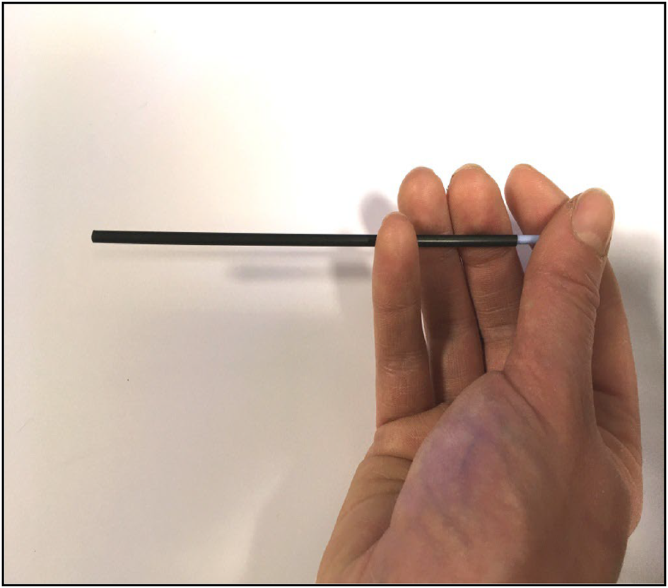
Fig. 1. Depicting how to hold the cautery stick covered by the straw.

Fig. 2. Demonstrating the protective mechanism of the straw covering the cautery stick within the nasal cavity.
Step four
When the covered cautery stick is within close proximity and directly over the bleeding point, advance the cautery stick forwards (Figures 3 and 4). Then, pull the cautery stick back to ensure the straw covers it once again. Observe the bleeding point and repeat again if further cautery is required. When removing the equipment from the nasal cavity, confirm the straw is covering the cautery stick end.
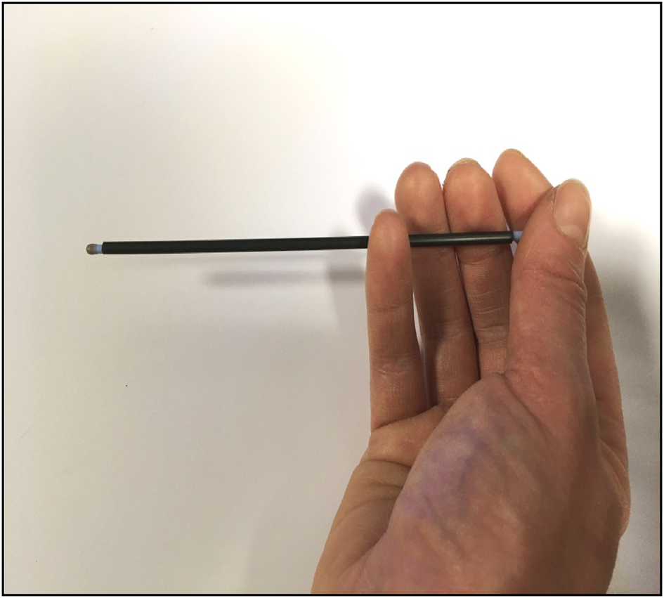
Fig. 3. Demonstrating how to advance the cautery stick from the straw.

Fig. 4. Displaying how the straw enables precise cauterisation without touching surrounding mucosa within the nasal cavity.
Conclusion
The use of a plastic straw to aid cautery of the posterior nasal cavity helps prevent damage to surrounding healthy nasal mucosa, which could result in infections, bleeding and adhesions. This technique simplifies a procedure such that a junior doctor with little otolaryngology experience could successfully cauterise the posterior nasal cavity. The straw enables greater fine control of the cautery stick given its rigidity and small diameter, providing an advantage over using suction tubing.
Competing interests
None declared


