


Introduction
Pathological air expansion of the paranasal sinuses is a rare cause of facial pressure symptoms and visual disturbance. An expansile, cystic lesion of the sinus was first described by Meyes in 1898, and was termed sinus pneumatocoele.Reference Benjamins1 The two predominant terms used in the literature are pneumosinus dilatans and pneumocoele, the distinction being based on the presence of bone erosion, as proposed by Benjamin in 1918.Reference Benjamins1 Other terms used include pneumatocoele, pneumosinus frontalis, sinus hypertrophy, aerocoele, hyperpneumatisation, sinus ectasia and sinus blistering. Over the years, inconsistencies in terminology definition have led to a tendency to use such terms interchangeably. It is quite possible that most of these lesions represent a spectrum of an essentially similar underlying pathology.
Most recent authors have adopted a classification system based on a spectrum of radiological and clinical findings, as described by Urken et al.,Reference Urken, Som, Lawson, Edelstein, Weber and Biller2 which uses the terms hypersinus, pneumosinus dilatans and pneumocoele.
A hypersinus (an abbreviated form of ‘hyper-pneumatised-sinus’) is an aerated sinus with normal walls confined within the normal boundaries of the sinus, and is asymptomatic.
Pneumosinus dilatans is an aerated sinus that expands beyond the normal boundaries (either focally or generally), with an intact and normal wall thickness, and is clinically symptomatic.
A pneumocoele is an aerated sinus with thinning of, or a defect in, the bony sinus walls (either focal or generalised), and loss of integrity of the bone.
In addition, pneumosinus dilatans affecting all sinuses has been described as pneumosinus dilatans multiplex.Reference Hwang, Lee, Lee and Lee3, Reference Kiroglu, Karabulut, Sabir, Yagci, Gakmak and Ozguler4 Furthermore, a pneumatocoele is distinguished as a subperiosteal collection of air on the external wall of the sinus, usually due to trauma, infection, tumour or radiotherapy.
Controversy over the correct nomenclature of these abnormalities has resulted in the use of the term ‘air cyst’ of a paranasal sinus, in order to cover all lesions of this type.Reference Tovi, Gatot and Fliss5
Case report
An 18-year-old, otherwise healthy man presented to an otolaryngology clinic with a three-month history of right facial pain. An initial dental review had led to a right upper molar extraction. However, the pain had progressed, with associated periorbital swelling and right nasal obstruction. There had initially been some bubbling through the extracted tooth socket, but this had settled?
Clinical examination revealed mild bony right cheek swelling, with no evidence of diplopia, paraesthesia or oroantral fistula. There was significant narrowing of the right nasal cavity due to medialisation of the right lateral wall complex posteriorly, including the inferior turbinate, with associated mild oedema.
A computed tomography scan of the paranasal sinuses (Figure 1) demonstrated pneumatic dilatation of the right maxillary sinus, with bony erosion and a small degree of air within the periorbital fat. The lesion was reported as pneumosinus dilatans, despite obvious bony erosion.

Fig. 1 (a) Axial and (b) sagittal computed tomography (CT) images of a right maxillary air cyst (arrow), reported as pneumosinus dilatans. (c) Coronal CT image showing thinning and erosion of the bone in the roof of the maxillary sinus (arrows), suggesting that this is a pneumocoele rather than pneumosinus dilatans.
A wide middle meatal antrostomy was performed endoscopically under general anaesthesia, including resection of the posterior inferior turbinate, without complication. There was no evidence of mucosal disease within the maxillary sinus or nasal cavity.
The patient made an uncomplicated recovery, with complete resolution of symptoms.
We conducted a literature search of the PubMed, Medline and Embase databases, using the key words ‘pneumosinus dilatans’, ‘pneumoc(o)ele’, ‘pneumatoc(o)ele’ and ‘maxillary sinus’. Articles were also hand-searched. Relevant articles published in English were reviewed. The results of this review are shown in Table I.
Table I Reported cases* of pneumosinus dilatans and pneumocoele
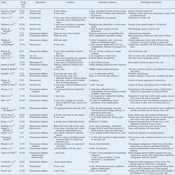
* Published in English. Pt = patient; yrs = years; M = male; R = right; max = maxillary; mths = months; inf = inferior; lat = lateral; F = female; med = medial; L = left; ant = anterior; bilat = bilateral; post = posterior; sup = superior; CT = computed tomography
Discussion
The maxillary sinus is filled with fluid in utero, and only becomes pneumatised at birth. It grows rapidly during the first three years of life, and again between the ages of seven and 12 years. The adult maxillary sinus is pyramidal in shape.
Enlargement of the maxillary sinus is rare, and can be due to air (pneumosinus dilatans or pneumocoele), mucus (mucocele) or tumour.Reference Lawson, Patel and Lin6
Pneumosinus dilatans and pneumocoeles can arise in any of the paranasal sinuses, and can be bilateral. However, they are relatively unusual in the maxillary sinuses, compared with the frontal and sphenoid sinuses.
In our literature review data, the mean age of presentation was 27 years (range, nine to 62 years), with a sex ratio slightly in favour of males (23 males to 13 females). We identified a total of 36 reported cases (including the present case) involving the maxillary sinus, with 19 cases reported as pneumosinus dilatans, 12 as pneumocoeles, two as pneumatocoeles, two as pneumosinus dilatans multiplex and one as an air cyst.
However, if the classification proposed by Urken et al. Reference Urken, Som, Lawson, Edelstein, Weber and Biller2 is strictly applied, the presence of bone erosion in the majority of these cases (including our own case) suggests that they should be reclassified as pneumocoeles. This implies that lesions at the more severe end of the spectrum are relatively more common in the maxillary sinus. However, an alternative explanation may be that because there is more room for expansion in the maxillary sinus, compared with the frontal and sphenoid sinuses, these lesions become more advanced before the onset of symptoms.
Aeration of the maxillary sinus most commonly results in expansion postero-medially into the infratemporal fossa and superiorly into the orbital floor. The right maxillary sinus is more commonly affected. Clinically, lesions become symptomatic when expansion results in pressure effects on local structures, or becomes cosmetically apparent. The commonest symptoms affecting the maxillary sinus are a cheek mass or swelling, pain, fullness, numbness, facial asymmetry, eye symptoms (e.g. proptosis), and nasal obstruction. Sphenoid lesions may present with increasing loss of vision and reduction of visual fields, and are often found in association with meningiomas. In fact, sphenoid pneumosinus dilatans can represent the first sign of a meningioma at the tuberculum sellae or the planum sphenoidale.Reference Hirst, Miller and Allen7, Reference Miller, Golnik, Zeidman and North8
The aetiology of paranasal sinus air cysts remains something of an enigma. Generally, they are described as primary or secondary. The proposed aetiologies of primary (idiopathic) cases include a one-way valve effect, gas-forming organisms, spontaneously discharged mucocele and dysregulation of hormones.
Many authors have suggested a one-way valve mechanism. This is supported by the presence of polypoid mucosa in the drainage pathway of the affected sinus in some cases.Reference Harrison and Young39 The concept may also explain increasing symptoms associated with air travel and atmospheric pressure changes, or during sneezing. Previous authors have documented elevation of sinus pressures;Reference Wolfensberger16, Reference Braverman38 the bubbling of gas through the extracted tooth socket in our patient's case would support this. However, most cases do not demonstrate sinus outflow obstruction, and the question as to why the sinus does not fill with mucus is still debated. The theory is also contradicted by the presence of focal expansions within the sinus in some cases.Reference Nayak, Pujary, Ramaswamy, Mahesh and Muddaiah35 The raised intra-sinus pressure may represent an end-stage effect of the process when the sinus has expanded to such an extent that it has occluded its own ostium, rather than representing a true causative phenomenon.
Other authors have suggested infection with a gas-forming organism as a possible aetiological factor; however, such an organism is yet to be identified.Reference Walker and Jones40 It is a feature of these abnormalities that the mucosa has a normal appearance, with functioning cilia.Reference Urken, Som, Lawson, Edelstein, Weber and Biller2, Reference Lombardi, Verdeja, Schellenberg and Blanchard37
Some authors have described symptoms of sudden, spontaneous nasal discharge, indicating the possibility of an air cyst forming from a discharged mucocele.Reference McNally, Stuart and Childe41, Reference Benedikt, Brown, Roth, Geyer and Ghaed42
Further theories relate to the dysregulation of sex and growth hormones, resulting in the stimulation of osteoblastic and osteoclastic activities, producing abnormal expansion.Reference Gardel and Maduro43 However, no hormonal alteration has thus far been recorded.Reference Smith, Maran and Von Haacke44
• Pathological air expansion of the maxillary sinus is relatively rare, with only 36 previously reported cases
• The nomenclature is confusing; the commonest terms are pneumosinus dilatans and pneumocoele
• The main difference between these two conditions is radiological, and does not alter management
• Use of the single term ‘air cyst’ to describe these lesions is supported
In syndromic (secondary) cases, observations particularly relating to the frontal sinus have noted over-development of the sinus occurring in association with under-development of the brain (e.g. in cranio-cerebral hemiatrophy),Reference Dyke, Davidoff and Masson45 or due to long-term shunting resulting in decreased intracranial pressure, hence allowing for expansion of the sinuses.Reference Van Schayck and Niedeggen46 Air cysts have also been described in association with Melnick–Needles syndrome,Reference Stretch and Poole19 Klippel–Trenaunay–Weber syndromeReference Spoor, Kennerdell, Maroon, Hepler and Krohel47 and arachnoid cysts.Reference Sener48, Reference Martin, Jarosz and Thomas49 Two of the five reported cases of pneumosinus dilatans multiplex were associated with mental retardation and facial deformity.Reference Hwang, Lee, Lee and Lee3, Reference Kiroglu, Karabulut, Sabir, Yagci, Gakmak and Ozguler4
The question remains as to whether pneumocoeles and pneumosinus dilatans represent a progression of a single pathological process, or whether they have separate aetiologies. Hitherto, there have only been two documented cases of pneumosinus dilatans progressing to pneumocoele: one in the frontal sinusReference Rossi, Fondelli, Piatelli, Gandolfo, Borasi and Tortori Donati50 and the other in the maxillary sinus.Reference Vlckova and White36 In general, the onset of pneumosinus dilatans tends to be chronic (developing over years), as opposed to pneumocoeles, the symptoms of which develop quickly over a number of months. However, this may simply be due to pneumocoele expansion reaching a critical size at which symptoms rapidly progress.
Computed tomography imaging is the most important modality in the diagnosis of these lesions. Magnetic resonance imaging should be used to exclude concomitant intracranial lesions in sphenoid and frontal lesions. Noyek et al. Reference Noyek, Zizmor, Morrison, Vines and Shaffer51 studied the radiological signs in four cases of pneumocoele; all showed an enlarged sinus, thinning of the sinus wall, hyperaeration, hyperlucency, bony dehiscence, expansion of recesses, effacement of the ethmomaxillary angle and displacement of the middle meatus. Three of the four cases demonstrated profiling of dental roots and displacement of the orbital floor. In our literature review, 12 cases showed thinning of the sinus wall, most commonly the anterior and orbital wall.Reference Tovi, Gatot and Fliss5, Reference Zizmor, Bryce, Schaffer and Noyek10, Reference Meyers and Burtschi13, Reference Wolfensberger16–Reference Chan, Chow and Sham18, Reference Omnell and Rohlin20, Reference Breidahl, Szwajkun and Chen21, Reference Dillard and Sillers23, Reference Juhl, Buchwald and Bollinger25, Reference Knapp and Klenzner32, Reference Viehweg and Hudson34 Only one case described bone thickening instead of thinning.Reference Lloyd14
Symptomatic lesions require surgical intervention. The commonest procedure performed is creation of a naso-antral window, aiming to equilibrate the intrasinus pressure. Thus far, no recurrence has been reported in treated lesions.
Conclusion
This report describes a case of pathological paranasal sinus expansion secondary to air, a rare but clinically significant type of sinus pathology. It also highlights the confusion regarding the nomenclature of such lesions. While it is possible that pneumosinus dilatans and pneumocoeles represent distinct pathological entities, the distinction is based more on radiological than clinical features. Furthermore, the distinction does not alter the management in the event of symptomatic cases, which comprises surgical decompression usually through enlargement of the anatomical ostium. We therefore support the use of a single term, such as ‘aircyst’, to describe these lesions.Reference Tovi, Gatot and Fliss5


