


Introduction
Piezoelectric technology has been used for many years as a surgical tool for soft tissue cavitation, and more recently for cutting bone using micro-vibrations. It has a widening range of surgical applications, as demonstrated by the increasing literature discussing its use in cranial and spinal surgery,Reference Bydon, Xu, Papdemetriou, Sciubba, Wolinsky and Witham 1 – Reference Kshettry, Jiang, Chotai and Ammirati 3 and in craniofacial,Reference Kshettry, Jiang, Chotai and Ammirati 3 – Reference Gleizal, Bera, Lavandier and Beziat 5 orbital,Reference Gonzalez-Lagunas and Mareque 6 – Reference Mostovych, Rabinowitz, Bilyk and Pritbitkin 8 handReference Hoigne, Stubinger, Von Kaenel, Shamdasani and Hasenboehler 9 and facial reconstructive surgery.Reference Robiony, Toro, Costa, Sembronio, Polini and Politi 10 – Reference Robiony, Polini, Costa, Torro and Politi 12
Ultrasonic aspirators operate on the converse piezoelectric effect, whereby application of an electric charge to certain crystals creates a reversible mechanical deformation. Piezoelectric crystals in the handpiece of the instrument rapidly expand and contract, causing high-frequency vibration of the instrument tip, which denatures proteins and emulsifies bone. The resulting emulsified tissue is removed by continuous irrigation and suction. The vibration frequency can be optimised for either bone or soft tissue removal,Reference Pritbitkin, Lavasani, Shindle and Greywoode 13 and the power can be modulated to achieve effective cutting across varying densities of bone.
The use of ultrasonic bone aspirators in otolaryngology has somewhat lagged behind its uptake in maxillofacial surgery and neurosurgery. This may be because, in the head and neck, vital structures are located in close proximity to each other, and accessing these areas requires instrument insertion via a narrow surgical corridor. Recent advances in handpiece and tip design have increasingly allowed ultrasonic aspirators to be applied in otological, rhinological, skull base and laryngological procedures.
Objectives
This study aimed to examine the role of ultrasonic bone dissectors in the field of otolaryngology, and head and neck surgery. Specifically, the current literature was examined to gain evidence for benefits and disadvantages of this technology and its application across different subspecialties. We also examined differences compared with traditional surgical methods, in terms of intra-operative blood loss, histological characteristics of cut bone and adjacent tissue, learning curve, operative time, and post-operative pain.
Materials and methods
We conducted a search of the databases Medline, the Cochrane Central Register of Controlled Trials (‘CENTRAL’), PubMed, Embase, Cambridge Scientific Abstracts and other resources for published literature pertaining to the use of ultrasonic aspirators in otolaryngology, and head and neck surgery. Key words included: ‘ultrasonics’, ‘piezosurgery’, ‘curettage’, ‘otolaryngology’, ‘otologic’, ‘laryngeal’ and ‘rhinoplasty’.
There were no limitations on the search. Abstracts were screened for relevance and reviewed once selected. The references of these articles were also screened for further relevant literature. Review articles, letters and correspondence papers were excluded. The most recent search was undertaken on 20 June 2016.
Piezosurgery in otology
Piezoelectric surgery has increasingly been applied in the field of otology.Reference Salami, Dellepiane, Proto and Mora 14 – Reference Golub, Weber, Leach, Pottschmidt, Zuccarello and Pensak 30 Since 2007, reports of the ultrasonic bone aspirator used for otological surgery have consistently shown both technique feasibility and safety, particularly in regard to hearing outcomes.
Salami et al. have published several series, including a large prospective study (n = 133), published in 2009, of post-operative outcomes after platinotomy, mastoidectomy, antro-atticotomy, posterior tympanotomy, facial nerve decompression and middle-ear tumour removal.Reference Salami, Dellepiane, Proto and Mora 14 They found no evidence of decline in pure tone audiometry (PTA), tympanometry, otoacoustic emissions (OAE), electronystagmography (ENG) or auditory brainstem responses (ABR) following piezosurgery.
In 2010, the same group compared the ultrasonic bone aspirator to the micro-drill for intact canal wall mastoidectomy, both primary and revision, and found that operative time and hearing outcomes were comparable.Reference Salami, Mora and Dellepiane 17 , Reference Crippa, Salzano, Mora, Dellepiane, Salami and Guastini 23 Their results with the ultrasonic bone aspirator for stapedotomy, and removal of osteomas and glomus tumours have similarly shown preservation of hearing and vestibular function, without intra-operative adverse events.Reference Salami, Mora, Dellepiane and Guastini 18 , Reference Salami, Dellepiane, Mora, Crippa and Mora 19 , Reference Salami, Mora, Dellepiane, Crippa and Guastini 21
The ultrasonic bone aspirator has also been used for facial nerve decompression. An initial cadaveric study of 17 temporal bones demonstrated the feasibility of piezosurgery to decompress the labyrinthine segment of the facial nerve.Reference Sami, Krishnamoorthy and Pensak 24 Only one specimen showed evidence of epineurial injury. Since then, several clinical studies have reported ultrasonic bone aspirator facial nerve decompression through all intratemporal segments, and to date no facial nerve injury has been reported, and no patients showed decline on PTA, ABR, OAE or ENG.Reference Salami, Dellepiane, Mora, Crippa and Mora 19 – Reference Dellepiane, Mora, Salzano and Salami 22
The current evidence suggests that the ultrasonic bone aspirator is a useful tool which potentially reduces surgical morbidity. The device's selectivity in aspirating bone, whilst preserving soft tissues, is particularly relevant in otology, where the lateral sinus, dura mater and facial nerve exist encased in bone, within a confined space. All are at higher risk of damage should they be inadvertently touched by conventional rotatory micro-drills. Further advantages cited by users include the availability of specifically designed handpieces to suit multiple approaches, including transcanal, transmastoid, translabyrinthine and endoscopic approaches.Reference Salami, Vercelloti, Mora and Dellepiane 25 , Reference Kakehata, Watanabe, Ito, Kubota and Furukawa 27 , Reference Presutti, Alicandri-Ciufelli, Cigarini and Marchioni 28
A clinically relevant advantage is the reduction in bone dust created by the ultrasonic bone aspirator when compared to the micro-drill. Bone dust is hypothesised to contribute to post-operative headaches in patients who have undergone retrosigmoid approaches, and the intra-operative reduction of bone dust may decrease morbidity. A cadaveric temporal bone quantitative study compared residual bone dust using the ultrasonic bone aspirator with that using the micro-drill during retrosigmoid approaches, and concluded that the ultrasonic bone aspirator resulted in a 25-times reduction in bone dust dispersal compared to the micro-drill, but a longer operative time was required.Reference Weber, Samy, Nahata, Zuccarello, Pensak and Golub 29 A follow-up clinical study of nine patients undergoing retrosigmoid vestibular schwannoma removal found that ultrasonic bone aspirator use practically increased operative time by only 5–15 minutes.Reference Golub, Weber, Leach, Pottschmidt, Zuccarello and Pensak 30 Further studies are needed to quantify whether the reduction in bone dust does in fact reduce post-operative headache, in order to justify a longer procedure time.
Piezosurgery and rhinoplasty
Piezosurgery was first applied to rhinoplasty in performing lateral osteotomies via a percutaneous approach, and later gained acceptance for medial osteotomies and dorsal hump reduction.Reference Robiony, Toro, Costa, Sembronio, Polini and Politi 10 – Reference Robiony, Polini, Costa, Torro and Politi 12 Three large studies have demonstrated the widening applications and potential advantages of piezosurgery in rhinoplasty.Reference Pritbitkin, Lavasani, Shindle and Greywoode 13 , Reference Gerbault, Daniel and Kosins 31 , Reference Greywoode and Pritbitkin 32
In one retrospective case review that included both primary (n = 55) and secondary (n = 5) rhinoplasties, all patients had their bony nasal dorsum sculpted using the Spetzler Claw tip of the Sonopet® ultrasonic bone aspirator.Reference Pritbitkin, Lavasani, Shindle and Greywoode 13 Outcome measures included visible and palpable dorsal irregularities, under- or over-resection of the dorsum, and asymmetry. The authors found no major short-term complications, and at six months only one patient had visible dorsal asymmetry.
The same authors published further results on 103 rhinoplasty patients, including smoothing of palpable osteotomy margins, nasal spine reduction, glabellar sculpting and turbinate reduction.Reference Greywoode and Pritbitkin 32 Complication rates were less than 2 per cent, comparable with the senior author's experience prior to the uptake of this technology. Advantages of use were precise cutting and sculpting of bone, even when fragments were mobile, which were possible because of the very light pressure required to achieve bony emulsification. Anecdotally, less intra-operative bleeding was observed, hypothesised to be the result of avoidance of disrupting intranasal mucosa with conventional osteotomes.
A more recent series of 82 primary and 103 secondary rhinoplasties using piezosurgical techniques revealed more specific technical considerations.Reference Gerbault, Daniel and Kosins 31 Of note, the authors recommended extensive subperiosteal dissection, including the release of pyriform aperture ligaments, in order to best visualise ultrasonic bone aspirator osteotomies and the shaping of bony asymmetries. Additional advantages included increasingly stable osteotomies, and the avoidance of radial and asymmetric fracture lines, and damage to the periosteum and underlying mucosa, when compared to conventional techniques. Disadvantages included cost, increased operating time, limited usefulness via an endonasal approach and the learning curve associated with using the ultrasonic bone aspirator (30 cadaver dissections were performed prior to clinical use). Limitations of the currently available studies include the lack of: controls, long-term results and objective data regarding operative time.
Piezosurgery in rhinology
Technological innovation has advanced rhinological techniques. Tissue removal has been facilitated by the development of the microdebrider, radiofrequency ablation and endoscopic drills.Reference Sindwani and Manz 33 The development of handpieces and tips (Figures 1 and 2) that allow the ultrasonic aspirator to be used within the narrow confines of the nasal cavity, under endoscopic visualisation, has seen its use increasingly reported.
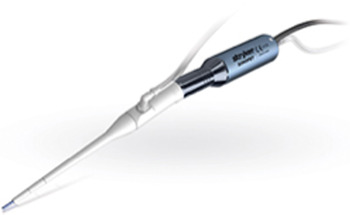
Fig. 1 Sonopet® ultrasonic bone aspirator handpiece. Image reprinted with permission from Stryker®.

Fig. 2 Sonopet® ultrasonic bone aspirator tips. Image reprinted with permission from Stryker®.
Dacryocystorhinostomy
Antisdel et al. first reported their experience using the ultrasonic aspirator in dacryocystorhinostomy (DCR).Reference Antisdel, Kadze and Sindwani 7 Their retrospective review of 16 patients undergoing DCR revealed that the ultrasonic aspirator was effective in the creation of a 10–12 mm bony rhinostomy in all cases. Since then, several studies have reported similar experiences, with particular reference to ‘blood-free operative fields, and no observed cases of damage to surrounding mucosa or inadvertent penetration of the sac’, which authors attributed to its non-rotational design and tissue selectivity.Reference Mostovych, Rabinowitz, Bilyk and Pritbitkin 8 , Reference Salami, Dellepiane, Salzano and Mora 34 – Reference Chappell, Moe and Chang 37 All patients had symptom resolution at 12–20 months post-operatively and normal functional dye test results, and, endoscopically, none had synechiae formation.
Two retrospective institutional reviews compared primary endoscopic DCR performed with the ultrasonic bone aspirator and traditional rotational drilling.Reference Murchison, Pritbitkin, Rosen and Bilyk 35 , Reference Steele, Wilson and Strong 36 In both studies, the two groups were demographically comparable, and there were no cases of cerebrospinal fluid leak, visual loss, diplopia, infection or epistaxis in either group. No statistically significant differences between surgical success or complication rates were found at a follow up of 2–26 months.
Septoplasty, turbinoplasty and piriform aperture enlargement
Turbinoplasty was initially reported in a small retrospective case review and prospective case series (n = 7) with a patient questionnaire.Reference Pritbitkin, Lavasani, Shindle and Greywoode 13 , Reference Greywoode, Van Abel and Pritbitkin 38 The inferior turbinate bone was dissected free of the overlying mucosa, and the aspirator was used to emulsify the conchal bone, before lateralisation. Similar findings were observed in a larger retrospective review of 30 patients undergoing septoplasty and inferior turbinate reduction, which compared a microdebrider with a microdebrider and ultrasonic bone aspirator.Reference Kim, Choi and Kwon 39 The ultrasonic bone aspirator was used to thin hypertrophied turbinate bone, and during septoplasty it was employed to reduce bony spurs and the maxillary crest, whilst preserving mucoperichondrium. In both studies, the authors reported no episodes of post-operative haemorrhage, synechiae, prolonged crusting or necrosis. Technical limitations included difficulties in reaching the posterior aspect of the turbinate. No significant improvement over traditional methods could be demonstrated.
Roy et al. recently published their retrospective chart review of 26 patients who underwent treatment for nasal obstruction, including enlargement of the piriform aperture using the ultrasonic bone aspirator via a 1.5 cm intranasal incision.Reference Roy, Iloreta, Bryant, Krein, Pribitkin and Heffelfinger 40 All patients had other surgical manoeuvres; therefore, this study represents a feasibility study of piriform aperture enlargement as an adjunctive procedure. No significant added morbidity or cosmetic change resulted (based on a review of peri-operative photography by a qualified, blinded evaluator).
Endoscopic sinus surgery
The first reported use of the ultrasonic aspirator in endoscopic sinus surgery was a case description of frontal sinus osteoma removal.Reference Pagella, Giourgos, Matti, Colombo and Carena 41 There are at least three further case reports detailing the use of ultrasonic aspiration in the management of fronto-ethmoidal osteomas.Reference Gotlib and Niemczyk 42 – Reference Villaret, Schreiber, Esposito and Nicolai 44 All reported no increase in operative time, a decrease in mucosal injury in adjacent areas and good visualisation around the curved handpiece. Other purported advantages included the lack of ‘blunting’, ‘skip’ or ‘chatter’ that may occur with rotatory drills, and subjectively decreased bone dust production using the ultrasonic aspirator.
The ultrasonic bone aspirator has also been used for maxillary antrostomy, and for opening the ethmoid and frontal recesses in chronic rhinosinusitis patients, in both primary and revision cases.Reference Bolger 45 , Reference Mancini, Buonaccorsi, Reale and Massimiliano 46 Both studies reported improved mucosal flap preservation, rapid healing time, and removal of thick bone without applying torque or significant pressure to the instrument. There are no large case series or comparative trials presently available to determine objective results of ultrasonic bone aspirator use in sinus surgery.
Endoscopic skull base surgery
There is a growing body of literature concerned with the use of ultrasonic aspiration in endoscopic skull base surgery, in both adults and the paediatric population.Reference Stapleton, Tyler-Kabara, Gardner and Snyderman 47 In 2003, the advent, and subsequent use of the handpiece for trans-sphenoidal pituitary surgery, was reported.Reference Yamasaki, Moritake, Nagai, Uemera, Shingu and Matsumoto 48 Since then, several case reports and small series of anterior skull base tumour excisions have demonstrated the feasibility of transnasal, endoscopic ultrasonic bone aspirator use.Reference Burgin, Porter, Mehrota and Welch 49 , Reference Lubbe, Fisher-Jeffes and Semple 50 Although the studies lack power and objective outcome measures, the authors felt that tissue selectivity, ‘bloodless dissection’ in the region of critical neurovascular structures and intra-operative visualisation around the handpiece were superior to traditional drilling.
Baddour et al. performed a prospective, randomised, single-blinded, controlled clinical trial comparing the efficacy of ultrasonic bone aspiration versus traditional rotational drilling in over 130 endoscopic pituitary tumour resections.Reference Baddour, Lupa and Patel 51 All cases were primary surgery, all participants were blinded to the technology used in their case and all operations were performed by the senior author. Primary outcomes were operative time and blood loss, both of which were found to be statistically significantly reduced (Table I).
Table I Use of sonopet® ultrasonic bone aspirator versus traditional instrumentation*

* During an endoscopic trans-sphenoidal approach in pituitary tumour resection (adapted from Baddour et al.Reference Baddour, Lupa and Patel 51 )
That study represents the only randomised controlled trial to compare the ultrasonic bone aspirator to traditional cold steel drilling. It is adequately powered and statistical analysis was rigorous. The authors concluded that use of the ultrasonic bone aspirator for an endoscopic approach to the skull base was safe (there were no adverse events reported for the ultrasonic bone aspirator group) and efficient. However, in a cost analysis, they found that the average cost per case using the ultrasonic bone aspirator was significantly higher than that of traditional instrumentation.Reference Baddour, Lupa and Patel 51
The experience of our institution is similarly satisfactory; we have used the ultrasonic bone aspirator in 24 cases of trans-sphenoidal pituitary tumour resections and 2 clival chordoma excisions. The ‘Superlong’ access tips allow access to the skull base, whilst the narrow, angled handpiece allows easy endoscopic visualisation of the tip within the surgical field. The range of tips that we have found useful in skull base surgery includes the Payner Superlong 360 and the Spetzler Superlong Open Angle tips, which allow bony evisceration using light handpiece pressure with superior tactile feedback. The 360-degree tip (Figure 3) is particularly useful for bone removal within the sphenoid sinus, without application of torque to the handpiece. The lack of a rotating tip allows precise bone cutting, with no concern of winding up cotton pads, and less ‘skip’ or ‘run on’. In areas where critical neurovascular structures are in close proximity, the claw provides the advantage of a non-traumatic side that may be kept facing the area of concern. Inadvertent contact with mucosa has not to our observation caused macroscopic soft tissue injury, including bleeding. This, combined with suction and irrigation at the tip, has improved field visualisation during skull base and tumour exposure.

Fig. 3 Payner™ 360 ultrasonic bone aspirator tip. Image reprinted with permission from Stryker®.
Piezosurgery in laryngology
Medialisation thyroplasty requires the removal of a window of thyroid cartilage, without disturbance of the perichondrium on the laryngeal surface, which is a known complication of traditional drilling methods. The ultrasonic bone aspirator shows promise in reducing this potential complication because of its tissue selectivity. A cadaveric study comparing laryngeal windows made with a standard surgical drill on one side versus the ultrasonic bone aspirator on the other has been performed.Reference Halum, Patel, Hoffman, Simpson and Merati 52 Outcome measures were: time required to create the window and perichondrium status. The ultrasonic bone aspirator compared favourably to the standard drill in terms of operative time, and there were fewer perichondrium perforations with the ultrasonic bone aspirator, although neither result reached statistical significance. These same outcomes have been tested in vivo, and the techniques were found to be comparable in terms of surgical success, complication rate and operative time.Reference Daniero, Spiegel, Brody and Fickes 53
The ultrasonic bone aspirator has also been described for endoscopic posterior cricoid split and cartilage graft laryngoplasty.Reference Yawn, Daniero, Gelbard and Wootten 54 Three adult patients underwent posterior cricoid split and cartilage grafting using the angled handpiece and Spetzler Micro Claw tip. This small series demonstrated feasibility of its use in endoscopic laryngeal procedures, and the authors commented on the ability of the ultrasonic bone aspirator to split even highly ossified laryngeal cartilage, which is frequently challenging to perform using traditional cold steel instrumentation, without causing inadvertent soft tissue injury.
Piezosurgery advantages
Intra-operative blood loss
The only randomised controlled trial to quantify intra-operative blood loss was that by Baddour et al.Reference Baddour, Lupa and Patel 51 Their study compared trans-sphenoidal pituitary surgery using the ultrasonic bone aspirator versus a traditional rotating drill. They found statistically significantly less intra-operative blood loss with the ultrasonic bone aspirator than the drill (16.55 ml vs 22.58 ml; p < 0.0001). Anecdotally, there is support for the ultrasonic bone aspirator providing good haemostasis in otological proceduresReference Salami, Dellepiane, Proto and Mora 14 – Reference Crippa, Salzano, Mora, Dellepiane, Salami and Guastini 23 and DCR.Reference Mostovych, Rabinowitz, Bilyk and Pritbitkin 8 , Reference Murchison, Pritbitkin, Rosen and Bilyk 35
Reduced tissue injury and necrosis
Several studies have examined histological characteristics of ultrasonic aspiration on bone and cartilage. One study examined nasal cartilage directly sculpted with the ultrasonic bone aspirator, and no loss of chondrocytes was observed.Reference Greywoode and Pritbitkin 32 Another study examined the bony defects left by the ultrasonic bone aspirator on different bony samples.Reference Claire, Lea and Walmsley 55 Cuts made in cortical bone were more precise than those in cancellous bone, a finding that has also been noted in vitro.Reference Romeo, Del Vecchio, Palaia, Tenore, Visca and Maggiore 56 Cut surfaces examined by other authors similarly showed reduced cellular damage and no evidence of thermal coagulative necrosis compared to rotative instruments, which authors hypothesise may accelerate healing.Reference Salami, Mora and Dellepiane 17 , Reference Daniero, Spiegel, Brody and Fickes 53 , Reference Salami, Dellepiane, Crippa and Mora 57 , Reference Rullo, Addabbo, Papaccio, D'Aquino and Festa 58
Wound healing and post-operative pain
No human studies have compared the tissue healing response following application of the ultrasonic bone aspirator versus conventional rotating drills. Animal studies have shown faster initial wound healing and decreased epineurial injury with the ultrasonic aspirator.Reference Metzger, Bormann, Schoen, Gellrich and Schmelzeisen 59
The reduction in heat generation with the ultrasonic bone aspirator offers a theoretical reduction in adjacent tissue injury and inflammation, and a potential reduction in post-operative pain. This has not been extensively studied, however, and currently available results have been inconclusive. A comparison of post-operative pain in patients undergoing intact canal wall mastoidectomy with either piezosurgery or micro-drills found that self-reported pain and analgesic use was lower on both days 1 and 3 post-operatively in the piezosurgery group, versus the micro-drill, but there was no statistical difference between groups at day 10. Similarly, data from maxillofacial and dental literature have not demonstrated a clear improvement in post-operative pain after ultrasonic bone aspirator use.Reference Rullo, Addabbo, Papaccio, D'Aquino and Festa 58
Piezosurgery limitations
Learning curve
As with any new technology, the learning curve for an instrument must be considered. A comparison of senior surgical staff and residents using both the ultrasonic bone aspirator and rotating drills for the creation of laryngeal windows suggested that the ultrasonic bone aspirator technique is ‘easily learned’, with resident performance times ‘closer to staff performance times using the aspirator for laryngeal window construction’ compared to the drill.Reference Halum, Patel, Hoffman, Simpson and Merati 52 , Reference Daniero, Spiegel, Brody and Fickes 53
Cadaveric studies in which the ultrasonic bone aspirator was used to decompress the facial nerve found that operative time halved between the first and second sessions of decompression, providing objective evidence for a short inherent learning curve.Reference Salami, Mora, Mora, Guastini, Salzano and Dellepiane 26 This has been anecdotally reported by several other authors,Reference Salami, Mora, Dellepiane, Crippa, Santomauro and Guastini 15 , Reference Chappell, Moe and Chang 37 , Reference Mancini, Buonaccorsi, Reale and Massimiliano 46 , Reference Wick, Rezaee and Zender 60 without quantification. Thus, several groups recommend an audited training period for surgeons adopting ultrasonic technology.
Operative time
Few studies have directly examined the impact on operative time. Several groups have measured operative time when using the ultrasonic bone aspirator versus rotating drills for mastoidectomy, and found no statistical difference.Reference Salami, Dellepiane, Proto and Mora 14 , Reference Salami, Mora, Dellepiane, Crippa, Santomauro and Guastini 15 , Reference Salami, Vercelloti, Mora and Dellepiane 25 , Reference Salami, Mora, Mora, Guastini, Salzano and Dellepiane 26 In fact, the only study to show a statistically significant reduction in operative time was the aforementioned randomised controlled trial comparing the use of the ultrasonic bone aspirator with rotating drills in trans-sphenoidal approaches.Reference Baddour, Lupa and Patel 51
Anecdotally, authors have discussed a longer operative time when the bone encountered is very thick, such as osteitic bone of diseased paranasal sinuses.Reference Villaret, Schreiber, Esposito and Nicolai 44 , Reference Mancini, Buonaccorsi, Reale and Massimiliano 46 This was also observed when opening the internal auditory canal with the ultrasonic bone aspirator; however, the authors did not find this result statistically (or clinically) significant, and reported reduced surgeon stress because the lack of a rotating tip allowed ‘good control of the cutting process, and no danger of winding up cotton pads or … inducing accessory nerve reaction because of heat development’.Reference Grauvogel, Scheiwe and Kaminsky 20 It remains contentious as to whether any clinically significant improvement in operative time can be expected through using the ultrasonic bone aspirator over traditional methods.
Economic feasibility
A significant limitation of piezosurgery is economic feasibility. The available analyses suggest that the ultrasonic systems are significantly more expensive than rotational drilling systems.Reference Sindwani and Manz 33 The system itself can cost up to $130 000 (depending on the supplier). Furthermore, each tip is single-use only, costing approximately $300. Given the applicability of this technology across several specialties, this expense may be more justifiable where the cost of a single system is shared amongst multiple subspecialties.
Conclusion
Piezoelectric technology has multiple applications in surgery; specifically in otolaryngology, its use is increasingly described. It has been successfully employed, particularly in the fields of otology and skull base surgery, where improved handpiece and tip designs have allowed manipulation within narrow surgical fields, guided by either microscopy or endoscopy. Technical advantages include ease of use, a short learning curve, visibility and tissue selectivity. In comparison to traditional rotating drills, ultrasonic technology does not ‘blunt’, and its use avoids problems associated with ‘skip’ and ‘chatter’, and injury to soft tissues that are inadvertently contacted. However, this technology is more expensive, and clinically significant improvements in operative time, blood loss and morbidity have not yet been shown. Rather, the evidence supports piezoelectric surgery as comparable in terms of outcomes when compared to traditional surgical methods. Further studies may define the evolving role of piezoelectric surgery in otolaryngology, and head and neck surgery.


