Introduction
The condition known as patulous eustachian tube is generally defined by a cluster of symptoms: autophony, hearing one's own breathing within the ear, a sensation of fullness in the ear most often described as a feeling of ‘being under water’, and sometimes a slight hearing loss. These symptoms typically decrease or disappear when the individual is in a recumbent position or when pressure is applied to the jugular veins, and they often increase after physical exercise. The discerning symptom of autophony can be present in superior semicircular canal dehiscence too.Reference Chi, Ren and Dai1 Common causative factors of a patulous eustachian tube include hormonal changes, rapid weight loss, and neurological or muscular factors.Reference Bluestone, Magit, Brackmann, Shelton and Arriaga2, Reference Olthoff, Laskawi and Kruse3 It can also occur following an episode of otitis media.Reference Tsuji, Yamaguchi and Moriyama4
Rather accidentally, we found that the application of a paper patch on the tympanic membrane has a beneficial effect on patulous eustachian tube symptoms in many patients. We present the effects of paper patching observed in our patients over the last five years.
Materials and methods
Between December 2006 and May 2011, 21 patients were diagnosed as having patulous eustachian tube syndrome (33 ears). Diagnosis was made on the basis of patient history. Patients were included in the study if they experienced autophony or heard their own breathing. In most patients, a feeling of fullness was present and sometimes other complaints as well, such as hyperacusis, tinnitus or slight hearing loss (Table I).
Table I Patient data and results summary

Patients 1–5 were considered non-responders, patients 6–13 were classed as short-term responders and patients 14–21 were long-term responders. *Patient considered a non-responder (see text). †Patient had previously experienced patulous eustachian tube symptoms, but had been symptom-free for some time prior to the new episode. No. = number; y = years; F = female; mth = months; += present; – = absent; OME = otitis media with effusion; M = male
Most patients had no otological history. There were no suspected cases of semicircular canal dehiscence. Some patients suffered from episodes of relapsing otitis media with effusion. In a few patients a tympanic drain had been placed for acute otitis media or for treatment in a hyperbaric oxygen chamber.
Clinical examination, audiometry and tympanometry revealed no abnormalities. Movement of the tympanic membrane with breathing can sometimes be observed but is not necessary for the diagnosis. In fact it was almost never seen, presumably because our patients were examined in a supine position.
The paper patching technique involved the use of 1–3 small, slightly wet, rectangular 2–4 mm paper patches (Rizla blue cigarette paper, 14.5 g/m²; L Lacroix, Wilrijk, Belgium). These were gently laid onto the tympanic membrane: typically, two smaller patches were laid on the upper half, one anterior and one posterior to the malleus (Figure 1a), or one larger patch was laid to cover the inferior half (Figure 1b). This was done during the clinical examination. In our department, otomicroscopy is always performed with patients in a supine position. During the examination, the paper patches were picked up and gently placed on the tympanic membrane without announcing to the patient what was going to happen. The doctor performing the procedure behaved simply as if some cerumen was being removed. Dialogue with the patient continued in a casual manner. Without changing the casual tone of the conversation and without explaining what had been done, patients were then asked to sit upright and to state whether their symptoms had changed for the better, worsened, or not changed at all.
Fig. 1 Typical positions of the paper patches: (a) two patches covering the superior half of the tympanic membrane, and (b) one larger patch, covering the inferior half.
Moderate reactions were considered to be a placebo effect; the paper patch was removed in such cases. Only when patients showed an immediate and obvious expression of surprise was the result recorded as positive. In those cases, patients invariably asked for the patch to be left in place. These patients were seen again one month later.
Generally, patients were seen again some months afterwards to assess the long-term effect of paper patching. Patients who did not make a follow-up appointment were contacted by telephone several months after the treatment.
This study was approved by the ethics committee of our institution.
Results
The series comprised 18 women and 3 men, aged between 19 and 72 years (mean age of 46 years). There was no difference in age distribution between the sexes (Table I).
Symptoms occurred bilaterally in 12 patients and unilaterally in 9 patients (5 left ears, 4 right ears). Of the 21 patients, 19 (90.4 per cent) experienced autophony, 11 (52.4 per cent) heard their own breathing, and 12 (57.1 per cent) had a sensation of fullness or pressure. In 10 patients (47.6 per cent), the symptoms lessened when in a supine position.
There were clinical grounds for the role of a causative factor in only 13 (61.9 per cent) of the 21 patients. In six of these patients (all female), the suspected causative factors were atrophy of the nasal mucosal membranes, hormonal changes or rapid weight loss. In four patients (one male), the symptoms developed after an episode of otitis media or grommet insertion. In three patients (one male), we suspected a muscular factor. The evidence for a muscular cause is rather indirect. In one male patient, a muscular factor was suspected because he had complaints only while exercising or when stressed. A female patient with cervico-brachial complaints and temporomandibular dysfunction experienced more patulous eustachian tube symptoms when hurrying or walking up stairs (this patient's symptoms appeared after she started sleeping in a new bed). The other female patient felt immediate partial relief with the paper patch, and experienced total relief a few days later when she stopped chewing gum (something she had previously been doing on a regular basis).Reference Bluestone, Magit, Brackmann, Shelton and Arriaga2
In most patients who underwent this procedure, the paper patches were placed in distinct locations. Over the years, we have found that the two locations described above give the best results. Typically, patients responded to patches placed in only one of these two locations, responding only slightly or not at all to placement at the other location. A drawback of this retrospective study is that we did not consistently note where the patches were placed in individual patients.
For patch placement at the superior location, we placed one or two paper patches on the posterosuperior quadrant of the tympanic membrane, and one smaller patch on the anterosuperior quadrant, anterior to the malleus. We feel that the application of the anterosuperior patch, small as it is, is essential for the result. The exact location of the posterosuperior patch is often important: shifting this patch by only a millimetre can have a dramatic effect on outcome. For the placement of the inferior patch, the exact location is not important as long as it is positioned quite low, so that the external part of the tympanic membrane adjacent to the annulus is well covered.
Short-term results
In 76.2 per cent of patients (16 of 21 patients), symptoms disappeared immediately. Our first patients only received one paper patch; in some of these patients symptoms were relieved partially, and it was necessary to place a larger patch or several smaller patches, which then gave complete relief. In one case, after several placements of single patches that did not produce any result, a female finally responded to the placement of a very thick layer of paper covered with ointment, but this made her feel as though she had earwax and so we removed it (counting her as a non-responder).
In one patient, the symptoms decreased but did not disappear completely; however, the combination of the paper patch and stopping chewing gum produced total relief a few days later. In all other cases, the symptoms disappeared completely and immediately.
When symptoms were present bilaterally, patches were applied bilaterally, and patients felt relief in both ears. Although we do not have the specific outcome data for each symptom type, in most patients all symptoms diminished or disappeared simultaneously.
The five non-responders (all female) had very prominent symptoms. Four of the females had bilateral complaints, and hormonal changes or atrophy were suspected; in the fifth case, we found no specific causative factor. None of the five non-responders reported the symptomatic feeling of fullness.
Long-term results
Paper patches are dislodged from the tympanic membrane by the action of epithelial migration. In most patients, the dislodged patches could be found on the medial canal wall, one to two months after the procedure.
In 50 per cent of the responders (8 of 16 patients, i.e. 38.1 per cent of all patients), the symptoms reappeared when the paper patch was spontaneously dislodged. The placement of a new paper patch on the tympanic membrane once again resulted in immediate relief. Some of these patients remained asymptomatic after three or four patch applications, with an interval of one to two months in between each treatment. Others regularly returned for repeat paper patching.
In the other 50 per cent of responders (i.e. 38.1 per cent of all patients), the symptoms disappeared for many months or indefinitely, long after the patch had been dislodged or had disintegrated.
The long-term responder group comprised the patients in whom a muscular factor was suspected, and the only three male patients.
Adverse effects
Some patients experienced slight discomfort due to the drying out of the patch some weeks after the procedure. This was easily remedied by instilling a drop of water or Terra-Cortril® suspension into the ear canal.
Discussion
These results of this study indicate that the simple procedure of paper patching is a safe treatment for patulous eustachian tube symptoms in many patients.
In half of the responders, the effects lasted long after the patch had been dislodged or disintegrated. In many cases, this may reflect the spontaneous resolution that is often the natural course of patulous eustachian tube syndrome, or the recovery from an underlying ailment such as middle-ear inflammation after otitis media. It is more difficult to explain the long-term results of patients with no accompanying or underlying ailment, who had sometimes suffered with the symptoms for several years. In our experience, individuals can be more prone to patulous eustachian tube syndrome during episodes of anxiousness or nervousness. Perhaps the knowledge that their problem is ‘only’ due to eustachian tube dysfunction, and that they can be helped in a simple way, has a soothing effect and helps to break a vicious anxiety-related cycle. In these patients, we speculate that a patulous eustachian tube is largely provoked by a (muscular) tension factor, possibly aggravated by stress and anxiety.
Although our patient group is too small to draw reliable conclusions, the results suggest that the patulous eustachian tube patients in the current study can be differentiated according to two distinct subgroups: In the long-term responder group allmost all patients complained of a feeling of fullness. This group comprised the patients in whom a muscular cause was suspected, as well as the two males in whom no cause was found. None of the patients in the non-responder group mentioned a feeling of fullness. This group comprised only female patients, in whom patulous eustachian tube was probably due to hormonal changes, mucosal atrophy or weight loss. It is possible that different mechanisms play a role in these groups.
This retrospective case series has several weaknesses. For instance, there was no quantification of patient responses to treatment and no placebo group. Furthermore, no dose–response relationship was established. We attempted to compensate for the lack of quantification by counting only the spontaneous and convinced patient reactions as positive. In addition, we tried to minimise the placebo effect by not telling patients about what we were doing, by continuing our casual conversation while applying the patch, and by asking patients to sit upright and comment on the effect (responses being ‘worse’, ‘no difference’ or ‘better’) without changing our casual tone. We noticed that the patients who returned for the application of a new paper patch, and in whom we tried placement at several locations without revealing the location, always preferred patch placement to be in the same location it was initially, reporting very little or no effect when the patch was placed elsewhere. We removed the patches from some patients without telling them, and found that the symptoms typically reappeared after around 15 minutes.
When we began using the paper patching technique in 2006, there was no other simple, safe method available to our patients. Before the paper patching technique was adopted, patients were frequently advised to wait for the spontaneous resolution of symptoms. Indeed, most existing therapies produced unsatisfactory results, were poorly tolerated or entailed surgery.Reference DiBartolomeo and Henry5
Some articles advocate paracentesis or the insertion of a tympanic drain as treatments for patulous eustachian tube syndrome. These treatments appear to ease symptoms in some patients. However, in our experience, patients often ask for removal of the drain, reporting that it aggravates their symptoms. There are several surgical procedures that focus on narrowing the eustachian tube lumen or altering the function of the palatal muscles. As these carry a significant risk of provoking subsequent otitis media with effusion, we have never proposed the procedures to our patients.
Murakami et al. describe a similar procedure to the one used in this study using Steri-strips™.Reference Murakami, Nakazawa, Watanabe, Takahashi, Honda, Goode, Gyo and Wada6 These were applied (after local anaesthesia) on the upper quadrants of the tympanic membrane. The findings indicated improvements in: autophony, for 33 per cent of the patients; hearing one's own breathing, for 44 per cent of patients; and fullness, for 86 per cent of patients. Murakami et al. reported long-term relief in 63 per cent of patients after several treatments.
In a paper by Bartlett et al., the authors describe a method involving the mass loading of the tympanic membrane, achieved via the application of clay.Reference Bartlett, Pennings, Ho, Kirkpatrick, van Wijhe and Bance7 In that study, 14 patients were asked to score their symptoms on a 0–10 scale. Patients' scores improved, dropping significantly after treatment. Average scores fell to: 4.47 for autophony, 4.09 for hearing one's own breathing and 3.82 for plugged sensation. Only two patients experienced long-term relief after disappearance of the clay. As discussed below, it is likely that both the Steri-strip and the clay methods act according to the same principles as the paper patching method.
We hope that future research will lead to a longer-lasting cure for those patients who suffer a relapse of symptoms when the patch is dislodged. Grafts (using fascia, a thin slice of cartilage or some synthetic material, for example) placed onto the mucosal surface of the tympanic membrane would be the next logical step to consider for patients who respond favourably to paper patching. However, before these more invasive steps are taken, additional basic research is needed to gain insight into the mechanisms by which paper patching and mass loading influence the complaints in patulous eustachian tube syndrome. More specifically, it is important to elucidate the effect that patching has on tympanic membrane vibration patterns as it relates to specific locations on the tympanic membrane.
Possible mechanism
The resonance frequencies of a vibrating membrane are mass and stiffness dependent. Increasing the stiffness of such a membrane results in an upward shift of the resonance frequency of the membrane; that is, a decrease in the admittance of lower frequencies at the membrane, and vice versa.
Some stiffness is exerted on the tympanic membrane by the middle-ear air cushion effect. Indeed, as the air in the middle ear cannot escape, it is compressed by every inward movement of the tympanic membrane. The compressed air acts as a spring on the tympanic membrane, causing an increase in the stiffness of the tympanic membrane and an upward shift of the resonance frequency. This air cushion effect is lost whenever air is permitted to flow freely through an opening in the middle-ear wall: the stiffness of the tympanic membrane decreases and the resonance frequency shifts downward, producing an increase in the admittance of lower frequency acoustic inputs.
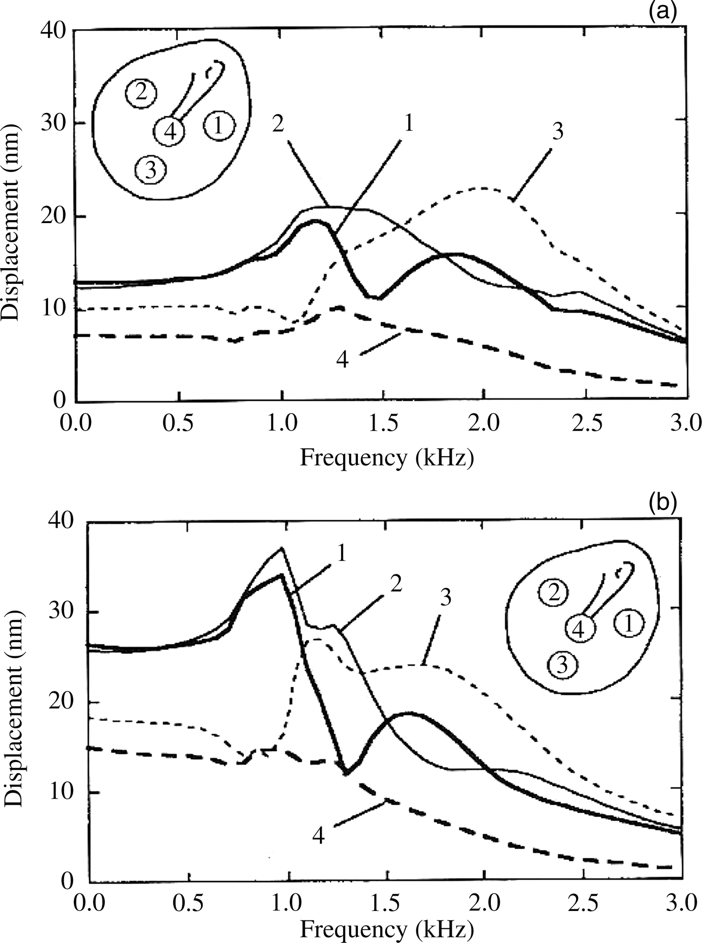
The influence of this middle-ear air cushion on tympanic membrane resonance frequency, and the effect of its elimination when an opening is made in the tympanic cavity, have been well documented. In models based on finite-element method analysis, opening the middle ear decreases the resonance frequency, or increases the vibrations of the tympanic membrane for low- to mid-frequency acoustic input.Reference Wada, Koike, Kobayashi and Hüttenbrink8 When considered in absolute terms, this opening affects the lower frequencies more (Figure 2), but in relative terms it has a more prominent effect on the mid-frequency range.Reference Koike, Wada and Kobayashi9 A study using an artificial middle ear revealed gradual lowering of the resonance frequency of the stapes movements when the eustachian tube diameter was progressively increased from 0 to 1.57 mm.Reference Kawase, Kano, Otsuka, Hamanishi, Koike and Kabayashi10 The opening of the middle-ear cavity in cadaver temporal bones has been shown to increase the admittance of low- to mid-frequency acoustic input.Reference Voss, Horton, Woodbury and Sheffield11, Reference Gyo, Goode and Miller12 The same effect has been demonstrated in the gerbil, using a small hole of only 1.2 mm in diameter (Figure 3).Reference Teoh, Flandermeyer and Rosowski13
Fig. 2 Frequency response of the tympanic membrane in a finite-element model: (a) middle-ear cavities intact, and (b) middle-ear cavities open. Opening the middle ear produces a downward shift of the peaks. With the open cavities, there is a small peak for the lower frequencies, connected with the upper quadrants of the tympanic membrane (1 and 2), and a broader peak for the mid-frequency range, connected with the lower half of the membrane (3). (Note: ‘4’ represents tympanic displacement at the umbo.) Reproduced with permission.8
Fig. 3 Input admittance of eight gerbil middle ears: (a) intact bulla, and (b) bullar hole (1.2 mm diameter) open. Opening the bulla produces a downward shift of the resonance frequency, resulting in an increase in admittance around 300–600 Hz, and a decrease of around 3000 Hz. Reproduced with permission.13
Based on the findings of these studies, it is reasonable to assume that the loss of the air cushion (associated with a wide open eustachian tube) in patulous eustachian tube patients produces a similar effect, in which there is a downward shift of tympanic membrane resonance frequency, or an increase in the admittance of low- to mid-frequency acoustic input at the tympanic membrane.
However, because of its particular anatomy, the tympanic membrane has not one but many vibration patterns, which are associated with distinctive membrane locations.Reference Wada, Koike, Kobayashi and Hüttenbrink8, Reference Koike, Wada and Kobayashi9, Reference Tonndorf and Khanna14–Reference Khanna, Decraemer and Hüttenbrink16 Maximum vibration patterns for the lower frequencies (those up to around 1000 Hz) are associated with the upper quadrants of the tympanic membrane, and those for the mid-frequency range (roughly between 1000 and 4000 Hz) implicate the lower half of the membrane. For the higher frequencies (above 4000 Hz), the vibration pattern is more complex (Figure 4). The tympanic membrane therefore has many resonance frequencies, each one connected to a specific location on the membrane.
Fig. 4 Finite-element model analysis, showing vibration patterns of the human tympanic membrane at the stimulus level of 80 dB SPL for: 850 Hz (left), 2000 Hz (middle) and 4000 Hz (right). Maximal vibration patterns for lower frequencies are situated in the upper quadrants, while those for mid-frequency range are mainly situated on the lower half of the tympanic membrane. Reproduced with permission.8
The application of a paper patch on a specific location of the tympanic membrane will increase the stiffness at that location, creating an upward shift of the particular resonance frequency associated with that location. Paper patches, Steri-strips, clay or any other material will therefore decrease admittance of the lower frequencies (up to around 1000 Hz) when applied to the upper half of the tympanic membrane, and likewise for the mid-frequency range (roughly 1000–4000 Hz) when applied to the lower half of the membrane. The application of material effectively counteracts the consequences of losing the middle-ear cushion effect (associated with the wide open eustachian tube), thereby eliminating the complaints.
• Paper patching was an effective treatment for patulous eustachian tube syndrome in 76 per cent of cases
• It was most effective when symptoms included a feeling of pressure or when caused by a suspected muscular factor
• Benefits continued after disappearance of the patch in 50 per cent of responders
• The patch appears to increase the resonance frequencies of the tympanic membrane by increasing membrane stiffness
In some patients, benefits were observed when the patch was placed on the upper half of the membrane, but not when applied to the lower half, and vice versa. This is in line with the theoretical data.Reference Wada, Koike, Kobayashi and Hüttenbrink8, Reference Koike, Wada and Kobayashi9, Reference Tonndorf and Khanna14–Reference Khanna, Decraemer and Hüttenbrink16 Specifically, two peaks form after the elimination of the middle-ear cushion, suggesting that patients may be disturbed either by the low-frequency peak or by the mid-frequency peak (Figure 2). The difference in the width of these peaks corresponds with our clinical experience, which highlights the importance of the precise location for the posterosuperior patch, but not for the lower patch. However, this observation is not in agreement with the results of a nasal audiometry study, in which only the lower frequencies (and not the mid-frequency range) were found to play a role in autophony.Reference Kano, Tawase, Baba, Sato and Kobayashi17
If correct, this hypothesis would imply that autophony in patulous eustachian tube is caused by a combination of two factors: an increase in sound intensity due to the free passage of pharyngeal sound waves through the eustachian tube, and an increase in the admittance of acoustic input at the tympanic membrane for the same frequencies. Both factors are needed for the complaint to arise. This implies that, as long as the admittance level of acoustic input at the tympanic membrane for these specific frequencies is low, a eustachian tube may be wide open without causing any symptoms. Such a situation may occur when the normal vibration pattern of a specific part of the tympanic membrane is hindered; for example, in the presence of retraction pockets or tympanosclerotic plaques at certain locations. Indeed, in a computed tomography study, bilateral eustachian tube opening was found in patients presenting with only unilateral patulous eustachian tube syndrome complaints.Reference Yoshida, Kobayashi, Takasaki, Takahashi, Ishimaru and Morikawa18
In clinical practice, one often notes movements of the tympanic membrane on breathing rhythm in patients who present no complaints at all. Such an asymptomatic open eustachian tube is present in around 5 per cent of the normal population, and in a much higher percentage of patients with middle-ear disease such as retraction pockets and cholesteatoma.Reference Mathew, Kuruvilla and Job19–Reference Flisberg and Ingelstedt23 In these patients, a wide open eustachian tube is not sufficient to cause autophony.
Conclusion
Paper patching of the tympanic membrane provided a symptomatic treatment that was temporary in some patients but lasting in others. The best results were obtained by those patients in whom a muscular cause was suspected and who complained of ear fullness, with a less impressive effect on those patients in whom a hormonal factor was involved.
With regard to the mechanism of action, we postulate that the complaints in patulous eustachian tube are (partly) due to the increase of the admittance of low- to mid-frequency acoustic input, caused by the loss of the middle-ear air cushion effect (associated with an open eustachian tube). This effect is counteracted by the application of a patch, which increases the stiffness of the tympanic membrane.
Additional basic research is needed. Although patulous eustachian tube is a rare condition, we predict that the study of autophony may provide us with valuable new insights into middle-ear acoustics.