


Introduction
Obstructive sleep apnoea syndrome (OSAS) is characterised by repeated episodes of complete or partial occlusion of the upper airways during sleep.Reference Langevin, Sukkar, Leger, Guez and Robert1 The development of OSAS has been related to a variety of predisposing factors, including obesity, nasal obstruction, and adenoidal and/or tonsillar hypertrophy.Reference Langevin, Sukkar, Leger, Guez and Robert1
Hypothyroidism has also been described as a risk factor for both obstructive and central sleep apnoea, such that hormone replacement therapy with thyroxine usually leads to amelioration or even cessation of the associated apnoea. Proposed mechanisms linking the two conditions include narrowing of the upper airway by deposition of mucopolysaccharides and protein extravasation into the tissues of the face, tongue and pharyngeal structures, as well as dysfunction of the upper airway dilator muscles due to hypothyroid myopathy or abnormalities of ventilatory control.Reference Langevin, Sukkar, Leger, Guez and Robert1, Reference Deegan, McNamara and Morgan2
However, little is known about whether large thyroid goitres occurring in the absence of hypothyroidism are a contributing factor to the development of OSAS. In such cases, OSAS may improve with thyroidectomy.Reference Deegan, McNamara and Morgan2–Reference De Felice, Fuschillo, Martucci, De Angelis and Balzano4 At present, there is very little literature on the underlying relationship between large goitres and OSAS.
Therefore, the objectives of this study were to analyse the experience of patients diagnosed with OSAS and large, multinodular goitre who underwent total thyroidectomy, to evaluate the types of goitre causing OSAS, to elucidate possible mechanisms of pathogenesis, and to determine whether thyroid surgery may be a curative treatment for these patients.
Subjects and methods
A retrospective review was undertaken of all patients with underlying OSAS who had undergone total thyroidectomy at the otorhinolaryngology and head and neck unit at Guy's and St Thomas' National Health Service Foundation Trust Hospital, London, between 2000 and 2010.
Obstructive sleep apnoea syndrome was diagnosed using body mass index, Epworth sleep score and polysomnographic index. All patients underwent thyroid function and thyroid antibody tests, and ultrasound-guided fine needle aspiration cytology. Patients' clinical thyroid status was graded based on the World Health Organization system.5 Cytological classification was based on the British Thyroid Association guidelines.6 Multiplanar computed tomography (CT) scanning was used to assess the anatomy of the multinodular goitre, for the purposes of anaesthetic and surgical planning.
All patients were given a complete description of the study before written, informed consent was obtained.
Results
Five consecutive patients with OSAS and large goitres were included in this study. All patients were women, with a median age of 58 years (range, 46–82 years). All patients had been diagnosed with OSAS and received continuous positive airway pressure therapy at specialist sleep disorder centres, guided by their body mass index, Epworth sleep score and polysomnographic index (Table I).
Table I Patient data summary

Y = years; ESS = Epworth sleep score; cytol = cytological classification; histopathol = histopathology; wt = weight; PI = polysomnographic index; desats/h = desaturation episodes per hour; min SpO2 = minimum oxygen saturation; F = female; NA = not available
Three patients were euthyroid at presentation, one was hypothyroid and the fifth had thyrotoxicosis. Fine needle aspiration cytology showed benign cytopathology (classification code THY2) in four patients, and was non-diagnostic (classification THY1) in one patient.
All patients were successfully intubated using flexible fibre-optic endoscope guided techniques. One patient had a pre-existing tracheal stent, which had been inserted at her referring hospital in an attempt to maintain airway patency.
Computed tomography scanning showed a multinodular goitre with retropharyngeal extension in all patients, plus significant retrosternal extension in two patients. In all patients, the retropharyngeal and retrolaryngeal goitre extension was so severe that there was complete anatomical contact between the two thyroid lobes at the posterior aspect of the larynx and pharynx.
All patients underwent total thyroidectomy. In addition, one patient underwent tracheal resection with primary end-to-end anastomosis and prophylactic tracheostomy, due to tracheal necrosis found at the time of surgery. (At the time of writing, decannulation of this patient had been unsuccessful due to severe suprastomal tracheomalacia.) No patient sustained vocal fold injury, and only one patient suffered post-operative hypocalcaemia requiring long-term replacement. All patients were adequately treated post-operatively with thyroxine replacement therapy.
Histological analysis of the goitres showed nodular hyperplasia in all specimens, without evidence of malignancy. The median multinodular goitre weight was 313 g (range, 56–450 g).
All patients had post-operative resolution of their OSAS and laryngeal oedema, and required no further continuous positive airway pressure ventilation. The median duration of hospital stay was 5 days (range, 3–17 days).
At 12 month follow up, all patients remained asymptomatic.
Patient characteristics are summarised in Table I, and two representative cases are described below.
Case study one
A 58-year-old woman known to suffer with OSAS was referred from the sleep apnoea clinic with one year's history of daytime fatigue, increasing dyspnoea, dysphonia and dysphagia. The patient had been dependent on continuous positive airway pressure ventilation for three years. She was a non-smoker and drank no alcohol.
Clinical examination demonstrated a body mass index of 40 and a World Health Organization grade III, multinodular goitre.
Flexible nasoendoscopy revealed a grossly oedematous larynx with anterior displacement of the aryepiglottic folds (Figure 1a).

Fig. 1 Investigations for patient one. (a) Pre-thyroidectomy flexible nasendoscopic view showing gross laryngeal oedema and distortion of the larynx, due to compression from the enlarged thyroid lobes wrapping around the larynx. (b) Pre-thyroidectomy, axial computed tomography scan showing enlargement of the thyroid gland with retropharyngeal extension and airway compression. (c) Laryngoscopic view six weeks post-thyroidectomy, showing normalisation of laryngeal anatomy, which corresponded to the patient's clinical improvement.
Thyroid functions tests indicated hypothyroidism, and a technetium scan showed normal thyroid gland uptake.
The patient's Epworth sleep score was 20/24, and polysomnography showed 50 marked desaturation events per hour, with a minimum oxygen saturation level of 48.2 per cent.
Computed tomography scanning of the neck and thoracic inlet confirmed multinodular enlargement of the thyroid gland, with retropharyngeal extension and laryngeal compression and oedema (Figure 1b).
Fine needle aspiration cytology was non-diagnostic (THY1 classification).
The patient underwent an uncomplicated total thyroidectomy and made an uneventful recovery.
The resected thyroid gland weighed 313 g. Histological analysis showed nodular hyperplasia.
On post-operative review, the patient had a normal appearance and mobility of both vocal folds, and normal laryngeal anatomy (Figure 1c). Respiratory evaluation revealed that OSAS symptoms had completely resolved.
Case study two
A 46-year-old woman was referred from another unit for an anaesthetic and surgical opinion. She had a six-year history of OSAS, treated with continuous positive airway pressure ventilation on a domiciliary basis. The patient initially complained of breathing difficulty with voice changes, as well as increasing anxiety attacks and a choking sensation. A previous attempt at thyroid surgery had failed, as she could not be intubated by conventional methods. Her past medical history included type II diabetes mellitus and asthma. She was a non-smoker.
On examination, the patient was obese, with a body mass index of 50, and had a large thyroid goitre (grade III).
Flexible laryngoscopy revealed significant supraglottic oedema, with gross distortion of the aryepiglottic folds (Figure 2a).
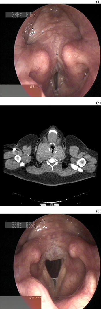
Fig. 2 Investigations for patient two. (a) Pre-thyroidectomy laryngoscopic view showing gross laryngeal oedema concentrated posteriorly, with distortion of the larynx, due to tracheal compression from the enlarged thyroid lobes. (b) Pre-thyroidectomy, axial computed tomography scan showing enlarged thyroid lobes compressing the trachea. (c) Laryngoscopic view six weeks post-thyroidectomy, showing improvement in airway and laryngeal anatomy, corresponding to the patient's clinical improvement, with minimal residual interarytenoid oedema.
Thyroid function testing showed a decreased thyroid-stimulating hormone level, a normal thyroxine level and a raised tri-iodothyronine level.
The Epworth sleep score was 18/24, and polysomnography showed 48 desaturation events per hour, with a minimum oxygen saturation level of 53 per cent.
Computed tomography demonstrated a grade III, retrosternal goitre with retropharyngeal extension and tracheal compression, and with the right lobe larger than the left, associated with some mediastinal lymphadenopathy (Figure 2b). Isotope scanning showed a large, multinodular goitre. Positron emission tomography was negative.
Fine needle aspiration cytology showed benign colloid nodules within a multinodular goitre (THY2 classification).
The patient underwent total thyroidectomy, resulting in an immediate improvement in her respiratory status (Figure 2c).
Follow up indicated that she had not required continuous positive airway pressure since her thyroidectomy.
Discussion
The development of obstructive sleep apnoea has been related to a myriad of predisposing factors, including obesity, nasal obstruction and adenotonsillar hypertrophy.Reference Langevin, Sukkar, Leger, Guez and Robert1 Large goitres occurring in the absence of hypothyroidism have been postulated as an additional causative factor in the development of OSAS; in such cases, thyroidectomy may relieve OSAS symptoms.Reference Deegan, McNamara and Morgan2–Reference De Felice, Fuschillo, Martucci, De Angelis and Balzano4 Given the scarcity of literature on this subject, it has previously been possible only to speculate about the precise relationship between large goitres and OSAS. The pathogenic relationship between large goitres and OSAS may be attributed to increased oedema with reduced patency of the upper airways, as a result of decreased venous return from the head and neck.Reference Deegan, McNamara and Morgan2 Moreover, tracheal displacement may also interfere with the upper airway stiffening reflex which normally occurs during inspiration.Reference Van de Graaff7 A large goitre may itself create a mass loading effect on the airways, which is further compounded by reduced activity of the pharyngeal dilator muscles.Reference Gardiner and Russell8, Reference van Lunteren, Haxhin and Cherniack9
All patients presenting with OSAS should undergo thorough clinical examination of the neck, followed by fibre-optic laryngoscopy to exclude a large thyroid goitre as a possible treatable cause. Conversely, patients presenting with large goitres and tracheal displacement should be questioned about symptoms of OSAS and investigated accordingly. Patients with large thyroid goitres associated with compression symptoms should be evaluated with flexible fibre-optic laryngoscopy in the clinic, to assess vocal fold function and the degree of laryngo-tracheal compression and oedema.Reference Shaha, Alfonso and Jaffe10
All our patients had significant laryngeal oedema and obvious compression clearly demonstrated on laryngoscopic assessment. Therefore, we recommend that such patients be discussed in advance with an anaesthetist experienced in the management of difficult airways, in order to enable systematic planning of the precise anaesthetic technique required to secure the airway. In our study, four patients were successfully intubated by the awake fibre-optic intubation technique. One patient had previously failed conventional intubation, and another patient had undergone emergency intubation necessitating guidance with a bougie.
The role of CT in patients with compressive goitres is well established in the literature. Patients with OSAS and a goitre should also be evaluated with multiplanar CT, to facilitate anaesthetic and surgical planning.
In patients with a retropharyngeal goitre, meticulous pre-operative planning of the surgical approach and technique is of paramount importance.Reference Shaha, Alfonso and Jaffe10, Reference Shaha11 The surgical incision should be generous, allowing an adequate approach to the goitre. Division of the pre-thyroid strap muscles will allow clear identification of the lateral and posterior aspects of the goitre, with appropriate access to and control of the carotid sheath, from which the goitre may need to be dissected. The dissection should proceed with a combination of sharp and blunt dissection of the goitre between the carotid sheath and the pre-vertebral fascia, where the capsule of the goitre is adjacent.
Once the superior pole is freed up and the retropharyngeal segment completely dissected, the goitre is mobilised medially and the recurrent laryngeal nerve and parathyroid glands are identified. Distortion of these structures may occur due to the size and anatomy of these goitres (Figure 3). In the present series, all recurrent laryngeal nerves were identified in the posterior aspect of the thyroid gland, with no ‘riding’ nerves, and there were no recurrent laryngeal nerve injuries. The parathyroid glands may be positioned anywhere on the surface of the goitre, due to the distorted anatomy. Therefore, the dissection should be performed very close to the gland, with careful attention paid to preserving the parathyroid glands. Only one of our patients became hypoparathyroid after her surgery, and required long-term calcium replacement.

Fig. 3 Post-resection surgical photograph of the bed of a retropharyngeal goitre, showing the degree of tracheal compression.
• The relationship between large goitres and obstructive sleep apnoea syndrome (OSAS) is poorly understood
• In this study, large, multinodular goitres with retrolaryngo-pharyngeal extension resulted in OSAS, in euthyroid patients, via laryngeal compression and oedema
• Total thyroidectomy relieved laryngeal compression and oedema and resolved OSAS
• Patients with OSAS should be screened for thyroid goitre
The precise extent of thyroidectomy required for large goitres is subject to much debate.Reference Shaha, Alfonso and Jaffe10 In our opinion, total thyroidectomy is the optimal approach for patients with large goitres with bilateral retrolaryngo-pharyngeal extension giving rise to severe compression symptoms with laryngeal oedema and OSAS. This procedure is curative as it completely alleviates the compression symptoms. In our series, all patients had complete post-operative resolution of their OSAS symptoms.
One of our patients had tracheal necrosis due to stent insertion and goitre-related tracheal compression, requiring tracheostomy. In this patient, resolution of OSAS symptoms could not be adequately assessed, and may well have been partly due to the presence of the tracheostomy (a well described treatment for severe, recalcitrant OSAS). However, this patient's laryngeal abnormalities returned to normal following total thyroidectomy, suggesting that this procedure made a significant contribution to symptom resolution.
The presence of a multinodular goitre as a likely underlying cause of OSAS has previously been reported only in three isolated case reports of patients whose respiratory status improved after goitre removal.Reference Deegan, McNamara and Morgan2–Reference De Felice, Fuschillo, Martucci, De Angelis and Balzano4 Indeed, the only previous documented case in the otorhinolaryngological literature comprises an acromegalic patient with OSAS and respiratory failure, who improved after thyroidectomy.Reference Stafford, Youngs, Waldron, Baer and Randall3 The size of the goitre has been questioned as the sole factor causing OSAS. It has been suggested that large goitres may obstruct venous return from the head and neck, resulting in engorgement and oedema of upper airway structures and reduced upper airway patency.Reference Deegan, McNamara and Morgan2 However, this is difficult to prove conclusively, due to inadequate clinical and radiological evidence.
We acknowledge that the main limitation of our study was the lack of objective post-operative parameters, such as apnoea–hypopnoea index, which could have definitively proven that the resolution of OSAS was indeed due to the total thyroidectomy procedure. Our patients noticed such a dramatic improvement in their respiratory symptoms and quality of life that they declined to undergo further investigations (e.g. polysomnography) which, although extremely helpful for our study purposes, would not have altered any aspect of their subsequent management.
Furthermore, three patients in our study went on to achieve significant weight reduction following their surgery, as they experienced an extraordinary improvement in their exercise tolerance. We believe this constitutes further evidence to support the fact that large goitres with retrolaryngo-pharyngeal extension cause significant venous congestion and laryngeal oedema.
It is also thought that an enlarged thyroid gland may impair the function of muscles attached to the hyoid bone, the anterior movement of which normally increases the patency of the pharynx and reduces upper airway resistance.Reference van Lunteren, Haxhin and Cherniack9
Whatever the pathophysiology, our study findings suggest that patients with OSAS and large, bilateral goitres with retrolaryngo-pharyngeal extension go on to have complete resolution of OSAS after total thyroidectomy, such that they require no further nasal continuous positive airway pressure ventilation.
Conclusion
Large, multinodular goitres with retrolaryngo-pharyngeal extension may be the cause of OSAS in euthyroid patients, due to laryngeal compression and oedema. Adequate CT imaging is necessary to assess goitre anatomy and facilitate surgical planning. Surgery, in the form of total thyroidectomy, relieves tracheal compression and resolves associated laryngeal oedema, leading to resolution of OSAS.
From the results of our study, we recommend that all patients presenting with OSAS be screened for underlying thyroid goitre as a possible treatable cause.
We also recommend that further studies be undertaken in order to validate our preliminary results with objective, post-operative polysomnographic parameters.


