Introduction
Viral croup (laryngotracheobronchitis) is an acute infection of the larynx, trachea and bronchi. It accounts for approximately 15 per cent of respiratory tract disease seen in paediatric practice, and is the most common cause of upper airway obstruction in children aged 6 months to 6 years.Reference Leung, Kellner and Johnson1, Reference Griffin, Ellis, Fitzgerald-Barron, Rose and Egger2 It is characterised by hoarseness, barking cough and stridor. Most viral croup is caused by the parainfluenza virus. There is seasonal variation, with a peak during the autumn and winter months. A prodromal upper respiratory tract infection is common. Infection rarely causes severe respiratory distress: hospitalisation occurs in 1.3–2.6 per cent of cases.Reference Denny, Murphy and Clyde3
Recurrent croup is less common. Hide and Guyer reported its prevalence as 6.4 per cent in children followed up for the first four years of life.Reference Hide and Guyer4 On first presentation, it can be clinically difficult to determine those children who will go on to develop recurrent croup. Recurrent croup is not a specific diagnosis in itself, but rather a descriptive term for an entity that may have a number of underlying causes.Reference Farmer and Wohl5 Asthma, allergy and bronchial hyper-responsiveness have all been implicated.Reference Konig6, Reference Zach, Erben and Olinsky7 More recent studies have highlighted the association between recurrent croup and upper airway disease processes, such as congenital or acquired subglottic stenosis, and gastrolaryngopharyngeal reflux.Reference Farmer and Wohl5, Reference Burton, Pransky, Katz, Kearns and Seid8, Reference Yellon, Coticchia and Dixit9 There should be a high index of suspicion regarding inhaled foreign bodies, which may also occasionally cause recurrent croup-like symptoms.
The prognosis of recurrent croup is not well established and in many children the exact cause of the symptoms is unclear. The objective of this study was to review the aetiology, diagnosis, treatment and clinical outcome of children with recurrent croup, and to identify which diagnostic procedures were of most value in these children.
Materials and methods
A retrospective case note review was carried out. We identified all the children referred to the otolaryngology service at our hospital with recurrent croup from November 2002 to March 2011. Children were identified from operating theatre records and from the individual records of the operating surgeons. The medical records of these patients were reviewed and clinical data were recorded. Recurrent croup was defined as two or more episodes of croup either confirmed medically or reported by the parent. Specific data collected included sex, age at onset, age at presentation, duration of croup prior to referral, number of episodes, comorbidities, number of hospital admissions, investigations performed, treatments given, duration of follow up and status at last follow up.
All children referred to the otolaryngology service with recurrent croup underwent microlaryngobronchoscopy as per unit policy. Laryngobronchoscopy findings considered to be consistent with pathological gastrolaryngopharyngeal reflux included cobblestone mucosa, oedema or erythema of the arytenoids, and posterior commissure, pachydermia and blunting of the carina. Where appropriate, the subglottis was sized with endotracheal tubes as described by Myer et al. Reference Myer, O'Connor and Cotton10 Some patients underwent blood sampling to examine levels of complement component proteins C3 and C4, and C1 esterase inhibitor. In addition, some patients were tested to evaluate total immunoglobulin E (IgE) levels, and radioallergosorbent testing (RAST) was carried out to identify common aeroallergens. A small number of children underwent oesophageal pH studies.
The treatment given to each patient was documented. Outcome was defined by resolution or improvement of symptoms at the last follow up.
Results
During the study period, 90 patients presented with a history of recurrent croup, consisting of 72 males and 18 females (a male to female ratio of 4:1). The median age of patients at the onset of croup was six months (ranging from birth to eight years; Figure 1). The median age of patients at presentation to ENT was 4 years and 6 months (ranging from 4 months, to 11 years and 10 months). Potential risk factors such as bottle rather than breast-feeding and parental smoking were not documented in enough detail to permit analysis.

Fig. 1 Ages of children at initial onset of croup. Y = years
The median duration of croup prior to referral was 2 years and 6 months (ranging from 2 months, to 11 years and 2 months). The median number of croup episodes per year was 4 (range of 1–16 episodes). Of the 90 patients, 42 (47 per cent) required hospital admission on at least one occasion, of which 8 (9 per cent) required intensive care unit admission. In 26 (29 per cent) of the patients the episodes of croup came on suddenly at night with no prior illness, whereas in 38 (42 per cent) of the patients typical attacks of croup were preceded by an upper respiratory tract infection. Seventy-three patients were given glucocorticoids during croup episodes, which resulted in immediately improved symptoms for all of those patients.
Nine patients (10 per cent) had a history suggestive of obstructive sleep apnoea in addition to recurrent croup. Thirty-two patients (36 per cent) had atopic disease, of which 24 patients (27 per cent) had asthma.
All patients underwent laryngobronchoscopy. Sixteen of the 90 patients (18 per cent) demonstrated various degrees of subglottic stenosis. Eleven (69 per cent) of these 16 patients had grade 1 stenosis (Figure 2), 4 patients (25 per cent) had grade 2 (Figure 3) and 1 patient (6 per cent) had grade 3 (Figure 4). The child with grade 3 stenosis underwent single-stage laryngotracheal reconstruction using anterior and posterior costal cartilage grafts with successful resolution of symptoms. One of the children with a grade 2 stenosis was successfully treated with endoscopic balloon dilatation. The remaining patients were all treated with observation only. With regards to other airway pathology requiring surgery, one patient underwent laser repair of a laryngeal cleft. Other significant findings on laryngobronchoscopy are shown in Table I.

Fig. 2 Laryngobronchoscopic view showing grade 1 subglottic stenosis.

Fig. 3 Laryngobronchoscopic view showing grade 2 subglottic stenosis.

Fig. 4 Laryngobronchoscopic view showing grade 3 subglottic stenosis.
Table I Laryngobronchoscopy findings

Twenty-five children had findings consistent with gastrolaryngopharyngeal reflux. Evidence of the reflux laryngitis was revealed on laryngobronchoscopy (23 patients) (Figure 5), pH studies (1 patient) or barium studies (1 patient). Subglottic stenosis was identified on laryngobronchoscopy in two of these patients. Fourteen patients had a history suggestive of reflux, with symptoms such as vomiting after feeds, heartburn and an acidic taste in the mouth. Of these, four patients showed no evidence on laryngobronchoscopy, pH studies or barium studies and so were not included in the gastrolaryngopharyngeal reflux diagnostic group.

Fig. 5 Laryngobronchoscopic view showing evidence of reflux laryngitis, in this instance, oedema of the supraglottic larynx.
Total IgE levels were measured in 39 patients, 9 of whom (23 per cent) had raised values. This group was further screened for common aeroallergens using RAST. The findings are shown in Table II.
Table II Rast findings

RAST = radioallergosorbent test
Serum levels of complement proteins C3 and C4, and C1 esterase inhibitor were measured in 37 patients. The C1 esterase inhibitor levels and C3 levels were found to be normal in all patients. However, C4 was low in three patients (8 per cent). In one of these patients the C4 level was borderline low at 0.10 g/l (the age-specific normal range is 0.12–0.4 g/l). This patient also had grade 1 subglottic stenosis which was thought to be the significant pathology. In another of these patients the C4 level was borderline low at 0.09 g/l (age-specific normal range is 0.12–0.4 g/l). This patient also had evidence of reflux which was thought to be the significant pathology. The remaining patient, however, had a significantly low C4 level at 0.07 g/l (age-specific normal range is 0.16–0.46 g/l). This patient had a history of episodes of lip swelling and abdominal pain in association with stridor, which was suggestive of angioedema. All other investigations were normal.
The patients' status at last follow up had been adequately documented in 78 patients. Nineteen patients (24 per cent) had received either omeprazole or ranitidine as anti-reflux medication, usually in addition to an alginate (Gaviscon Infant, Reckitt Benckiser Healthcare (UK), Kingston upon Thames, UK). Of these, 11 patients (58 per cent) had laryngobronchoscopy findings suggestive of reflux and 10 patients (53 per cent) had a history suggestive of reflux. A comparison of these patients' outcomes with patients who did not receive anti-reflux medication is presented in Table III. Clinical improvement was defined as decreased symptom severity, decreased frequency of episodes or resolution of symptoms. Laryngobronchoscopy findings that indicated reflux were predictive of benefit from anti-reflux medications: 10 patients (91 per cent) found to have positive laryngobronchoscopy findings showed improved symptoms with anti-reflux medication; only 2 (25 per cent) of the patients in this group who did not use anti-reflux medication showed improved symptoms (p = 0.006, two-tailed Fisher's exact test). However, a history suggestive of reflux was not predictive of benefit from anti-reflux medications: six patients (60 per cent) with a history suggestive of reflux showed improved symptoms with anti-reflux medication compared with only one patient (33 per cent) who did not use anti-reflux medication (p = 0.56, two-tailed Fisher's exact test).
Table III Patient outcome following anti-reflux medication

Med = medication; pts = patients; Y = yes; N = no
Table IV shows the final diagnosis of the cause of the recurrent croup for the 90 patients after all investigations and treatments had been completed. In 41 patients (45 per cent), no cause for the recurrent croup was found. Of those with an adequately documented last follow up, it was found that 20 patients (59 per cent) with a preceding viral upper respiratory tract infection were well or improving after a median follow up of 7 months. In addition, 14 patients (70 per cent) with a sudden nocturnal onset and no prodromal illness were well or improving after a median follow up of 8 months. The final diagnoses of these two groups of patients are shown in Table V. No significant differences were found between the two groups in terms of the final diagnoses (p > 0.05, chi-square test).
Table IV Cause of recurrent croup

Table V Final diagnosis of patients with sudden night onset or preceding viral URTI

*Total n = 26. †Total n = 38. URTI = upper respiratory tract infection
Fourteen patients had only two episodes of croup. The final diagnoses of these patients were similar to those of patients with more than two croup episodes. The final diagnoses of these two groups of patients are shown in Table VI. No significant differences were found in terms of the final diagnoses (p > 0.05, chi-square test).
Table VI Final diagnosis of patients with history of two or more croup episodes

*Total n = 14. †Total n = 76.
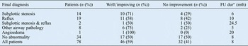
Table VII shows the number of patients with each diagnosis whose symptoms were improving or resolved at last follow up. Of the group of patients with a final diagnosis of reflux, those with evidence of reflux on laryngobronchoscopy who received treatment had the best prognosis, both within their group and out of all patients. The children with no identifiable cause for their recurrent croup were the group most likely to have continuing symptoms. This group also included the only death in our series. A child who had previously been discharged after a normal laryngobronchoscopy continued to have episodes of croup with sudden nocturnal onset and no preceding illness over a two-year period until he had a particularly severe episode of stridor and died of respiratory failure on arrival at hospital. No post-mortem examination was performed so the exact cause of death remains unknown.
Table VII Patient status at last follow up

*Median average. FU = follow up; dur = duration; mth = months
Discussion
Recurrent croup is a relatively common childhood disease, but surprisingly little has been published on its epidemiology, aetiology, natural history and management. Our retrospective, observational study involved 90 children and is therefore the largest study to date on the investigation and management of recurrent croup.
Recurrent croup was associated with a male predominance, which is consistent with other studies.Reference Hide and Guyer4, Reference Farmer and Wohl5, Reference Zach, Erben and Olinsky7, Reference Hoa, Kingsley and Coticchia11–Reference Waki, Madgy, Belenky and Gower13 In our study, the median age at onset of croup was six months. This is a little younger than the reported age at onset in previous studies, which ranged from 9 to 18 months.Reference Zach, Erben and Olinsky7, Reference Waki, Madgy, Belenky and Gower13, Reference Arslan, Cipe, Ozmen, Kondolot, Piskin and Yoney14
It is advised that children with recurrent croup undergo an endoscopic airway evaluation to establish whether there is any underlying airway disorder.Reference Cressman and Myer15 All patients in our study underwent endoscopic evaluation. We identified a total of 25 patients (27.7 per cent) with anatomical airway abnormalities, which is in line with the findings of previous, smaller studies.Reference Waki, Madgy, Belenky and Gower13, Reference Chun, Preciado, Zalzal and Shah16 Sixteen (17.7 per cent) of these patients demonstrated various degrees of subglottic stenosis on laryngobronchoscopy, a minority of which were severe enough to require intervention. A further nine patients (10 per cent) had no subglottic stenosis but did have other significant airway pathology. Children with airway pathology are predisposed to recurrent croup.Reference Tan and Manoukian17–Reference Baker19 We therefore recommend that all children with recurrent croup undergo laryngobronchoscopy.
Laryngobronchoscopy can also provide diagnostic and prognostic information regarding reflux. Reflux has been reported in as many as 47 to 87 per cent of children with recurrent croup in other series.Reference Hoa, Kingsley and Coticchia11–Reference Waki, Madgy, Belenky and Gower13, Reference Yellon and Goldberg20 Our lower prevalence of 28 per cent may reflect the fact that our department rarely uses pH studies and relies solely on clinical assessment. While some may consider pH monitoring the investigation of choice, it is often difficult to perform in children and only detects reflux events with a pH of less than 4. Refluxed material with a pH above 4 may result in desquamation of the tracheal mucosa and delayed regeneration, and may therefore be pathologic.Reference Wynne, Ramphal and Hood21 We have not routinely used pH studies because we remain unconvinced of their value in predicting the benefit of anti-reflux medication. Our experience indicates that laryngobronchoscopy findings are the best predictor, and that children with no visual evidence of reflux laryngitis are unlikely to benefit from anti-reflux medication, which is in keeping with previous findings.Reference Myer, O'Connor and Cotton10 We are now in discussion with colleagues in gastroenterology about the potential value of oesophagoscopy in these children to identify further evidence of reflux as well as eosinophilic oesophagitis.Reference Yellon, Coticchia and Dixit9
Previous studies have suggested that recurrent croup is associated with reactive airway disease and allergy,Reference Konig6, Reference Zach, Erben and Olinsky7, Reference Zach, Schnall and Landau22–Reference Contencin and Narcy25 although they have been unable to establish causality. Our experience suggests that routine allergy testing is unhelpful in children with recurrent croup. The positive RAST results did not correlate with clinical symptoms in a way that suggested the allergen in question was triggering the croup attacks, for any of our patients. The RAST findings were therefore thought to be incidental. We suggest that RAST be reserved for cases where there is a suggestive history of croup episodes being associated with a particular allergen (such cases are rare). Similarly, routine measurement of complements C3 and C4, and C1 esterase inhibitor levels appeared to be unhelpful, but may have a place where angioedema is suspected due to lip or tongue swelling in association with episodes of stridor.
Historically, croup attacks preceded by an upper respiratory tract infection have been labelled as viral croup, while it has been suggested that attacks with a sudden night-time onset could be labelled as spasmodic croup. We found no significant differences in the final diagnoses of these groups compared with the final diagnoses of all patients.
We were intrigued to find apparent differences in prognosis in children with different causes for their recurrent croup. Surprisingly, the children with anatomical abnormalities of the airway had a very good prognosis. Seventy-one per cent of patients with subglottic stenosis were well or improving after a median follow up of 6 months, and 75 per cent of patients with other airway abnormalities were well or improving after a median follow up of 5 months. The best prognosis, however, was in the children with treated reflux diagnosed on laryngobronchoscopy. Ninety-one per cent of those with laryngobronchoscopy findings suggestive of reflux who were receiving anti-reflux medication were reported to be well or improving after a median follow up of nine months. Although this response rate may seem dramatic, our results, which are based on a small sample size with limited follow up, are supported by Hoa et al. Reference Hoa, Kingsley and Coticchia11 These authors demonstrated symptomatic improvement in 31 of 35 patients (88.6 per cent) who had endoscopic findings consistent with gastroesophageal reflux and were treated with anti-reflux medications. In comparison, only 50 per cent of the children with no identifiable cause for their recurrent croup were well or improving after a median follow up of 8 months. It was found that a history suggestive of reflux was not predictive of benefit from anti-reflux medications. A laryngobronchoscopy is therefore required prior to commencing anti-reflux medication. A Cochrane review has shown glucocorticoids to be effective in the acute management of an episode of croup,Reference Russell, Liang, O'Gorman, Johnson and Klassen26 and our experience is that they are very effective regardless of the underlying cause of the croup.
Due to the limitations of a retrospective study, it is possible that some variables were not accounted for. Questions concerning history of reflux, night-time onset or occurrence of a preceding upper respiratory tract infection may not have been addressed in all patients.
There is no consensus (some debate) in the literature regarding how many episodes of croup constitute labelling of the condition as recurrent. Some studies have suggested a threshold of three or more croup episodes,Reference Van Bever, Wieringa, Weyler, Nelen, Fortuin and Vermeire27 while others, like our study, have used a threshold of two episodes.Reference Farmer and Wohl5, Reference Waki, Madgy, Belenky and Gower13 More than one episode of croup symptoms with a normal airway is uncommon, unless there is immunodeficiency or reinfection with a different viral strain.Reference Farmer and Wohl5, Reference Hoa, Kingsley and Coticchia11 We found no significant differences in terms of the final diagnoses of patients with only two episodes of croup compared with patients with more than two episodes. At our institution, we investigate after only two episodes of croup. The results for two or three or more episodes of croup are comparable, and our threshold therefore appears appropriate for this demographic. As our service is a tertiary service, we are reliant on onward referrals from other services. Our preference for earlier investigation may reflect a referral bias, as evidenced by the greater number of children with a history of three or more croup episodes versus those with only two episodes. The appropriate number of croup episodes required to progress to endoscopic investigation therefore remains unsettled. At the minimum, we would recommend endoscopic investigation after two croup episodes in those with atypical symptoms, or after three episodes in patients with no atypical symptoms.
• Recurrent croup may have a number of underlying causes and is associated with upper airway disease
• Anatomical airway anomalies should always be suspected in children with recurrent croup; most anomalies can be managed conservatively
• Laryngobronchoscopy (but not clinical) findings that indicate reflux are predictive of benefit from anti-reflux medications
• Laryngobronchoscopy is helpful for diagnosis and prognosis, whereas measurement of immunoglobulin E and complement proteins is not
• Cases of recurrent croup with no obvious cause should be observed closely long-term; recurrent croup may not always be a benign diagnosis
An initial laryngobronchoscopy under general anaesthesia is recommended in all children with recurrent croup, as the subglottis and trachea cannot be adequately assessed by any other means. This will allow subglottic stenosis and other airway pathology to be identified and treated where appropriate. It can also provide useful information regarding prognosis, as well as reflux. If laryngobronchoscopy is positive for reflux, we recommend that anti-reflux medication is started. In our patients, this resulted in a 91 per cent improvement over a median follow up of 9 months, compared with 25 per cent of children (2 out of 8) in whom laryngobronchoscopy demonstrated reflux but who were not started on anti-reflux medications. We recommend that those children with no obvious cause for their recurrent croup on laryngobronchoscopy continue to be observed for at least one year to exclude worsening of their condition. This group of patients have the worst prognosis. Based on the results of our study, around half of these patients will have continuing symptoms one year later. In addition, our only death occurred in this group. Recurrent croup should not always be presumed to be a benign diagnosis.