Introduction
The technique of stapedotomy is well established as a minimally invasive, highly successful surgical intervention to improve hearing in patients with otosclerosis. In a few cases, hearing improvement is present but not satisfactory; more rarely, post-operative hearing remains identical to pre-operative hearing. Revision surgery is usually undertaken, but in about 8 per cent of this refractory population no pathological surgical findings explaining the failure can be found.Reference Lippy, Battista, Berenholz, Schuring and Burkey1, Reference Vincent, Rovers, Zingade, Oates, Sperling and Deveze2
Computed tomography (CT) of the temporal bone can demonstrate coincidental pathology before proceeding to revision surgery.
We report the case of a high-riding jugular bulb with an associated jugular bulb diverticulum, which was dehiscent towards the vestibular aqueduct, in a patient with confirmed otosclerosis who did not experience hearing improvement after stapedotomy.
Case report
A 48-year-old woman was diagnosed with bilateral suspected otosclerosis. She had: a history of bilateral progressive hearing loss, without trauma or chronic middle-ear infection; apparently normal tympanic membranes', ipsilateral negative Rinne tuning fork test; bilateral conductive hearing loss with an air–bone gap of 30 dB (pure tone average at 0.5, 1 and 2 kHz); a type A tympanogram; and absent stapedial muscle tendon reflexes bilaterally. She had never experienced tinnitus, a feeling of pressure or vertigo.
This patient underwent a middle-ear inspection on the right side. Stapes fixation was confirmed by palpation, and a stapedotomy with interposition of a Teflon® prosthesis was performed.
Post-operatively, she did not experience hearing improvement, in fact her hearing was identical to the pre-operative audiogram (for both bone and air conduction thresholds). She did not report any additional symptoms such as vertigo, tinnitus or hearing deterioration.
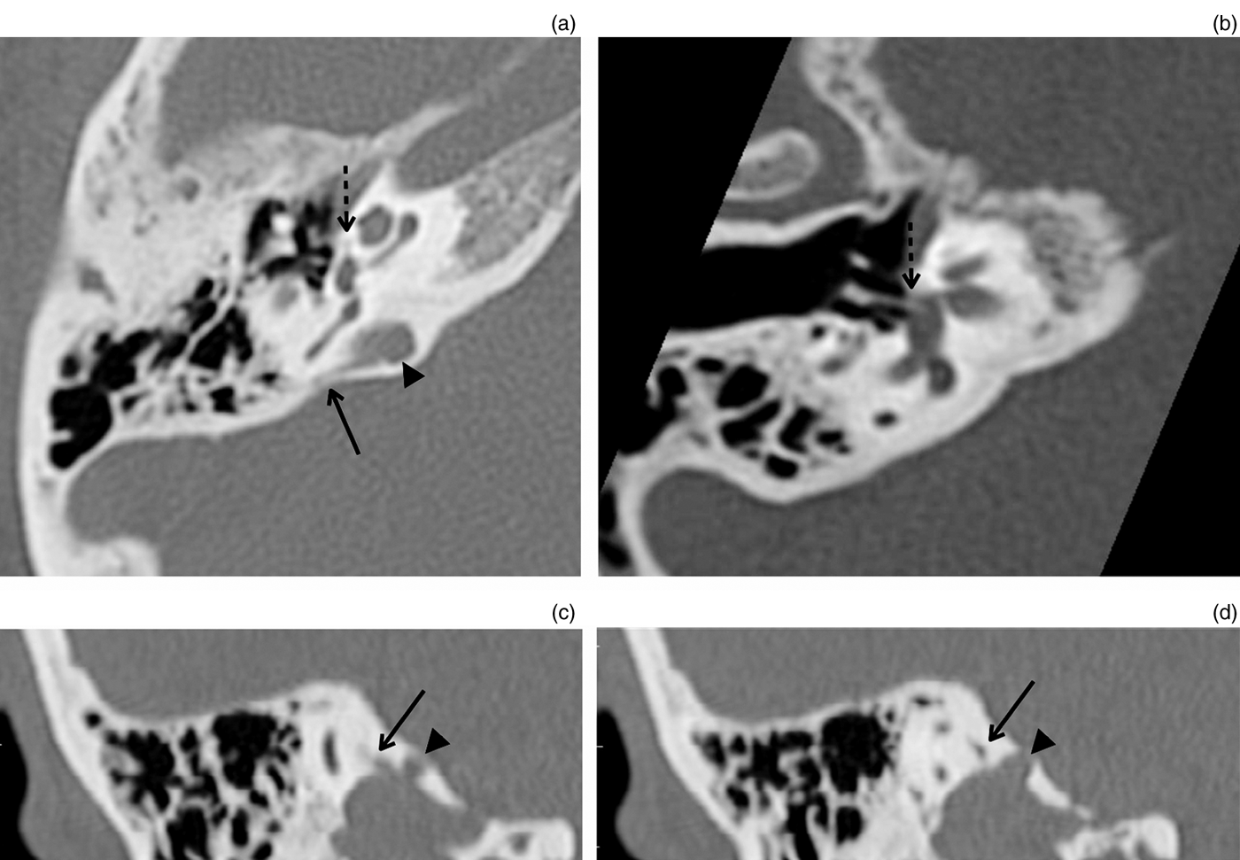
A temporal bone CT scan was performed to evaluate coincidental pathology and the position of the prosthesis. We found the unilateral, right-sided presence of a high-riding jugular bulb with an associated jugular bulb diverticulum, which protruded posteromedially and was dehiscent towards the vestibular aqueduct (Figure 1).Reference Vincent, Rovers, Zingade, Oates, Sperling and Deveze2, Reference Somers, Govaerts, de Varebeke and Offeciers3 A focal hypodensity in the fissula ante fenestram was also found bilaterally. The Teflon prosthesis had a central position in the stapedotomy and did not protrude into the vestibulum (Figure 1b). No other causes of conductive hearing loss of middle- or inner-ear origin were found.

Fig. 1 High-resolution computed tomography images of the right temporal bone. (a) Axial image: the jugular bulb diverticulum (dotted arrow) is seen at the posteromedial surface of the temporal bone (arrowhead), along with the vestibular aqueduct (arrow). (b) Axial image showing the high jugular bulb, vestibular aqueduct and centrally positioned prosthesis (dotted arrow) in one slice. (c) Coronal image: the high jugular bulb and associated diverticulum are seen (arrowhead), along with the dehiscence towards the vestibular aqueduct (arrow). (d) Coronal image: the high jugular bulb and associated diverticulum are seen (arrowhead), along with the vestibular aqueduct (arrow).
Discussion
Persistent hearing loss after stapes surgery in otosclerotic patients is unusual. This type of surgical failure has to be distinguished from recurrent hearing loss, which involves a different set of causes. The causes of persistent hearing loss most frequently cited are: malleus ankylosis, short or long prosthesis, incomplete previous operation, and negative findings. In 5.8–37.7 per cent of patients, no abnormalities are detected during middle-ear inspection (i.e. negative findings).Reference Lippy, Battista, Berenholz, Schuring and Burkey1–Reference Somers, Govaerts, de Varebeke and Offeciers3
A temporal bone CT can demonstrate coincidental pathology before revision surgery is undertaken. Pathological findings acting as potential mimickers include ‘third-window’ lesionsReference Merchant, Rosowski and McKenna4–Reference Van Rompaey, Potvin, van den Hauwe and Van de Heyning6 (e.g. semicircular canal dehiscence (most frequently of the superior or posterior canal), large vestibular aqueduct syndrome, and dehiscence between the cochlea and carotid canal) and inner-ear, Mondini-like malformations (e.g. Ménière's disease and intralabyrinthine schwannomas).Reference Merchant, Rosowski and McKenna4
Recently, attention has been drawn towards jugular bulb abnormalities eroding inner ear structures.Reference Friedmann, Le, Pramanik and Lalwani7 These include the high-riding jugular bulb and the jugular bulb diverticulum potentially impinging upon the posterior semicircular canal or the vestibular aqueduct. Clinical symptoms can include sensorineural hearing loss, vertigo and/or conductive hearing loss. Again, this is explained by a pathological third-window effect.
• A temporal bone computed tomography scan is useful to screen for third mobile window lesions in otosclerosis cases with persistent or recurrent conductive hearing loss, even if stapes fixation was confirmed
• This paper describes the first clinical case with a jugular bulb diverticulum dehiscence towards the vestibular aqueduct in a patient with otosclerosis, with confirmed stapes fixation
In the presented case, we diagnosed a high-riding jugular bulb with an associated diverticulum, which was dehiscent towards the vestibular aqueduct, in a patient with confirmed otosclerosis who did not experience hearing improvement after stapedotomy. In this patient, revision surgery was cancelled because of the potential risk of sensorineural hearing loss.
Conclusion
This case of a high-riding jugular bulb with an associated jugular bulb diverticulum, which was dehiscent towards the vestibular aqueduct, demonstrates the usefulness of a temporal bone CT scan in the evaluation of patients with otosclerosis in whom stapedotomy has not improved hearing. Revision surgery might have imposed a risk on the inner ear, and this can be avoided by making the diagnosis pre-operatively.


