Introduction
Acute mastoiditis is a suppurative infection of the mastoid air cells and is the most common complication of acute otitis media. It is subdivided into two stages: acute mastoiditis with periostitis (also known as incipient mastoiditis), which is defined by the presence of purulent material in the mastoid cavity; and coalescent mastoiditis (also known as acute mastoid osteitis), which is characterised by the destruction of the bony septa between the air cells of the mastoid cavity.Reference Bluestone, Klein, Bluestone, Casselbrant and Stool1
Acute mastoiditis is rare, with an incidence of approximately 1.2 per 10 000 child-years.Reference Groth, Enoksson, Hultcrantz, Stalfors, Stenfeldt and Hermansson2 The epidemiology of mastoiditis is similar to that of acute otitis media, with patients under two years of age exhibiting the highest incidence.Reference Groth, Enoksson, Hultcrantz, Stalfors, Stenfeldt and Hermansson2–Reference Thompson, Gilbert, Long, Saxena, Sharland and Wong4 The literature does not agree on whether there has been an increase in incidence in recent years, and it is unclear whether this represents an actual increase in disease prevalence or not.Reference Ghaffar, Wördemann and McCracken5–Reference Choi and Lander8
The diagnosis of acute mastoiditis is made clinically, with well-documented characteristic findings,Reference Cherry, Calzada, Shapiro, Cherry, Demmler-Harrison, Kaplan, Steinbach and Hotez9–Reference Wald, Conway, Long, Pickering and Prober11 and imaging is often not necessary when these findings are present.Reference Tamir, Schwartz, Peleg, Perez and Sichel12 Computed tomography (CT) may be used to confirm the diagnosis in patients who do not present with the typical clinical findings, and is an important adjunct in the identification of coalescent mastoiditis and when determining intra-cranial extension.Reference Stähelin-Massik, Podvinec, Jakscha, Rüst, Greisser and Moschopulos13
The opacification of mastoid air cells is a commonly reported radiological finding, and patients are often erroneously diagnosed with acute mastoiditis when this finding is present, despite the absence of the characteristic clinical findings. This issue has been previously studied by examining incidental findings on magnetic resonance imaging (MRI) in childrenReference Singh, Rettiganti, Qin, Kuruva and Hegde14–Reference Abbas, Yuen, Trinidade and Watters18 and on CT in the adult population.Reference Paluska, Sise, Sack, Sise, Egan and Biondi19 To the authors’ knowledge, CT findings in the paediatric population have not been studied.
This study aimed to quantify the incidental finding of mastoid opacification on CT in the asymptomatic paediatric population, and to further contribute to epidemiological data on this condition. Such data may be used by primary care physicians, emergency physicians and otolaryngologists in the diagnosis, prognostication and management of patients presenting with this finding.
Materials and methods
Data were obtained retrospectively from the electronic medical records of all paediatric patients who underwent relevant CT imaging. Patients who underwent imaging for otological indications and those who presented with temporal bone fractures were excluded.
Patients aged 0–17 years were included in the study. The exact timing of mastoid air cell pneumatisation is not agreed upon, although it has been stated to occur between 24 weeks’ gestation and birth,Reference Anson and Donaldson20 and air cells are readily visible by age 2 years.Reference Schillinger21
The data collected included: age; gender; the presence, location and severity of mastoid opacification; the existence of middle-ear opacification; and the presence of clinical signs of mastoiditis. These data were analysed using Microsoft Excel® spreadsheet software and R statistical software.
Results
Data were collected from 767 patients. Prevalence by age group and severity (complete vs partial opacification, and unilateral vs bilateral opacification) are recorded in Table 1.
Table 1. Prevalence of mastoid opacification by age and severity

Opacification of mastoid cells was reported in 82 patients (prevalence = 10.7 per cent). Of the patients with mastoid opacification, a total of 34 had radiological evidence of middle-ear effusion (prevalence = 41 per cent). The severity of the opacification was recorded as partial opacification (n = 48, prevalence = 6.2 per cent) or complete opacification (n = 34, prevalence = 4.4 per cent), and as unilateral opacification (n = 45, prevalence = 5.9 per cent) or bilateral opacification (n = 37, prevalence = 4.8 per cent).
A total of 480 males and 287 females were recruited. The Fisher's exact test was performed on the gender data, which showed no statistically significant difference in the prevalence of: overall opacification (male = 9.7 per cent, female = 12.1 per cent; p = 0.3342), partial opacification (male = 6 per cent, female = 6.6 per cent; p = 0.7597), complete opacification (male = 3.8 per cent, female = 5.5 per cent; p = 0.277), unilateral opacification (male = 5.8 per cent, female 5.9 per cent; p = 1) or bilateral opacification (male = 4 per cent, female = 6.3 per cent; p = 0.1649).
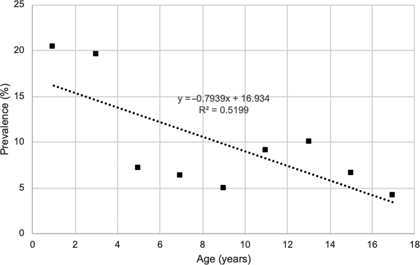
Data were analysed by age, dividing the population studied into age brackets of two years. The prevalence was highest in patients aged zero to one year (n = 25, prevalence = 20.3 per cent), followed by those aged two to three years (n = 17, prevalence = 19.5 per cent).
The Fisher's exact test was performed to determine if there were significant differences between the age groups and the total population studied. The prevalence rates for mastoid opacification in those aged zero to one year (p = 0.0006) and two to three years (p = 0.0088) were shown to be statistically significant. The prevalence rates in these two age groups were shown to be statistically significant for complete mastoid opacification (zero to one year, p = 0.0001; two to three years, p = 0.0452) and for bilateral mastoid opacification (zero to one year, p = 0.0008; two to three years, p = 0.0278). There was no statistically significant difference observed between the other age groups examined.
A linear regression analysis was performed to examine the total prevalence of mastoid opacification according to age groups (Figure 1). The incidence of mastoid opacification was found to be inversely correlated to increasing age, and this was statistically significant on regression analysis (effusion rate = (0–0.07939) × age + 0.1614, slope co-efficient p = 0.0284). The linear regression fitted the data moderately well (R2 = 0.5199).

Fig. 1. Total prevalence of opacification.
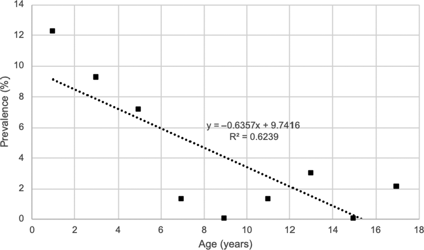
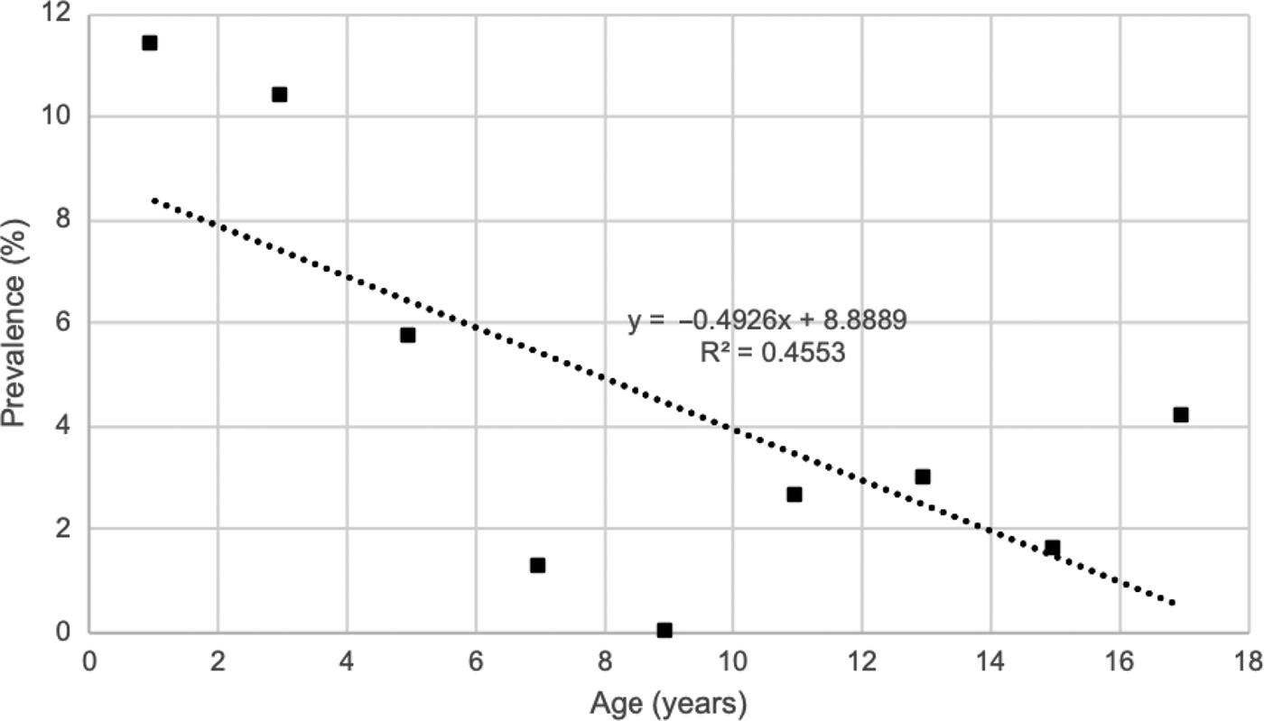
The prevalence of complete mastoid opacification by age group was examined with a linear regression analysis (Figure 2). This showed that the incidence of complete mastoid opacification was inversely correlated to increased age (complete opacification rate = (0–0.01271) × age + 0.1038, slope co-efficient p = 0.0113). Linear regression analysis additionally revealed that the incidence of bilateral mastoid opacification was inversely correlated to increased age (bilateral opacification rate = (0–0.009853) × age + 0.09382, slope co-efficient p = 0.0462; Figure 3). Linear regression analyses were also performed to examine the prevalence of partial mastoid opacification and unilateral mastoid opacification in the same age groups, but these showed no statistical significance.

Fig. 2. Prevalence of complete opacification.

Fig. 3. Prevalence of bilateral opacification.
Discussion
Mastoid opacification is a common incidental finding in the asymptomatic paediatric population, with prevalence rates between 5 per cent and 20 per cent, depending on age. The prevalence peaks in patients aged zero to four years (19–20 per cent), and is inversely correlated with increasing age. This is likely due to the incidence of otitis media in this age group.Reference Groth, Enoksson, Hultcrantz, Stalfors, Stenfeldt and Hermansson2–Reference Thompson, Gilbert, Long, Saxena, Sharland and Wong4
Younger patients (aged zero to four years) had higher prevalence rates of more severe mastoid opacification (complete opacification vs partial, and bilateral vs unilateral opacification). The prevalence rates of bilateral mastoid opacification and complete mastoid opacification were shown to inversely correlate with increasing age.
This study indicates that incidental findings of mastoid opacification are common in the asymptomatic paediatric population, especially in those aged less than four years.
Our results suggest a prevalence rate lower than those previously published. Singh et al. studied a population of 515 paediatric patients who underwent MRI, and found an incidence rate of between 21 per cent and 47 per cent.Reference Singh, Rettiganti, Qin, Kuruva and Hegde14 von Kalle et al. prospectively studied 147 patients with MRI and showed a prevalence rate of 25–40 per cent.Reference von Kalle, Fabig-Moritz, Heumann and Winkler16 These rates are significantly higher than those seen in our population group. Previous studies have shown that up to 82 per cent of MRI-diagnosed mastoiditis cases do not show any clinical mastoiditis, potentially explaining the increased prevalence found in studies using the MRI modality.Reference Polat, Aksoy, Serin, Yıldız and Tanyeri17
Abbas et al. conducted a retrospective case series evaluating radiological mastoid opacification on MRI scans in the adult population, and showed an incidence of 5.8 per cent.Reference Abbas, Yuen, Trinidade and Watters18 Wilkinson et al. investigated incidental findings of mastoid opacification on CT imaging of the adult population, and showed a prevalence of 11 per cent, which is higher than expected given the decreased prevalence of middle-ear disease in the adult population.Reference Wilkinson, Sahota, Constable, Harper and Judd15
• Mastoiditis is a suppurative infection of mastoid air cells, typically diagnosed clinically
• Computed tomography (CT) and magnetic resonance imaging scans are commonly ordered as adjuncts to aid diagnosis of patients with atypical presentations and evaluate for complications
• Mastoid opacification on CT is a common finding in the asymptomatic paediatric population
• There are no published data on the prevalence of mastoid opacification on CT in the paediatric population
Limitations of this study include the retrospective design and the reliance on accurate documentation of patients’ presenting complaints. The radiological diagnosis of mastoiditis may not always be clinically relevant. Further longitudinal studies are needed to determine whether treatment is indicated or not.
Acknowledgement
The Townsville Hospital and Health Service, Australia.
Competing interests
None declared


