Introduction
Superior semicircular canal dehiscence is characterised by the absence of part or all of the bone overlying the canal, exposing the membranous labyrinth to the middle cranial fossa. This condition was first described by Minor in 1998.Reference Minor, Solomon, Zinreich and Zee1 It presents clinically with the classical symptom of noise- or pressure-induced vertigo or disequilibrium. The diagnosis is confirmed by demonstration of the dehiscence in the coronal images of high resolution computed tomography (CT) scans of the temporal bones. Most of the published literature describes such dehiscence as an isolated finding confined to the canal.
We present three cases in which the superior semicircular canal dehiscence was accompanied by multiple tegmen defects, forming a characteristic ‘honeycomb’ pattern.
Case reports
Patient one
A 34-year-old woman presented with a history of the sudden onset of a feeling of imbalance while shouting as a spectator at a rugby match. Following this, she had become unsteady, with short-lived episodes of vertigo, whenever carrying heavy objects, climbing up a ladder, straining on the toilet or even getting out of a car. She had sustained a whiplash injury three years before presentation. Hearing was muffled in her left ear. There was no tinnitus or ear discharge, and she was otherwise fit and well.
Otoneurological examination did not reveal any abnormality.
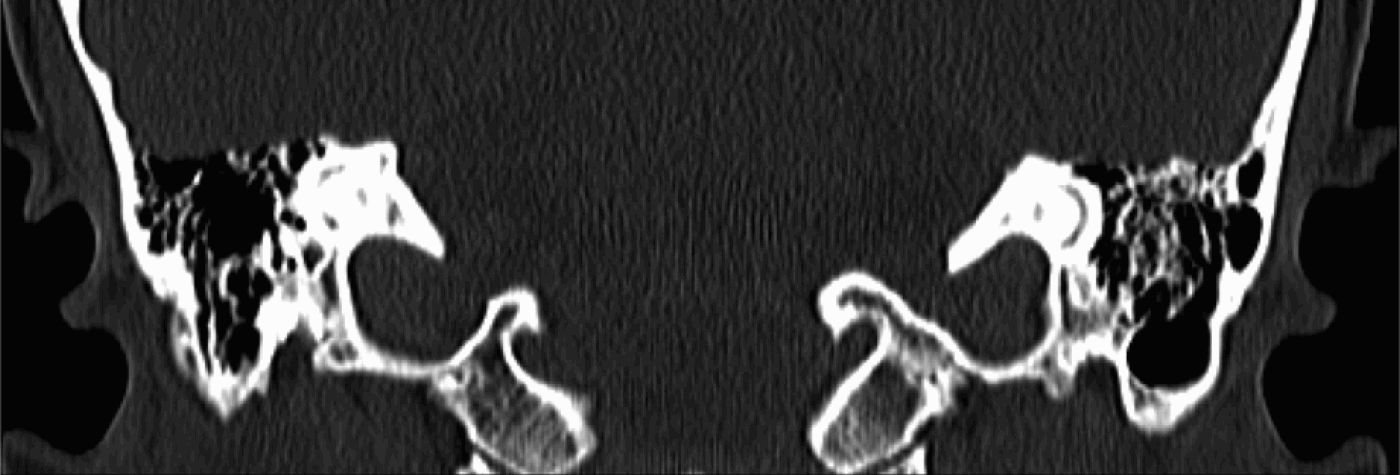
Pure tone audiometry showed a mild, low frequency, conductive hearing loss on the left side, with a normal tympanogram. The Weber test localised to the opposite ear. Caloric testing did not reveal any significant canal paresis. Magnetic resonance imaging of the brain and internal auditory canal was normal. Computed tomography scanning of the temporal bones showed bilateral superior semicircular canal dehiscence (Figures 1 and 2).

Fig. 1 Coronal CT image of patient 1 showing multiple tegmen defects.

Fig. 2 Coronal CT image of patient 1 showing multiple tegmen defects.
The patient underwent obliteration of her left superior semicircular canal dehiscence via a middle fossa craniotomy, during which the honeycomb tegmen was noted. The dehiscence was covered with bone dust and bone cement.
Patient two
This 64-year-old man was referred with a history of hearing vibrations in the ears when singing or speaking. This was worse in the right ear but also occurred a little in the left ear, and had been happening for the previous five years. The symptom was so disturbing that the patient had had to cease singing, and had reduced the volume of his conversations to a minimum. He also reported feeling dizzy when shouting.
Otoscopy was normal. The patient was able to hear a tuning fork placed on his ankle. Neurotological examination did not reveal any abnormality.
Audiography showed moderate to severe, bilateral, high frequency, sensorineural hearing loss. Computed tomography scanning of the temporal bones showed bilateral dehiscence of the superior semicircular canals, more marked on the right than the left (Figures 3 and 4).

Fig. 3 Coronal CT image of patient 2 showing the superior semicircular canal dehiscence.

Fig. 4 Coronal CT image of patient 2 showing multiple tegmen defects.
The patient underwent repair of his right-sided dehiscence via a middle fossa approach. Again, a honeycomb tegmen was noted. Repair was performed using temporalis fascia and bone pâté.
Patient three
A 53-year-old woman presented with a 20-year history of right-sided, pulsatile tinnitus, hyperacusis, and disorientation and unsteadiness, with increasing intra-aural or intracranial pressure. All these symptoms had appeared after a road traffic accident.
Neurotological examination was normal. The patient's symptoms were triggered by tympanometry. Her pure tone thresholds were normal. High resolution CT scanning of the temporal bones demonstrated right superior semicircular canal dehiscence.
The patient underwent middle fossa approach craniotomy, and multiple tegmen defects (honeycomb tegmen) were found (Figure 5). Both superior semicircular canal dehiscence and tegmen defects were closed with bone pâté and temporalis fascia.

Fig. 5 Peroperative image taken during middle fossa craniotomy showing the multiple tegmen defects.
Discussion
The tegmen defects described in the literature are mainly characterised by one or two isolated areas of bony dehiscence.
In a study of 1000 temporal bones, Carey et al. found bony dehiscence over the superior semicircular canal in five (0.5 per cent) specimens (0.7 per cent of individuals).Reference Carey, Minor and Nager2 Of these five specimens, the dehiscence was found between the canal and the middle cranial fossa in one, while in the other four the defect lay between the canal and the superior petrosal sinus. These authors also found markedly thin (less than 0.1 mm) bone covering the superior semicircular canal in another 1.4 per cent of specimens. The majority of the patients with thin bone covering did not report any vestibular symptoms. Such thin bone could easily be missed by imaging, unless ultra high resolution CT scanning was used; such cases would probably be shown as having a dehiscence by a normal scanner.
Single tegmen defects have also been found by various other authors, indicating an incidence that varies from below 1 per cent to as high as 34 per cent.Reference Ahren and Thulin3–Reference Tsunoda and Terasaki6
Tsunoda and Terasaki examined 69 cadaveric middle cranial fossae and found only two bony defects over the superior semicircular canal.Reference Tsunoda and Terasaki6 Both mastoids were well developed, with thin bone over the air cells. This study also included 244 dry bones, four of which showed a bony defect over the superior semicircular canal.
However, none of these temporal bone studies documented the multiple tegmen defects, forming a honeycomb pattern, noted in our patients.
Carey et al. postulated that the thinning and dehiscence of bone over the superior semicircular canal may result from failure of postnatal development of the outer and/or middle layer of bone over the canal.Reference Carey, Minor and Nager2 This conclusion was based on three facts. First, the dehiscence was similar in appearance to the findings in infant temporal bones, in which the bone over the canal was thin and did not reach adult dimensions until three years of age. It is however interesting to note that, despite this finding, there are no reports of symptoms in children. Second, the thin bone over the canal was found to be lamellar bone, indicating that long-standing processes were responsible for the thinning. Third, the fact that these defects are often bilateral also suggests a developmental cause.
Our finding of a honeycomb tegmen coexisting with a dehiscent superior canal would also support the above argument, indicating that the failure of bone formation need not be limited to the superior semicircular canal but could involve the entire tegmen plate.
Despite the presence of thin or dehiscent bone, many patients are asymptomatic.Reference Minor, Solomon, Zinreich and Zee1 However, extra force, in the form of direct trauma or barotrauma, may breach the thin bone, leading to the development of superior semicircular canal syndrome.
Sound-induced vertigo (Tullio phenomenon) or oscillopsia is often the symptom that prompts suspicion of superior semicircular canal dehiscence. The cause can be just loud noise, or sound of a specific frequency such as a telephone dial tone, a child's scream or a particular note on a church organ.Reference Banerjee, Whyte and Atlas7–Reference Ostrowski, Byskosh and Hain9 Clinically, the vestibular symptom can be elicited by a tone between 500 and 2000 Hz played at an intensity of 110 to 120 dB.Reference Minor10 Any circumstance causing changes in middle-ear pressure or intracranial pressure can produce vestibular symptoms. This includes performing a fistula test or Valsalva manoeuvre, nose-blowing, lifting heavy weights, straining of any nature, and air travel. The vestibular symptoms are attributed to increased compliance and abnormal movement of the endolymph within the canal, due to the presence of a third window.Reference Minor, Solomon, Zinreich and Zee1 Occasionally, chronic disequilibrium can result.Reference Minor10 The evoked eye movement is generally upward, torsional nystagmus in the plane of the affected superior canal.Reference Minor11
Patients with superior semicircular canal dehiscence can develop hearing loss with a characteristic low to mid-frequency air–bone gap, alongside bone conduction thresholds that are often better than 0 dB HL.Reference Minor10, Reference Mikulec, Poe and McKenna12 The air–bone gap can be as high as 60 dB at the lower frequency.Reference Mikulec, Poe and McKenna12 This large air–bone gap is due to the fact that some fraction of the fluid volume displaced by the oscillating stapes is shunted through the superior canal away from the cochlea.Reference Mikulec, Poe and McKenna12 The air–bone gap improves with surgical correction of the dehiscence.Reference Minor, Carey, Cremer, Lustig, Streubel and Ruckenstein13 Bone conduction thresholds are elevated due to the presence of a third window, resulting in greater fluid motion than normal, when the skull is made to vibrate by bone-conducted sound.Reference Mikulec, Poe and McKenna12 This elevated bone threshold has reportedly enabled patients to hear their own heel strike when running, or to hear a tuning fork placed on the lateral malleolus.Reference Watson, Halmagyi and Coltebach8, Reference Minor10
Some patients may present with mild to moderate sensorineural hearing loss.Reference Minor10 Their speech discrimination scores and tympanometric pressures are normal. Caloric test results are often normal, except when the dehiscence is large enough for the brain to press on the membranous labyrinth. Electronystagmography using a three-dimensional scleral coil is ideal to study torsional nystagmus, but video-occulography may also be helpful. The direction of nystagmus can give important clues to help determine which semicircular canal is defective.
Superior semicircular canal dehiscence may be misdiagnosed as otosclerosis when it presents with hearing loss alone, without vestibular symptoms. Even stapedectomy has been performed erroneously.Reference Mikulec, Poe and McKenna12 Bone conduction thresholds, being better than 0 dB (−5 to −15 dB), give a clue. In addition, a normal stapedial reflex can help to differentiate the condition from otosclerosis. The diagnosis is also likely to be missed in the presence of a coexisting condition such as chronic otitis media, in which case the symptoms will be attributed to the more obvious disease. Ramsey et al. detected superior canal dehiscence incidentally on a CT scan performed for chronic disequilibrium in a patient with persistent otorrhoea after mastoidectomy.Reference Ramsey, McKenna and Barker14 In such a situation, the clinician is more likely to suspect a fistula in the lateral semicircular canal, as did these authors. Tullio phenomenon can also be associated with syphilis, Ménière's disease, perilymph fistula and Lyme's disease.Reference Banerjee, Whyte and Atlas7 Interestingly, one patient with bilateral dehiscence in Carey and colleagues' study also had a large vestibular schwannoma on one side.Reference Carey, Minor and Nager2
• Superior semicircular canal dehiscence is characterised by the absence of part or all of the bone overlying the canal, exposing the membranous labyrinth to the middle cranial fossa
• This report describes three cases in which superior semicircular canal dehiscence was accompanied by multiple tegmen defects forming a characteristic ‘honeycomb’ pattern
• This finding has not previously been reported, and supports the theory of a developmental defect as the origin of superior semicircular canal dehiscence
Surgical treatment of superior semicircular canal dehiscence leads to resolution of symptoms. Plugging with bone pâté has been shown to be more efficacious than resurfacing.Reference Minor11, Reference Mikulec, Poe and McKenna12 Surgical closure of superior semicircular canal dehiscence carries a risk of sensorineural hearing loss, probably of similar magnitude to that of posterior canal plugging for benign positional vertigo. The middle fossa approach with temporal lobe retraction adds a further risk of epilepsy and this risk needs to be well understood by the patient, along with its implications for driving.Reference Aggarwal, Green and Ramsden15


