


Introduction
The human adult vocal fold has a layered structure consisting of the epithelium, the lamina propria (superficial, intermediate and deep layers) and the vocalis muscle.Reference Hirano1 The vocal ligament consists of the intermediate and deep layers of the lamina propria.Reference Hirano1 The layered structure is essential for vocal fold vibration and phonation.Reference Hirano1 On the other hand, at birth, the layered structure seen in adult vocal folds is not present.Reference Hirano, Kurita, Nakashima, Bless and Abbs2 After birth, the human vocal fold grows and develops, and its layered structure matures during adolescence.Reference Hirano, Kurita, Nakashima, Bless and Abbs2
Our previous research shows there is growing evidence that the cells in the maculae flavae located at both ends of the lamina propria of the vocal fold mucosa are tissue stem cells of the human vocal fold mucosa, and the macula flavae are a candidate for a stem cell niche.Reference Sato, Umeno and Nakashima3–Reference Sato, Chitose, Sato, Kurita, Sato and Umeno10
Tissue stem cells (tissue-specific stem cells, adult stem cells, somatic stem cells) have the capacity to self-renew and generate functionally differentiated cells that replenish cells lost throughout an organism's lifetime.Reference Sato8 Therefore, the cells in the maculae flavae participate in the growth and development of the human vocal fold as a vibrating tissue after birth.
The mitochondria, one of the intracellular organelles where many vital metabolic reactions take place, are the major source of cellular adenosine triphosphate.Reference Ghadially11 Because the mitochondria supply the cell with most of its usable energy, they are considered to be the powerhouse or powerplant of the cell.Reference Ghadially11 The features of the tissue stem cells in the newborn maculae flavae, including energy metabolism, before growth and development of the human vocal fold are of interest.
In this study, the energy metabolism of the cells in the maculae flavae of the human newborn vocal fold was investigated from the aspect of mitochondrial microstructure.
Materials and methods
The authors assert that all procedures contributing to this work comply with the ethical standards of the relevant national and institutional guidelines on human experimentation (Kurume University) and with the Helsinki Declaration of 1975, as revised in 2008.
Five normal human newborn vocal folds obtained from autopsy cases were investigated. The mitochondria in the cytoplasm of the cells in the maculae flavae of the human newborn vocal fold mucosa were observed using transmission electron microscopy.
For transmission electron microscopy, the specimens were fixed in 2.5 per cent glutaraldehyde at 4°C for 2 hours, rinsed with cacodylate buffer solution and post-fixed in 2 per cent osmium tetroxide with cacodylate buffer solution at 4°C for 2 hours. After rinsing with cacodylate buffer solution, the specimens were dehydrated in graded concentrations of ethanol and embedded in epoxy resin. Semi-thin sections were prepared with an ultramicrotome, stained with 1 per cent toluidine blue and examined with a light microscope. Thin sections were made with an ultramicrotome. Thin sections were stained with uranyl acetate and lead citrate. Observation was conducted with an H-7650 (Hitachi High-Tech Corporation, Tokyo, Japan) transmission electron microscope.
In order to assess the number of mitochondria per cell in the maculae flavae, their numbers were calculated in 50 random cells in the electron micrographs of the specimens. The size of each mitochondrion was measured in each of the 50 cells.
Results
Mitochondrial morphology
In the newborn maculae flavae, the nucleus-cytoplasm ratio of the cells was large, and cytoplasm occupied a small area around the nucleus. Out of the mitochondria that were present, not many were randomly distributed. Their shape was oval (Figure 1), and they were 420–880 nm (626 ± 159 nm, mean ± SD) in diameter. Their numbers per cell ranged from 0 to 3 (0.8 ± 1.0) in the electron micrographs of the specimens.

Fig. 1. Transmission electron micrograph (uranyl acetate and lead citrate stain) showing mitochondria in the cytoplasm of the cells in the maculae flavae of the human newborn vocal fold. Generally, the mitochondrial cristae of the cells in the newborn maculae flavae were sparse. Mitochondria consisted of a double membrane bounded body containing matrices. However, both membranes of many mitochondria were ambiguous. Close association between mitochondria and rough endoplasmic reticulum in the cytoplasm of the cells was observed. The rough endoplasmic reticulum fused with the mitochondrial membrane and outer and inner membranes adjacent to the rough endoplasmic reticulum had disappeared (circle with white dotted line). rER = rough endoplasmic reticulum
Generally, some mitochondria consisted of a double membrane bounded body containing matrices and a system of cristae. And some mitochondria were limited by smooth-countered outer and inner membranes. However, both membranes of many mitochondria were ambiguous, indicating the mitochondria were immature (Figure 1).
The inner membrane ran parallel to the outer membrane where double membrane bounded bodies were not ambiguous. In a few mitochondria, cristae, the inner membrane forming thin folds that project into the interior of the mitochondria (lamellar cristae), were observed (Figure 1).
The mitochondrial outer and inner membranes formed two compartments. One was a smaller membrane space (outer chamber) comprising the narrow cleft between the outer and inner membranes and extending inward between the membranes of cristae. Another was a large intercristal space consisting of all of the area within the inner membrane.
The intercristal space was occupied by a mitochondrial matrix which was as electron dense as the contents of the membrane space. Mitochondrial DNA was observed as a loose aggregation of slender filaments in the electron-lucent area of the mitochondrial matrix (Figures 1, 2 and 3). Ribonucleoprotein granules, approximately 12 nm in diameter, were distributed throughout the matrix (Figures 1, 2 and 3). Some mitochondrial dense granules (Figure 2) and some mitochondrial inclusions (Figure 3) were observed in the mitochondrial matrix.

Fig. 2. Transmission electron micrograph (uranyl acetate and lead citrate stain) showing mitochondria in the cytoplasm of the cells in the maculae flavae of the human newborn vocal fold. Mitochondria consisted of an ambiguous double membrane bounded body containing matrices. Mitochondrial dense granules were observed.
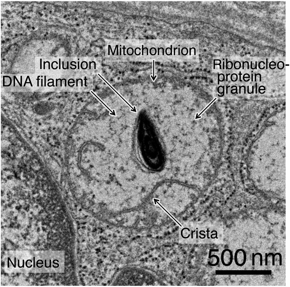
Fig. 3. Transmission electron micrograph (uranyl acetate and lead citrate stain) showing mitochondria in the cytoplasm of the cells in the maculae flavae of the human newborn vocal fold. Mitochondria consisted of an ambiguous double membrane bounded body containing inclusion.
Concentration of cristae
Some, though not many, cells had mitochondrial cristae as a system of membranous laminae or plate-like structures lying in the mitochondrion (Figures 1, 2 and 3). They arose from the inner membrane and traversed a variable distance across the organelle. This characteristic feature of the mitochondrial cristae of the cells in the human newborn maculae flavae was sparse.
Mitochondrial division and fusion
Mitochondrial profiles suggesting impending division or fusion were present (Figure 4). The static electron micrographs could not on their own indicate the direction in which the process was moving.

Fig. 4. Transmission electron micrograph (uranyl acetate and lead citrate stain) showing impending division or fusion of the mitochondria in the cytoplasm of the cells in the maculae flavae of the human newborn vocal fold. (a) Each mitochondrial outer and inner membrane adjacent to the membrane of another mitochondrion had disappeared (arrowhead). (b) Each mitochondrial membrane had been attached (arrowhead).
Mitochondrial associations
A few lipid droplets were present in the cytoplasm of the cells in the newborn maculae flavae. Close association between mitochondria and lipid droplets in the cytoplasm of the cells was present. In some cells, mitochondrion fused to the surface of a lipid droplet in the cytoplasm (Figure 5). Additionally, both the mitochondrial outer and inner membranes adjacent to the lipid droplets had incarcerated and disappeared (Figure 5).

Fig. 5. Transmission electron micrograph (uranyl acetate and lead citrate stain) showing close association between mitochondria and lipid droplets in the cytoplasm of the cells in the human newborn maculae flavae. A single mitochondrion fused to the surface of a lipid droplet in the cytoplasm. Both the mitochondrial outer and inner membranes adjacent to the lipid droplet had incarcerated and disappeared (circle with white dotted line).
Some close association between mitochondria and rough endoplasmic reticulum in the cytoplasm of the cells in the newborn maculae flavae was observed (Figure 1 and 6). Curved profiles of rough endoplasmic reticulum with cisternae (moderately distended rough endoplasmic reticulum with proteinaceous secretary product) had wrapped around and attached to mitochondria (Figure 1 and 6). The rough endoplasmic reticulum partially or almost completely encircled the mitochondrion. The rough endoplasmic reticulum fused with the mitochondrial membrane, and outer and inner membranes adjacent to the rough endoplasmic reticulum had incarcerated and disappeared (Figures 1 and 6).

Fig. 6. Transmission electron micrograph (uranyl acetate and lead citrate stain) showing close association between mitochondria and rough endoplasmic reticulum in the cytoplasm of the cells in the human newborn maculae flavae. The curved profiles of rough endoplasmic reticulum wrapped around mitochondria. The rough endoplasmic reticulum fused with the mitochondrial membrane, and mitochondrial outer and inner membranes adjacent to the rough endoplasmic reticulum had incarcerated and disappeared (circle with white dotted line. rER = rough endoplasmic reticulum
Discussion
Physiologically, the latest research shows that the maculae flavae located at both ends of the lamina propria of the human vocal fold mucosa are involved in the metabolism of extracellular matrices, which are essential for the viscoelastic properties of the human vocal fold mucosa as a vibrating tissue, and they are responsible for maintaining the characteristic layered structure of human vocal fold mucosa.Reference Sato, Umeno and Nakashima12,Reference Sato13 Furthermore, maculae flavae are considered to be an important structure in the growth, development and aging of the human vocal fold mucosa.Reference Sato, Umeno and Nakashima14–Reference Sato16 Moreover, our previous research showed there is growing evidence that maculae flavae are a stem cell niche containing tissue stem cells of the human vocal fold mucosa.Reference Sato, Umeno and Nakashima3–Reference Sato, Chitose, Sato, Kurita, Sato and Umeno10
The mitochondria, one of the intracellular organelles where many vital metabolic reactions take place, are the major source of cellular adenosine triphosphate and supply the cell with most of its usable energy. In living cells, these organelles are seen to make slow sinuous movement accompanied by changes in size and shape.Reference Ghadially11
In this study, energy metabolism of the cells in the maculae flavae of the human newborn vocal fold was investigated from the aspect of mitochondrial microstructure.
Variations of mitochondria
Mitochondria show many variations in size, shape and fine structures; however, they are sufficiently characteristic to be distinguishable from other organelles in electron micrographs.Reference Ghadially11
Although the basic morphology of the mitochondrion is very characteristic, innumerable variations occur. Such variations can be considered in terms of species differences, tissue or organ differences, differences in physiological and functional activity and differences in pathological states.Reference Ghadially11
Numbers of mitochondria and cristae
In general, the number of mitochondria per cell and the number of cristae per mitochondrion are related to the energy requirement for the function carried out by that cell type.Reference Fawcett17 There is a positive correlation between the metabolic activity of a tissue and the number and size of mitochondria.Reference Ghadially11
The present study showed that the number of mitochondria per cell and the number of cristae per mitochondrion were small in the cells of newborn maculae flavae. These electron microscopic findings were the same as in adults.Reference Sato, Chitose, Sato, Sato, Kurita and Umeno18
Double membrane bounded body and a system of cristae
In general, the main morphological features can be summarised by defining a mitochondrion as a double membrane bounded body containing matrices and a system of cristae.Reference Ghadially11
The cristae are the main sites of mitochondrial energy conversion.Reference Kühlbrandt19 The inner membrane and its spheres are the site of oxidative phosphorylation.Reference Ghadially11 Most of the tricarboxylic acid cycle enzymes are located in the matrices, and electron transport and oxidative phosphorylation enzymes form molecular assemblies in or on the inner mitochondrial membrane covering the wall and cristae.Reference Ghadially11 The shallow proton gradient between the inter membrane space and the matrix drives adenosine triphosphate production by adenosine triphosphate synthase in the membrane of the cristae.Reference Kühlbrandt19
There is a positive correlation between the metabolic activity of a tissue and the number, size, surface area and concentration of cristae.Reference Ghadially11 The number of cristae per mitochondrion is much greater in cells with high-energy requirements than in those with a lower rate of metabolism.Reference Fawcett20
Regarding the cells in the maculae flavae of the human adult vocal fold, our previous investigation showed that the main morphological features of the mitochondria were double membrane bounded bodies containing matrices and a system of cristae.Reference Sato, Chitose, Sato, Sato, Kurita and Umeno18 However, in each mitochondrion, the lamellar cristae were sparse.Reference Sato, Chitose, Sato, Sato, Kurita and Umeno18
The present study showed that the mitochondria of the cells in the newborn maculae flavae not only had sparse lamellar cristae but also had ambiguous double membrane bounded bodies. This indicates that the mitochondria are immature, and their metabolic activity and oxidative phosphorylation are low.
Matrix of mitochondria
The present study showed that the mitochondrial matrices in mitochondria of the cells in the newborn maculae flavae contained DNA filaments, ribonucleoprotein granules and mitochondrial dense granules, which are observed in normal mitochondria.
The previous studies on isolated dense granules indicated that they contain calcium, magnesium, phosphorus and inorganic material.Reference Ghadially11,Reference Pasquali-Ronchetti, Greenawalt and Carafoli21
Division and fusion of mitochondria
Mitochondria divide and multiply, and old, effete or damaged mitochondria can suffer regressive changes and be removed in various ways in the cells.Reference Ghadially11 Regarding the cells in the maculae flavae of the human adult vocal fold, our previous investigation showed mitochondrial profiles suggesting impending division or fusion.Reference Sato, Chitose, Sato, Sato, Kurita and Umeno18 The present study showed that the mitochondria of the cells in the newborn maculae flavae showed mitochondrial profiles suggesting impending division or fusion, although the static electron micrographs could not indicate the direction in which the process was moving.
Mitochondrial associations with other organelles
A close association between mitochondria and other organelles provides correlation between structure and function. Mitochondria are often located near a supply of substrate or at sites in the cell known to require the adenosine triphosphate generated by the mitochondria.Reference Ghadially11
The human maculae flavae contain vocal fold stellate cells that are stellate in shape and possess lipid droplets containing vitamin A in their cytoplasm.Reference Sato13,Reference Sato, Hirano and Nakashima22,Reference Sato, Hirano and Nakashima23 The roles of lipid droplets in the cytoplasm of vocal fold stellate cells have been unclear.
In the present study, some mitochondria were closed to the surface of a lipid droplet in the cytoplasm of the cells in the newborn maculae flavae. Additionally, both the mitochondrial outer and inner membranes adjacent to the membrane of lipid droplets had incarcerated and disappeared. These electron microscopic findings are also observed in the cytoplasm of the cells in the adult maculae flavae.Reference Sato, Chitose, Sato, Sato, Kurita and Umeno18 These fine features suggested that the lipid droplets in the cytoplasm had supplied the fatty acid that was degraded by beta-oxidation in the mitochondria. Because the mitochondria are known to contain many of the enzymes (fatty acid oxidases) necessary for the metabolism of triglycerides,Reference Ghadially11 these fine features suggested that the close association between mitochondria and a lipid droplet brings the mitochondrial enzymes into close association with the lipidic substrate. The cells in the newborn maculae flavae, like those in adults,Reference Sato, Chitose, Sato, Sato, Kurita and Umeno18 may utilise lipids for their metabolic needs to some extent.
Another close association was seen between the mitochondrion and the rough endoplasmic reticulum. Curved profiles of rough endoplasmic reticulum with cisternae wrapped around mitochondria and the rough endoplasmic reticulum fused with the mitochondrial outer and inner membrane indicated that mitochondria provide energy for the rough endoplasmic reticulum.
Energy metabolism
The major source of DNA damage in stem cells, besides that associated with cell proliferation, is endogenously generated genotoxic agents produced as signalling molecules or metabolic by-products, among which reactive oxygen species are the most common threat.Reference Riz, Hawley, Rajasekhar and Vemuri24 Oxidative stress shortens the life span of stem and progenitor cells.Reference Nesti, Pasquali, Mancuso, Siciliano, Rajasekhar and Vemuri25 Reactive oxygen species accelerate aging through random and sequential damage to cell components.Reference Nesti, Pasquali, Mancuso, Siciliano, Rajasekhar and Vemuri25
• Mitochondria in the maculae flavae of human newborn vocal fold consisted of a double membrane bounded body containing matrices and a system of cristae
• Microstructural features of mitochondria in maculae flavae of the human newborn vocal fold suggested that metabolic activity and oxidative phosphorylation was low
• This may result in favourable metabolism to maintain the stemness and undifferentiated states of the cells after birth
• Energy metabolism of the cells in the human newborn maculae flavae may have shifted to other metabolic systems
Reactive oxygen species are continuously generated by normal metabolic processes such as oxidative phosphorylation in mitochondria.Reference Riz, Hawley, Rajasekhar and Vemuri24 The inner mitochondrial membrane covering the wall and cristae are the site of oxidative phosphorylation.Reference Ghadially11 The oxidative phosphorylation in the mitochondria is the major source of endogenous reactive oxygen species.Reference Riz, Hawley, Rajasekhar and Vemuri24 There is usually a good correlation between the metabolic rate and the level of reactive oxygen species generated by mitochondria.Reference Nesti, Pasquali, Mancuso, Siciliano, Rajasekhar and Vemuri25
Reactive oxygen species, the most significant mutagens in stem cells, activate the protective mechanisms blocking self-renewal of the stem cells when elevated, and at the same time serve as a signal stimulating stem cell differentiation.Reference Riz, Hawley, Rajasekhar and Vemuri24
Quiescence (G0 phase) is critical for protecting the stem cell compartment.Reference Nesti, Pasquali, Mancuso, Siciliano, Rajasekhar and Vemuri25 The quiescent state is generally viewed as a mechanism for avoiding accumulation of damage resulting from physiological stress, including oxidative stress.Reference Suda, Takubo and Semenza26 Most of the cells in the maculae flavae of the vocal fold are resting cells (G0 phase).Reference Sato8,Reference Sato, Kurita, Chitose, Sato, Umeno and Yano9 Consequently, it is suggested that the cells in the maculae flavae avoid accumulation of damage resulting from oxidative stress.
In this study, fine features of the mitochondria suggested that the metabolic activity and oxidative phosphorylation of the cells in the human newborn maculae flavae were low, indicating that the intracellular reactive oxygen species production is suppressed. After birth, the cells in the newborn maculae flavae seem to rely more on glycolysis than oxidative phosphorylation for energy supply. The cells in the newborn maculae flavae may utilise lipids to some extent for their metabolic needs.
The energy metabolism of the cells in the human newborn maculae flavae seems to be favourable to maintaining the stemness and undifferentiated states of the tissue stem cells after birth. Further investigation using other experimental methods is necessary in regard to the energy metabolism of the cells in the maculae flavae of human newborn vocal fold mucosa.
Conclusion
The microstructural features of the mitochondria of the cells in the maculae flavae of the human newborn vocal fold suggested that metabolic activity and oxidative phosphorylation was low, resulting in favourable metabolism for maintaining the stemness and undifferentiated states of the cells after birth. Consequently, energy metabolism of the cells in the human newborn maculae flavae may have shifted to other metabolic systems.
Acknowledgements
This study was supported by a Grant-in-Aid for Scientific Research (number 18K09362) from the Japanese Ministry of Education, Culture, Sports, Science and Technology.
Competing interests
None declared


