Introduction
Transoral endoscopic procedures are generally preferred to open surgery where possible, as they are associated with fewer risks and complications.Reference Van Eeden, Lloyd and Tranter1 Their usage in the management of early laryngeal cancers is increasing throughout the UK according to successive annual Data for Head and Neck Oncology (‘DAHNO’) reports.2
The literature shows that there can be up to a 30 per cent conversion rate from an endoscopic to an open surgical approach,Reference Sen and Bhattacharyya3 or abandonment for a non-surgical approach. This is mainly due to anatomical problems, including the presence of teeth, prominent incisor teeth, retrognathia, and other contributing factors such as a long neck or poor neck extension.Reference Sen and Bhattacharyya3
A novel method of overcoming this problem is to electively extract the maxillary incisor teeth to create sufficient access to undertake the transoral surgical procedure. Subsequent dental replacement can be easily undertaken with dental implant based restorations, with the dental implants being placed at the same time as the transoral surgery or at a later date once the extraction sockets have healed. This approach provides a clear advantage for selected patients when managed in a multidisciplinary team environment in a dedicated head and neck surgical centre.
This paper describes two cases which illustrate this approach. The first case relates to the management of a pharyngeal pouch which was complicated by a previous failed endoscopic and open treatment. The second case relates to the management of glottic carcinoma by means of transoral laser microsurgery which was compromised by poor visualisation of the tumour.
Case report
Case one
A 68-year-old gentleman had a pharyngeal pouch that required endoscopic stapling. As well as causing significant pouch-related symptoms, the pouch was hindering the general surgeons from performing regular routine flexible gastroscopies, which were being used as part of an upper gastrointestinal cancer surveillance programme. The patient had a retrognathic mandible, dental crowding and limited mouth opening (Figure 1), which had made a transoral approach impossible.

Fig. 1 Pre-operative situation, with crowded incisor teeth and poor access due to retrognathia.
Multiple attempts were made to manage the pouch in another centre, including an open approach; however, these attempts were unsuccessful. The patient suffered post-operative mediastinitis following the open surgical approach, resulting in a four-week stay in the intensive care department.
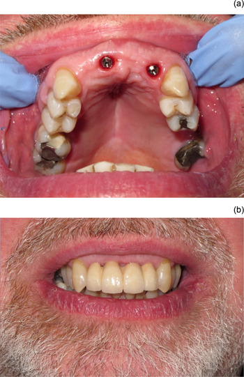
An endoscopic approach was suggested, with extraction of four maxillary incisor teeth to facilitate transoral endoscopic stapling of the pouch. Following extraction, the access was good and the procedure was undertaken within a few minutes (Figure 2). Immediate single-stage, co-axis dental implants (Southern Implants, Irene, South Africa) were placed into two of the extraction sites, with excellent primary stability (Figure 3). A temporary, removable partial denture was fitted immediately following surgery. This was followed by the fitting of the definitive screw-retained implant bridge three months later (Figure 4).

Fig. 2 (a) & (b) Excellent access following dental extractions.

Fig. 3 (a) & (b) Immediate placement of co-axis dental implant fixture to facilitate restoration with a screw-retained bridge.

Fig. 4 (a) & (b) Implants and screw-retained fixed bridge, with excellent aesthetics.
The patient made a rapid post-operative recovery, with significant improvement in his symptoms. He subsequently required an additional procedure on a partially reformed pouch. This was facilitated by removal of the implant-supported bridge prior to surgery and replacement immediately afterwards. The patient experienced complete cessation of all dysphagia and regurgitation symptoms.
Case two
A 72-year-old gentlemen opted for transoral laser resection to manage a T3N0 (tumour–node stage) glottic carcinoma; however, he had reduced mouth opening. This prevented full access to the tumour, which extended up to the anterior commissure.
The patient's maxillary central and lateral incisors were extracted to create the necessary space to facilitate the transoral laser resection. A temporary partial denture was provided in the interim. Unfortunately, because of the pathology findings, it was necessary to treat him with post-operative chemoradiotherapy, from which he had a complete response and remains disease free and with laryngeal preservation over five years later. Twenty months following the chemoradiotherapy, he underwent dental implant placement under local anaesthesia and a screw-retained implant-supported bridge was constructed.
Three years post-operatively, the patient reported some tightness of the airway. As he was at risk of total airway obstruction if a respiratory tract infection were to have occurred, the decision was made to carry out microlaryngoscopy and dilation of the stenotic larynx. Oral access was again required for this procedure, and therefore the implant-supported bridge was unscrewed and temporary healing caps were placed immediately prior to the procedure. The bridge was then screwed back on post-operatively, in the out-patient clinic.
Overview
In both cases, the patients were assessed by a maxillofacial prosthodontist prior to undergoing definitive surgery in order to assess their overall dental status and their suitability for immediate dental implant placement. Dental extractions should be carried out extremely carefully to preserve the thin buccal alveolar bone plate. The alveolus should be protected as much as possible with a gauze swab during the transoral ENT surgical procedure to prevent any trauma to the extraction site, which is of particular importance for immediate implant placement.Reference Buser, Martin and Belser4
Following completion of the transoral procedure, dental implants can be placed immediately into the extraction sockets to facilitate a quicker prosthetic rehabilitation for the patient following surgery. Immediate implant placement is also believed to maintain the vertical alveolar bone height, and therefore preserve what is left and create a more aesthetic appearance.Reference Esposito, Grusovin, Polyzos, Felice and Worthington5 The authors prefer to use a co-axis osseointegrated dental implant (Southern Implants). This allows the final restoration to be screw-retained and easily removable should the patient require a further transoral procedure. A temporary removable denture can be provided for the patient immediately following the surgical procedure. Following successful implant integration, the definitive prosthesis can be provided in an out-patient setting by the prosthodontic specialist.
Discussion
We report a novel technique for facilitating transoral surgical access, with high-quality fixed tooth replacement following surgery. This technique has not previously been reported in the literature. The two cases highlighted show how such an approach can facilitate transoral surgery for patients with poor anatomical access for instrumentation.
Dental implants have an extremely high success rate, with the facility to provide high-quality, durable and aesthetic restoration of the teeth. Such treatment requires an additional 30 minutes of operating time when implants are placed simultaneously, together with a small number of out-patient clinic visits post-operatively. These cases are funded by the National Health Service at a cost of around £3000 (approximately US$4500 at the time of writing). The authors believe this is minimal compared to the costs of managing any serious complications of open surgery, such as intensive or high-dependency care services.
With appropriate patient positioning, the authors feel that most endoscopic procedures can be performed without the need for any extractions, and the technique described remains a rare approach within our unit. Often patients have missing molars and pre-molars, facilitating a lateral approach. In the rare cases where endoscopy is truly impossible because of dental obstruction, we perceive there to be a reluctance to even consider maxillary incisor extractions in many units, seeing it as too high a price to pay for the patient. We hope that these cases illustrate that with appropriate specialist prosthodontic input early on in the patient's management, the sacrifice of the maxillary incisor teeth to facilitate transoral surgery can be a low morbid procedure. It is well tolerated by patients and allows an alternative to open surgery (which has increased risks). The importance of providing screw-retained retrievable implant-supported restorations for this group of patients cannot be over-stressed; repeat transoral surgery may be required at a later stage, as was demonstrated in both the cases described.