


Introduction
Tympanic membrane perforations have been shown to be responsible for a certain degree of hearing impairment.Reference Mehta, Rosowski, Voss, O'Neil and Merchant1–Reference Ribeiro Fde, Gaudino, Pinheiro, Marcal and Mitre3 The scope of the problem is evident, especially in children. It has been evidenced that otitis media, a major cause of tympanic membrane perforation, is the most common specifically treated disease in children and the second most common disease of childhood.4
Some controversies persist regarding the determinants of hearing loss severity subsequent to tympanic membrane perforation. In general, the larger the perforation, the greater the hearing impairment. This relationship is, however, not constant or consistent in clinical practice.Reference Ludman, Ludman and Wright5, Reference Irwin, Thomas and Katz6 Additionally, authors do not have a univocal opinion on the effect of tympanic membrane perforation site on hearing.Reference Voss, Rosowski, Merchant and Peake7, Reference Berger, Finkelstein, Avraham and Himmelfar8
Modern technology enables the objective and precise evaluation of: the exact site and surface area of the tympanic membrane perforation, and the proportion of tympanic membrane perforation in relation to the total surface area of the tympanic membrane.
Certain factors specific to sub-Saharan Africa could have an impact on the characteristics of tympanic membrane perforation, making the results of such a study differ greatly from those obtained in developed countries. For instance, Ibekwe et al. showed that tympanic membrane perforations in Africa are mostly long-standing.Reference Ibekwe, Ijaduola and Nwaorgu9 The paucity of otolaryngologists makes early access to specialised services difficult, hence chronic cases become more frequent.Reference Ijaduola and Okeowo10–Reference Benitez12 High humidity and wet climatic conditions in West Africa predispose to allergies and upper respiratory tract infections.Reference Ibekwe, Ijaduola and Nwaorgu9 Inhabitants, especially children, will therefore suffer frequent episodes of otitis media, with a propensity toward perforation and chronicity.
Therefore, it appears highly relevant to obtain precise information using objective methods in this specific setting. Foreseeing the impact of tympanic membrane perforation on hearing, and adapting treatment modality choices, could aid patient management. This study was conducted to describe the pattern of tympanic membrane perforation, and determine the relationship between tympanic membrane perforation characteristics and resulting hearing defects, in our setting.
Materials and methods
Study design and procedure
A cross-sectional study was conducted from September 2016 to June 2017 in five referral hospitals of Yaounde, the capital city of Cameroon. These centres have well-structured ENT units with similar equipment, and hence were suitable for performing pure tone audiometry.
We included consenting patients aged over 10 years who presented with tympanic membrane perforations, as evidenced by otoscopy. This age limitation permitted easy and reliable audiometric evaluation. We excluded marginal and attico-antral tympanic membrane perforations in order to eliminate potential cases of ossicular damage.Reference Ludman, Ludman and Wright5 We also excluded all cases with air–bone gaps greater than 50 dB, on the basis that hearing loss solely caused by tympanic membrane perforation, with no ossicular involvement, cannot exceed 50 dB.Reference Voss, Rosowski, Merchant and Peake7, Reference Voss, Rosowski, Merchant and Peake13, Reference Voss, Rosowski, Merchant and Peake14 Cases with continuous or recurrent ear discharge after two weeks of adequate treatment were also excluded.
Prior to initiating the study, our research protocol was submitted and validated by the Ethical Review Board of the Faculty of Medicine and Biomedical Sciences of the University of Yaounde I, Cameroon. Administrative authorisations were obtained from the various authorities of our study sites. Eligible patients were briefed on the study objectives. Information forms were handed out, after which a consent form was given to the subject or their guardian (for minors) to be signed.
For each participant, we collected sociodemographic data and medical history details using a pre-tested questionnaire. Next, we carried out a thorough otoscopic examination of the ear using a video otoendoscope, coupled to a digital camera with computer digital capture, to ascertain that the perforation was dry. This permitted the precise calculation of tympanic membrane perforation and tympanic membrane surface areas, using appropriately calibrated integrated software. The proportion of the perforation in relation to the total surface area of the tympanic membrane was obtained by dividing the tympanic membrane perforation surface area by the tympanic membrane surface area, expressed as a percentage. Tympanic membrane perforation site was defined in terms of four tympanic membrane regions: anterior-superior, anterior-inferior, posterior-superior and posterior-inferior regions. These regions were morphologically demarcated by a vertical line passing through the handle of malleus and another passing perpendicularly to the first line. Pure tone audiometry was carried out in each case to confirm hearing loss and to determine severity. Each ear was considered as an independent case.
The predictor variables considered were: size, site, duration and aetiology of tympanic membrane perforation. Duration was considered as the time elapsed since the first symptom assumed to be linked to the tympanic membrane perforation. Tympanic membrane perforation aetiology was determined by taking a thorough history and consulting patient records.
The outcome variable was hearing loss severity. The ears were tested at frequencies of 0.5, 1, 2 and 4 kHz on air and bone conductions. Mean conductive hearing loss was evaluated by calculating the average of the air–bone gaps obtained at the four frequencies, based on World Health Organization guidelines. The degree of hearing loss was classified according to the Bureau International d'Audiophonologie (‘BIAP’) classification.15
We defined a central tympanic membrane perforation as a pars tensa perforation overlapping three quadrants, as evidenced by otoendoscopy. A small tympanic membrane perforation was any perforation with a surface area proportion less than 25 per cent of the tympanic membrane. A medium tympanic membrane perforation was one with a surface area proportion between 25 and 50 per cent of the tympanic membrane. A large or subtotal tympanic membrane perforation was defined as a perforation with a surface area proportion greater than or equal to 50 per cent of the tympanic membrane.
Statistics
Statistical analysis was conducted using SPSS® version 24 software. Comparisons of means were performed using t-test or analysis of variance, as required. Predictors of hearing loss severity were analysed using Pearson's correlation coefficients and a binomial logistic regression model for multivariate analysis. Hearing loss was the dependent variable, analysed in terms of air–bone gap categories of: 40 dB HL or less, and more than 40 dB HL. Values of p < 0.05 were considered statistically significant.
Results
Tympanic membrane perforation pattern
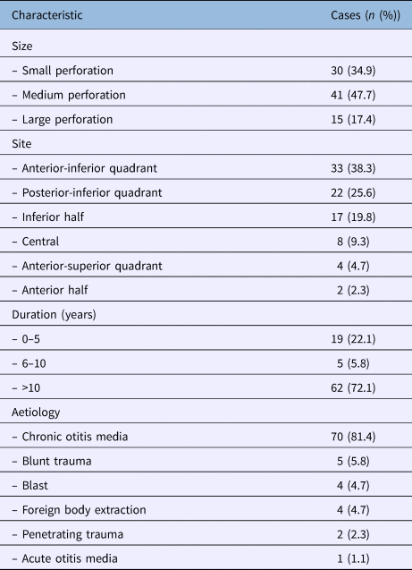
Eighty-six cases of tympanic membrane perforation were identified, in 67 patients (37 males (55.2 per cent) and 30 females), with 19 patients (28.4 per cent) having bilateral perforations. The mean patient age was 25.1 ± 9.4 years. The most affected quadrants were the anterior-inferior quadrant (33 cases; 38.3 per cent) and the posterior-inferior quadrant (22 cases; 25.6 per cent) (Table 1). The mean tympanic membrane perforation surface area proportion was 34.1 ± 18.4 per cent (range, 2.3–83.1 per cent). The leading cause of tympanic membrane perforation was chronic otitis media (70 cases; 81.4 per cent). The median duration of the condition was 240 months.
Tympanic membrane perforation impact on hearing
The mean air–bone gap was 33.1 ± 9.1 dB (range, 13.8–50 dB), with predominantly mild hearing loss (70.9 per cent). The greatest hearing loss was found at low frequencies, with 0.5 kHz having the highest average loss (39.4 dB), followed by 1 kHz (36 dB) (p < 0.001). The lowest average hearing loss was at 2 kHz (26.1 dB). The low frequencies had the greatest air–bone gaps, irrespective of perforation size (Figure 1).

Fig. 1. Effect of tympanic membrane perforations (TMP) of various sizes on hearing according to sound frequency. ABG = air–bone gap
Hearing loss severity predictors
Mean hearing loss appeared to increase with tympanic membrane perforation size and duration. Posterior tympanic membrane perforations and perforations of infectious origin were associated with more severe hearing loss (Table 2). There was a significant positive correlation between tympanic membrane perforation proportion and mean hearing loss (r = 0.47; p < 0.001) (Figure 2). On multivariate logistic regressions, size was the only statistically significant predictor of hearing loss severity. Site, aetiology and duration of tympanic membrane perforation did not significantly influence hearing loss severity (Table 3).

Fig. 2. Scatterplot for tympanic membrane perforation (TMP) proportion versus mean hearing loss (HL).
Table 2. Hearing loss according to tympanic membrane perforation characteristics

*Includes anterior-superior and anterior-inferior tympanic membrane perforations. †Posterior-inferior tympanic membrane perforations
Table 3. Multivariate logistic regression model for predictors of hearing loss

*Total n = 86. †Includes anterior-superior and anterior-inferior tympanic membrane perforations. ‡Posterior-inferior tympanic membrane perforations. OR = odds ratio; CI = confidence interval
Discussion
A total of 86 tympanic membrane perforations were included in our study. There was no particular predilection for any age group or sex. The pattern of tympanic membrane perforations was consistent with worldwide literature when considering site and aetiology. Data concerning tympanic membrane perforation size and duration were comparable to those obtained in similar settings. Tympanic membrane perforation size seemed to be a predictor of hearing loss severity in our setting.
Tympanic membrane perforation size was calculated and expressed in terms of surface area and proportion. Medium tympanic membrane perforations were most frequent (47.7 per cent). Ibekwe et al.Reference Ibekwe, Ijaduola and Nwaorgu9 similarly found 40.5 per cent of tympanic membrane perforations between 25 and 50 per cent. They reported a significant correlation between the duration and size of the tympanic membrane perforation (p = 0.042). Therefore, the frequency of moderately large tympanic membrane perforations could be due to long-standing infections in our setting, leading to larger perforation sizes. The distribution of hearing loss according to frequency significantly showed the greatest loss at low frequencies, with the lowest average hearing loss at 2 kHz. Studies have shown that the resonance frequency of the tympanic membrane and ossicular chain is around 2 kHz; therefore, loss of sound transmission is least around this frequency.Reference Lokberg, Hogmoen and Gundersen16, Reference Gyo, Aritomo and Goode17
Tympanic membrane perforation size was considered the main predictor of hearing loss severity. There was a statistically significant positive correlation between tympanic membrane perforation size and mean hearing loss. Multivariate analysis showed that the risk of more severe hearing loss was 2.5 times greater for a medium as compared to a small tympanic membrane perforation (95 per cent confidence interval = 1.02–6.13, p = 0.04) (Table 3). This confirms that tympanic membrane perforation size is a predictor of hearing loss severity, with hearing impairment increasing with tympanic membrane perforation size.
Many similar studies agree with these findings, both in developing and developed countries. Lerut et al.Reference Lerut, Pfammatter, Moons and Linder18 and Voss et al.Reference Voss, Rosowski, Merchant and Peake14 also reported that hearing loss increased with tympanic membrane perforation size. This can be explained by the fact that larger perforations result in greater loss of middle-ear and mastoid volume, hence there is a drop in the pressure difference across the tympanic membrane.Reference Mehta, Rosowski, Voss, O'Neil and Merchant1 However, this was contradicted by Ribeiro et al.,Reference Ribeiro Fde, Gaudino, Pinheiro, Marcal and Mitre3 who instead concluded that factors such as ossicular chain disjunctions or fixations, and not perforation size, compromise auditory acuity. They arrived at this conclusion using correlations that did not take into account other independent variables.
Anterior tympanic membrane perforations had the lowest average mean air–bone gap. This could suggest that posterior tympanic membrane perforations lead to greater hearing impairment than anterior tympanic membrane perforations. Berger et al.Reference Berger, Finkelstein, Avraham and Himmelfar8 found that tympanic membrane perforations involving the posterior-inferior quadrant were associated with the largest air–bone gap. They suggest that when the perforation overlies the round window (in posterior-inferior tympanic membrane perforations), there is a phase effect cancellation. Our multivariate analysis showed contrary results, concurring with Ibekwe et al.Reference Ibekwe, Nwaorgu and Ijaduola19 and Voss et al.,Reference Voss, Rosowski, Merchant and Peake7 who found that the tympanic membrane perforation position does not affect the resultant magnitude of conductive hearing loss. We excluded posterior-superior tympanic membrane perforations, which are recognised to be prone to complications. Hence, we agree with authors who suggest that hearing loss severity is not dependent on site but on size, and on the state of other components of the conductive pathway.
Interestingly, our study showed no relation between tympanic membrane perforation duration and hearing impairment. Ibekwe et al.Reference Ibekwe, Ijaduola and Nwaorgu9 found a positive relationship between symptom duration and the tympanic membrane perforation size (r = 0.459, p = 0.04). One might therefore infer that hearing loss is worsened by tympanic membrane perforation duration. If there is any effect of tympanic membrane perforation duration on hearing loss, it could be linked to associated infectious complications of chronic otitis media. This eventuality in our study was addressed by excluding cases with possible ossicular damage that potentially develops over time.Reference Varshney, Nangia, Bist, Singh, Gupta and Bhagat20
• The most common cause of tympanic membrane perforation is chronic otitis media
• Tympanic membrane perforation has been shown to cause some degree of hearing loss
• This hearing loss is determined by: perforation size, site, cause and other sound conduction pathway components
• This study informs on the tympanic membrane perforation profile in sub-Saharan Africa, regarding size, site, duration and aetiology
• The data suggest that perforation site and duration do not have a significant influence on hearing loss severity
• The effect of other perforation characteristics on hearing loss may be linked to inflammatory complications of the sound conduction pathway
Aetiologies of tympanic membrane perforations were grouped into infectious and traumatic causes. There was a slightly greater effect of infectious tympanic membrane perforations on hearing loss, but this was not significant. Studies have reported the effect of chronic infection on hearing as a result of ossicular discontinuity and cholesteatoma,Reference Haidar, Sheikh, Larem, Elsaadi, Abdulkarim and Ashkanani21 conditions we excluded. In addition, the relative rarity of cholesteatoma in AfricansReference Kuo, Shiao, Yung, Sakagami, Sudhoff and Wang22 would explain why we observed little effect of tympanic membrane perforation aetiology on hearing loss severity, given that ossicular discontinuity and cholesteatoma are the major purveyors of severe hearing loss in chronic otitis media.
Some studies have highlighted the role of middle-ear volume and umbo involvement in hearing loss severity. These studies reported that hearing loss varied inversely with middle-ear volume.Reference Mehta, Rosowski, Voss, O'Neil and Merchant1, Reference Voss, Rosowski, Merchant and Peake14 Umbo involvement in the tympanic membrane perforation was also found to worsen subsequent hearing loss.Reference Lerut, Pfammatter, Moons and Linder18
Limitations of our study include the fact that middle-ear volume and umbo involvement were not considered. In addition, tympanic membrane perforation duration and the upper cut-off value for mean hearing loss set at 50 dB (aimed at excluding cases of ossicular damage) were presumptive.
Conclusion
Our study provides information on the profile of tympanic membrane perforation and factors influencing hearing loss in our setting. It suggests that clinicians should predominantly consider the effect of tympanic membrane perforation size on hearing loss; site, duration and aetiology of tympanic membrane perforation, in the absence of inflammatory complications, seem to have no influence on hearing loss severity. Given that most patients present with long-lasting tympanic membrane perforations, a factor that possibly predisposes to larger tympanic membrane perforations, specialists should consider non-conservative approaches more often.
Acknowledgements
The authors are grateful to the administrative authorities of the institutions that granted permission for this study and all the patients who voluntarily participated in the study.
Competing interests
None declared


