Introduction
Otosyphilis or luetic osteitis of the temporal bone is a rare but important cause of deafness as it remains one of the few treatable causes. Its occurrence has been increasing with the rising prevalence of acquired immunodeficiency syndrome. Early diagnosis of this condition, whilst often challenging, is known to be associated with improved treatment outcomes. In the report below, we highlight key computed tomography (CT) imaging features that can aid the diagnosis of otosyphilis. We also emphasise specific imaging features that enable differentiation from other, more common conditions presenting with deafness and otic capsule abnormalities.
Case report
A 58-year-old woman with a 15-year history of progressive, bilateral, asymmetrical, moderate to severe, mixed hearing impairment which was worse on the left was referred for further specialist investigation. Other symptoms included increasing and intermittently intrusive tinnitus, vestibular migraines, and vertigo.
The patient had a past medical history of recurrent ear infections and left chronic suppurative otitis media, and had been previously treated with grommet insertions and bilateral tympanic steroid injections.
There was no family history of hearing loss.
On otological examination, she had a permanent left tympanic membrane perforation and wore a hearing aid in her right ear.
Pure tone audiometry revealed a right-sided moderate to severe hearing loss and a left-sided profound, mixed hearing loss. Tympanometry showed normal traces on the right side but flat traces and a large canal volume on the left, consistent with a left tympanic membrane perforation.
High resolution CT of the petrous temporal bones was performed, as the patient was unable to tolerate magnetic resonance imaging due to claustrophobia.
Computed tomography findings
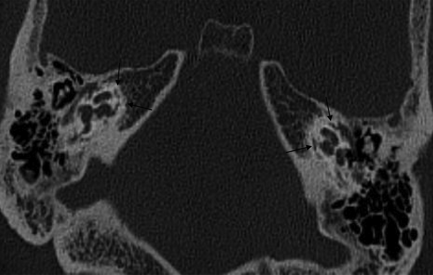
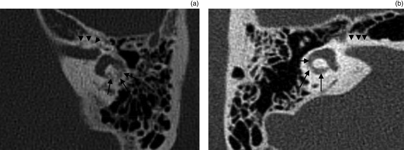
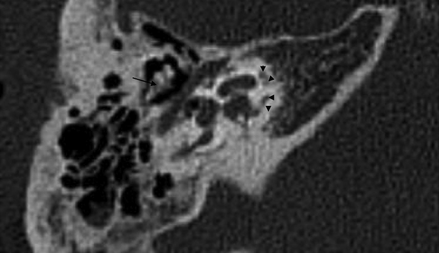
The high resolution CT scan of the petrous temporal bones showed extensive, bilateral lucent areas in the otic capsule, particularly around the cochlea (Figure 1) but also involving the vestibule and semicircular canals (Figure 2). Importantly, there was ossicular involvement and sparing of the fissula ante fenestram (Figures 3 and 4). The remainder of the skull base was normal.

Fig. 1 Axial, unenhanced computed tomography image at the level of the cochlea, showing a symmetrical, permeative, ‘moth-eaten’ pattern of demineralisation (arrows) primarily involving the otic capsule.

Fig. 2 (a) Magnified axial computed tomography (CT) image of the left ear, demonstrating loss of definition around the lateral semicircular canal (arrows). Note also the patchy demineralisation within the otic capsule (arrowheads). (b) Magnified axial CT image of the right ear, demonstrating a normal otic capsule (arrowheads) and lateral semicircular canal (arrows).

Fig. 3 Magnified axial computed tomography image of the left ear. Note sparing of the fissula ante fenestram (arrow).

Fig. 4 Axial computed tomography image of the petrous temporal bone demonstrating ill-defined lucencies within the ossicles (arrow) and patchy lucencies around the cochlear capsule (arrowheads).
These findings were atypical for the more common otodystrophies, and the suggestion of otosyphilis was raised based on radiological appearances. On revisiting the clinical history, we discovered a history of congenital syphilis, treated at the age of 12 years. Subsequent serology results were consistent with a previous treponemal infection.
Discussion
Otosyphilis is a rare disease the incidence of which has only recently been rising due to the increased prevalence of human immunodeficiency virus infection.Reference d'Archambeau, Parizel, Koekelkoren, Van De Heyning and De Schepper1–Reference Little, Gardner, Acker and Land4 It nonetheless remains an important differential to consider in patients presenting with bilateral sensorineural deafness and extensive otic capsule lucencies, as it is one of the few potentially treatable causes.Reference Guttenplan and Hendrix5 This is particularly true where the pattern of lucencies is not typical for otosclerosis.
Whilst otosyphilis is typically accompanied by systemic manifestations of syphilis, involvement of the inner ear is recognised in the late congenital, late latent and tertiary stages of syphilis.Reference Swartz2, Reference Heimert, Lin and Yousem3, Reference Alkadhi and Rissman Kollias6, Reference Fayad and Linthicum7
To our knowledge, the differentiating CT appearances of otosyphilis have only rarely been described in the literature.Reference Swartz2, Reference Sonne, Zeifer and Linstrom8
Otosyphilis can manifest as a gummatous lesion of the internal auditory canal, a labyrinthitis or a luetic osteitis producing the typical permeative, moth-eaten appearance of the temporal bone.Reference Swartz2, Reference Heimert, Lin and Yousem3, Reference Sonne, Zeifer and Linstrom8 Histopathologically, otosyphilis involves an obliterative endarteritis and multinucleated giant cell and round cell infiltration, leading to varying degrees of bony resorption and replacement with fatty marrow.Reference Alkadhi and Rissman Kollias6–Reference Sonne, Zeifer and Linstrom8
In the case presented above, the differential diagnosis for the CT findings included the otodystrophies, defined as a group of primary osseous lesions of the temporal bone that result in hearing loss.Reference d'Archambeau, Parizel, Koekelkoren, Van De Heyning and De Schepper1 The differentiating features of the otodystrophies have been described elsewhere in the literature.Reference d'Archambeau, Parizel, Koekelkoren, Van De Heyning and De Schepper1, Reference Swartz2, Reference Wycherly, Berkowitz, Noone and Kim9 A brief summary of the key differentiating CT features of the commoner conditions is provided below.
Otosclerosis
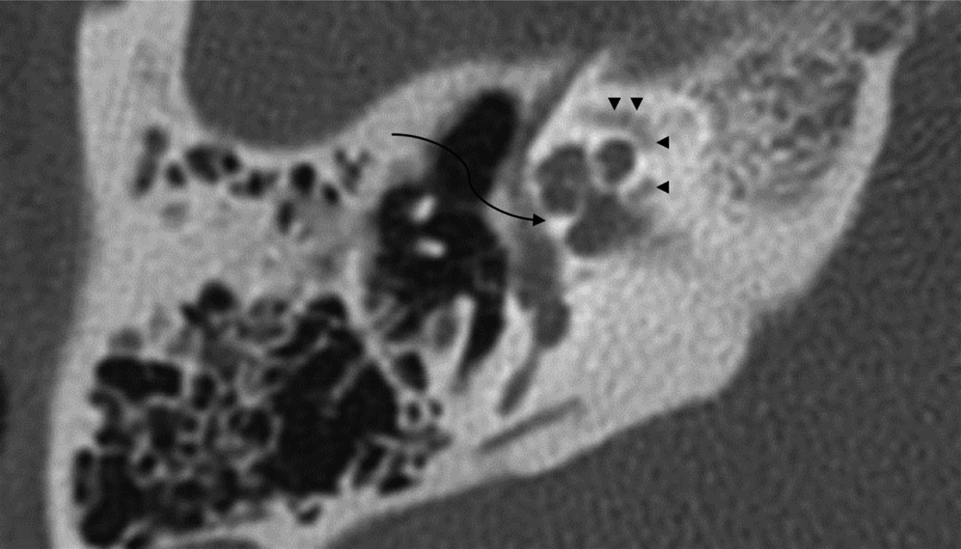
This is the most common cause of bilateral otic capsule lucencies, with bilateral lesions described in up to 90 per cent of cases.Reference d'Archambeau, Parizel, Koekelkoren, Van De Heyning and De Schepper1, Reference Heimert, Lin and Yousem3 It is sometimes hereditary (autosomal dominant) and occurs predominantly in females, with a female to male ratio of 2:1.Reference d'Archambeau, Parizel, Koekelkoren, Van De Heyning and De Schepper1, Reference Swartz2 The disease usually manifests early, commonly within the second or third decade of life.Reference Swartz2 Typically, there is plaque-like demineralisation of the otic capsule, with a high preponderance for involvement of the fissula ante fenestram (62 per cent in one study) (Figure 5).Reference Swartz2, Reference Sonne, Zeifer and Linstrom8, Reference Wycherly, Berkowitz, Noone and Kim9 Ossicular involvement is not a feature.Reference Alkadhi and Rissman Kollias6, Reference Sonne, Zeifer and Linstrom8

Fig. 5 Axial computed tomography image demonstrating otosclerosis, with plaque-like demineralisation of the fissula ante fenestram (curved arrow). Note also the pericochlear involvement (arrowheads).
Osteogenesis imperfecta
This is a rare genetic disorder of collagen synthesis characterised by bone fragility and hearing loss. It can present with bilateral, symmetrical otic capsule lucencies and deafness. The pattern of otic capsule demineralisation can be indistinguishable radiographically from otosyphilis (Figure 6).Reference Sonne, Zeifer and Linstrom8 However, the findings are usually more extreme and a relevant clinical history is always present.Reference Swartz2, Reference Alkadhi and Rissman Kollias6, Reference Sonne, Zeifer and Linstrom8

Fig. 6 Axial computed tomography image demonstrating osteogenesis imperfecta, with gross lytic change involving the entire otic capsule bilaterally. The cochleae are particularly affected and are difficult to delineate (arrows).
Paget's disease
Paget's disease is a generalised bone disorder that usually affects elderly male patients. It primarily involves the axial skeleton though the skull is commonly affected. When the skull is involved, the disease can present with an extensive pattern of otic capsule demineralisation (Figure 7).Reference Sonne, Zeifer and Linstrom8 The findings are often asymmetrical and there is always involvement of the remainder of the calvarium.Reference d'Archambeau, Parizel, Koekelkoren, Van De Heyning and De Schepper1, Reference Swartz2, Reference Alkadhi and Rissman Kollias6 Ossicular chain lucencies are not a feature.Reference Alkadhi and Rissman Kollias6

Fig. 7 Axial computed tomography image demonstrating Paget's disease. Note the diffuse, homogeneous demineralisation of the entire skull base. Only small areas of normal otic capsule remain (black star).
Fibrous dysplasia
This is an inherited, indolent and progressive disorder of unknown aetiology. There is a female predominance, with monostotic and polyostotic forms described. Involvement of the temporal bone, though rare, is characteristically monostotic (Figure 8). This typically manifests on CT as expansion and increase in the temporal bone volume. There is often an associated cholesteatoma of the external ear canal and middle ear.Reference Swartz2 Inner ear involvement and otic capsule demineralisation are not seen.Reference d'Archambeau, Parizel, Koekelkoren, Van De Heyning and De Schepper1, Reference Swartz2

Fig. 8 Axial computed tomography image demonstrating monostotic fibrous dysplasia, with gross expansion of the petrous temporal bone and the classical ‘ground glass’ pattern of demineralisation.
Osteopetrosis
This is an inherited disorder of childhood characterised by a generalised increase in bone density which can involve the temporal bones and skull base (Figure 9).Reference d'Archambeau, Parizel, Koekelkoren, Van De Heyning and De Schepper1, Reference Swartz2 In cases of temporal bone involvement there is marked sclerosis, but without the expansion typical of fibrous dysplasia. The progressive, bony sclerosis eventually encroaches on the internal auditory canal resulting in a sensorineural hearing loss.Reference d'Archambeau, Parizel, Koekelkoren, Van De Heyning and De Schepper1

Fig. 9 Axial computed tomography image demonstrating osteopetrosis, with diffuse sclerosis involving the otic capsule and adjacent temporal bone. Note the generalised loss of normal cortico-medullary differentiation.
The demineralised appearance of the temporal bone produced by the otodystrophies can be mimicked by the resorptive form of otosyphilis.
The present case
In our patient's case, the main differential diagnosis was extensive otosclerosis, osteogenesis imperfecta and otosyphilis. However, as discussed above, osteogenesis imperfecta is unlikely in the absence of an associated history. Furthermore, whilst ossicular involvement can be seen, the changes are usually far more extensive than those demonstrated in our patient. Extensive otosclerosis is likewise unlikely in the setting of sparing of the fissula ante fenestram; in addition, ossicular involvement is not a feature.
• Otosyphilis is a rare but increasingly prevalent cause of sensorineural hearing loss
• Syphilitic involvement of the otic capsule often manifests as lytic temporal bone lesions on computed tomography (CT)
• The presented case emphasises the importance of otosyphilis as a potentially treatable cause of sensorineural deafness
• In this case, CT helped to exclude more common, less treatable causes of otic capsule lucencies
Our patient had extensive bilateral and symmetrical demineralisation of the otic capsule, involvement of the ossicles, and sparing of the fissula ante fenestram (thus excluding extensive otosclerosis); therefore, a diagnosis of otosyphilis required consideration. Furthermore, the pattern and location of involvement, with permeative resorption of the otic capsule and involvement of the ossicular chain, is highly specific for otosyphilis.Reference Swartz2, Reference Alkadhi and Rissman Kollias6, Reference Sonne, Zeifer and Linstrom8 In such cases, a diagnosis of otosyphilis can be confirmed with positive serology results.Reference Sonne, Zeifer and Linstrom8


