Introduction
Cochlear implantation has emerged as the treatment of choice for hearing rehabilitation in paediatric and adult patients with severe-to-profound sensorineural hearing loss (SNHL). Clear visualisation of the round window niche and round window membrane through the facial recess is essential for an ENT surgeon when inserting the electrode into the cochlea during implantation. Adequate intra-operative visualisation of the round window niche can become difficult because of variations in the position of the round window.
A posterior tympanotomy approach is commonly used for cochlear implantation. It exposes the facial recess – a triangular intratemporal area bordered by the fossa incudis superiorly, a vertical segment of the facial nerve medially and the chorda tympani laterally – and provides secure access to the basal turn through the round window niche. Pre-operative prediction of round window niche visibility is important, as successful cochlear implantation by this approach depends greatly on conclusive identification of the round window niche, which may be difficult in patients with limited round window niche visibility. Axial, high-resolution computed tomography (CT) scans of the temporal bone provide radiographic information about internal ear structures, which can be helpful in predicting the round window niche visibility and accessibility pre-operatively.
Jeffery and SpoorReference Jeffery and Spoor1 suggested that there is a significant age-related reduction in the angle of the cochlea relative to the sagittal plane during fetal life, although the post-natal changes in this angle were not found to be statistically significant in their study. Li et al.Reference Li, Wang and Northrop2 suggested that the bone around the round window niche should be removed, and that a cochleostomy immediately anterior and inferior to the round window membrane should be performed, as the critical structures of the inner ear are at their greatest distance from the cochleostomy site in this position and access to the scala tympani is optimal. Pendem et al.Reference Pendem, Rangasami and Arunachalam3 showed that the distance between the short process of the incus and the round window niche, and the distance between the oval window and the round window niche, can help in determining the extent of visibility of the round window seen during posterior tympanotomy.
These measurements can be used to guide surgeons in locating the round window niche and in planning surgery. The present study was conducted to compare the mean angles of the cochlear basal turn relative to the mid-sagittal plane, using endoscopic visualisation and categories of round window niche visibility during a posterior tympanotomy (facial recess) approach.
Materials and methods
An observational study was conducted in patients with severe-to-profound SNHL, with no congenital anomalies, who were scheduled for cochlear implantation. Prior clearance from the institutional ethical committee was obtained.
High-resolution temporal bone CT images were acquired on a 128 slice multi-detector CT scanner; the images underwent multiplanar reformatting and were then opened using MicroDOM software. Axial scans through the temporal bone were acquired in planes parallel to the infraorbitomeatal line. The most representative axial slice through the basal turn of the cochlea was identified. A line was drawn through the midline and through the long axis of the basal turn. The midline was defined by a line drawn through the junction of the face of the sphenoid bone and the nasal septum anteriorly, and through the internal occipital protuberance posteriorly. The angle between these bisecting lines was then measured using radiological software (Figure 1).

Fig. 1. Technique for measuring angles between the cochlear basal turn relative to the mid-sagittal plane pre-operatively on axial, high-resolution temporal bone computed tomography scans.
All angles (in the first angle measurement) were measured by a single observer. However, to check intra-observer error, the measurements for the 19 consecutive patients planned for cochlear implantation were repeated by the same observer a week later (second angle measurement). The mean of the first and second angle measurements was then calculated for each patient (Table 1).
Table 1. Angles measured pre-operatively one week apart

Data represent angles, in degrees. Round window niche visibility grade: *type I, †type IIa and ‡type IIb. Pt no. = patient number
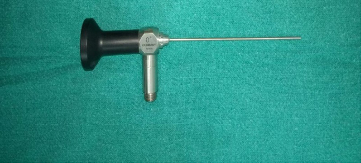
Endoscopic evaluation of the round window niche was performed using a rigid, 0°, 1.9 mm diameter Hopkins otoendoscope (Figure 2) inserted via posterior tympanotomy. The St Thomas’ Hospital ClassificationReference Leong and Jiang4 was used to assess visibility of the round window niche, according to four types (Figure 3): type I = 100 per cent of the round window niche was visible; type IIa = more than 50 per cent of the round window niche was visible; type IIb = less than 50 per cent of the round window niche was visible; type III = the round window niche was not visible (0 per cent). The averages of the mean angle measurements were calculated for all patients with endoscopic visualisation types I, IIa and IIb.

Fig. 2. Otoendoscope (0°, 1.9 mm diameter) used for endoscopic evaluation of the round window niche.

Fig. 3. Endoscopic images of round window niche visibility intra-operatively: (a) type I, (b) type IIa, (c) type IIb and (d) type IIb.
Statistical analysis
The collected data were entered into Microsoft Excel® spreadsheet software, and were analysed and statistically evaluated using SPSS® software, version 17. Data were expressed as means ± standard deviations; a p-value of less than 0.05 was considered statistically significant. The paired t-test was used to analyse differences in intra-operative round window niche visibility and radiographic measurements. Differences in radiographic measurements among the types of intra-operative round window niche visibility were analysed by the one-way analysis of variance (ANOVA) test. The Pearson test was used to calculate intra-observer reliability; p < 0.001 was considered statistically significant.
Results
Of the 19 patients who underwent cochlear implantation in our study, 8 (42 per cent) showed type I round window niche visibility, 8 (42 per cent) showed type IIa and 3 (16 per cent) showed type IIb.
The angles were measured pre-operatively on axial, high-resolution temporal bone CT scans, on two occasions one week apart, for each individual, by a single observer. The Pearson test was used to check for intra-rater reliability. The Pearson r value was calculated as 0.986, which was found to be statistically significant (p < 0.001). This demonstrated very high consistency between the measurements made by the same observer.
The mean angle between the long axis of the cochlear basal turn and the mid-sagittal plane for types I, IIa and IIb visibility were 64.06°, 63.81° and 56.48°, respectively (Table 2). A one-way ANOVA revealed a statistically significant difference (p < 0.05) in the mean angle according to each round window niche visualisation type.
Table 2. Distribution of intra-operative endoscopic grading and averages of mean angle measurements

Sociodemographic profile
The patients' age ranged between 2 and 20 years. Fourteen of the 19 patients were aged 3–8 years; 2 patients were post-lingual, aged 17 years and 20 years. Ten of the patients were male and nine were female. All were scheduled to undergo endoscopic-assisted cochlear implantation under general anaesthetic.
Discussion
Cochlear implant surgery has become the standard of care for patients with severe-to-profound SNHL. Severely deaf patients currently can regain at least partial auditory function by using a cochlear implant. Whereas hearing aids deliver amplified sound to a damaged sensory system, often resulting in louder but non-interpretable information, a cochlear implant can improve speech understanding. Surgical implantation via the transmastoid approach has been well standardised.
There are three techniques to place a cochlear implant: via cochleostomy, in which the promontory is drilled to fixate the implant, via the round window, or via the extended round window, antero-inferior to round window niche. Cochleostomy was first described in the 1980s.Reference Karatas, Aud and Baglam5 Less drilling is required in the round window technique, thus reducing: trauma, loss of perilymph, and bone powder in the scala tympani.Reference Roland, Wright and Isaacson6 Preservation of residual hearing has been viable and beneficial given the combination of electrical and acoustic stimulation, but it requires non-traumatic electrode insertion to minimise damage to inner-ear structures and reduce neural tissue degeneration.Reference Francis and Niparko7
The bony round window orientation (at 90° to the oval window) and variations in the amount of bony overhang are such that, when viewed through the facial recess (without any further bone work), visibility of the membrane itself is restricted to a maximum of approximately 30 per cent of the inferior part of the vertical segment.Reference Takahashi and Sando8 It has previously been suggested that the perceived angle of the round window affects electrode insertion trauma: the more posteriorly oriented the round window membrane, the greater the likelihood of atraumatic electrode insertion, with inherent implications for hearing preservation.Reference Shapira, Eshraghi and Balkany9
In contrast to the standard bony promontory cochleostomy, the round window membrane route has several distinct advantages: (1) a reduced risk of intracochlear trauma (no drilling and no bone dust into the scala tympani);Reference Pau and Just10 (2) the array will necessarily be introduced into the scala tympani;Reference Briggs, Tycocinski and Stidham11 and (3) it utilises the entire basilar turn for electrical stimulation (likelihood of stimulating residual dendrites).Reference Briggs, Tycocinski and Stidham11
Round window membrane insertion has previously been found to increase the spiral lamina length available for stimulation by approximately 2 mm, relative to standard bony cochleostomy insertion.Reference Paprocki, Biskup and Kozłowska12 Clear visualisation of the round window niche through the facial recess during cochlear implantation is a prime requisite for the operating surgeon while inserting the electrode into the cochlea. Obtuse angulation of the basal turn relative to the sagittal plane may make cochlear implantation more difficult.Reference Lloyd, Kasbekar and Kenway13 Variations in the round window position can hinder visualisation of the round window niche.
Chen et al.Reference Chen, Lyu, Xie, Yang, Zhang and Dai14 conducted a study to measure and compare the round window niche in: congenital aural atresia, congenital aural stenosis and control groups. They found that the wall, length and depth of the round window niche tended to be longer, with more severe aural malformations. Bettman et al.Reference Bettman, Appelman and van Olphen15 stated that pre-operative CT measurements, such as the width of the facial recess, and the angle between the facial recess and the cochlear basal turn, were not useful in predicting the problems encountered during surgery. They also claimed that advances in CT, such as multi-slice CT, could improve diagnostic accuracy. Pendem et al.Reference Pendem, Rangasami and Arunachalam3 conducted a study to determine the accuracy of high-resolution CT measurements of the temporal bone in predicting the actual visualisation of the round window niche as viewed during posterior tympanotomy (i.e. facial recess). The authors calculated the distance between the short process of the incus and the round window niche, and the distance between the oval window and the round window niche. They found that pre-operative high-resolution CT measurements had a sensitivity of 92.3 per cent and a specificity of 96.2 per cent in determining actual visualisation of the round window, as compared with the different categories of round window niche visibility determined intra-operatively.
The present study assessed whether the rotation of the cochlear basal turn affects round window niche visibility. We measured the angle of the cochlear basal turn relative to the mid-sagittal plane using the slice containing most representative cross-section of the basal turn. We found that higher grades of round window niche visibility were associated with a more acute angle between the long axis of the cochlear basal turn and the mid-sagittal plane. Hence, the pre-operative high-resolution CT measurements of the angle between the basal turn long axis and the mid-sagittal plane can be useful for determining the actual visualisation of the round window niche during surgery, as viewed during posterior tympanotomy using a 0° otoendoscope.
• This study compared round window niche visibility seen endoscopically during cochlear implant surgery with pre-operative high-resolution computed tomography (CT) of the temporal bone
• There was a significant difference in the mean angles measured pre-operatively on CT and round window niche visibility seen intra-operatively using endoscopes
• The mean angle of the cochlear basal turn relative to the mid-sagittal plane was more acute with higher grades of round window niche visibility
• Pre-operative CT measurements are useful in predicting endoscopic round window niche visualisation
• Otoendoscopes can aid round window niche localisation, and prevent large posterior tympanotomy and surgical complications
Use of an otoendoscope in cochlear implant surgery (via posterior tympanotomy) can improve access to the round window membrane, allow successful insertion into the scala tympani without injuring intracochlear structures, and prevent the need for cochleostomy. The operation should be carried out using smooth, atraumatic electrode carriers, employing adapted surgical procedures. In order to avoid surgical trauma, insertions through the round window membrane might be effective. This was also indicated in a study undertaken to evaluate cochlear implant electrode insertion through the round window membrane in a human temporal bone model; that study concluded that smooth implantations via the round window membrane resulted in deep, atraumatic insertions into the scala tympani.Reference Adunka, Unkelbach and Mack16
Conclusion
The mean angle of the cochlear basal turn relative to the mid-sagittal plane becomes more acute with a higher grade of round window niche visibility (seen intra-operatively). There was a statistically significant difference in the mean angles measured pre-operatively on the high-resolution temporal bone CT scans and round window niche visibility as seen intra-operatively using endoscopes. The mean angle for type I window niche visibility was 64.06° (range, 61.16–69.37°). There was a statistically significant difference between the distribution of round window membrane classifications performed using a microscope or endoscopy. Additional advantages of otoendoscopes over microscopes include: their use as a guide for locating the round window niche (mainly in visibility types IIb and III) on microscopy, the avoidance of large posterior tympanotomy and the prevention of surgical complications.
Competing interests
None declared