


Introduction
Granular myringitis is a chronic inflammation of the surface of the tympanic membrane, characterised by de-epithelialisation and granulation of the involved area.Reference Stoney, Kwok and Hawke1–Reference Blevins and Karmody3 The condition is usually confined to the outer epithelial layer, but sometimes also affects the underlying fibrous layer. In serious cases, it may extend to the adjacent external meatal skin (Figure 1), but it never invades the middle ear. Although this disease can be associated with local trauma, poor hygiene, impacted cerumen, infectious agents (e.g. bacteria and fungi), allergy, inflammation, irritation and dysfunctional epithelialisation, its underlying pathogenesis remains unclear.Reference Stoney, Kwok and Hawke1–Reference El-Seifi and Fouad5
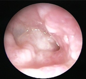
Fig. 1 Right ear showing diffuse granulation of the tympanic membrane and surrounding external meatus.
A variety of treatment options has been proposed, including: irrigation with antibiotic steroid eardrops; chemical cauterisation; and surgical curettage of the polypoid granulation on the tympanic membrane.Reference Stoney, Kwok and Hawke1, Reference Blevins and Karmody3, Reference Khalifa, Fouly, Bassiouny and Kamel6, Reference Jang, Kim, Cho and Wang7 All the available therapeutic regimens have been proved to be generally effective for mild cases, but not for severe cases (with diffuse granulation tissue over the tympanic membrane, or even involvement of the external ear canal), as indicated by the high recurrence rate.Reference Khalifa, Fouly, Bassiouny and Kamel6
In the present study, which focussed on severe cases, we developed a surgical technique which combined tympanic epithelial avulsion with overlay myringoplasty. Our results indicate that this new surgical procedure represents improved treatment for severe, chronic granular myringitis.
Materials and methods
Patient selection
Cases of chronic granular myringitis diagnosed in the otolaryngology department of the second Affiliated Hospital of Sun Yat-Sen University were retrospectively analysed. There were 107 cases of granular myringitis, of which 21 involved the diffuse form, giving a prevalence for the latter of 19.62 per cent (21/107). According to Wolf and colleagues' classification system, 18 cases of diffuse granular myringitis were grade III (six men and 12 women) and three cases were grade IV (one man and two women).Reference Wolf, Fever, Bershack, Charcon and Kronenberg8 Patients' ages ranged from 17 to 58 years, with a mean age of 34.2 years. All the cases involved unilateral disease, with 17 cases involving the right ear and four the left ear. Disease duration ranged from two to seven years, with a mean of 3.5 years. None of the patients had any history of ear trauma, tympanic membrane perforation, profuse mucous otorrhoea or myringoplasty.
In all patients, high resolution computed tomography showed a normal tympanic cavity and well pneumatised mastoid cavity, while audiometry indicated an air–bone gap of less than 20 dB (at 500, 1000, 2000 and 4000 Hz).
Surgical technique
Surgery was performed under general anaesthesia.
The procedure began with the usual postauricular incision, in order to completely expose the granulation tissue of the anterior tympanic membrane and the adjacent skin of the external auditory canal (Figure 2). Following the method of Portmann and Portmann, after complete exposure of the entrance to the bony external canal an endaural semi-ring incision was made from the 12 o'clock to the 6 o'clock position, at a distance from the tympanic annulus which varied from case to case.Reference Portmann and Portmann9 Through the endaural incision on the posterior canal wall, a piece of cotton gauze was inserted into the external auditory canal to hold the pinna and posterior meatal flap upwards and forward, so that the lesions could be thoroughly exposed using an endaural retractor (Figure 2).

Fig. 2 Retroauricular incision showing granulation of the tympanic membrane and surrounding external auditory meatus.
After the lesion dimensions had been explored and determined using a operating microscope, a cut was made along the lesion's cutaneous margin, and then a meatal skin flap was elevated from the bone of the external auditory canal toward the tympanic annulus. A cut was made in the tympanic epithelial layer along the tympanic annulus, using a sickle knife in a rounded scalpel handle. The tympanic epithelial layer was then peeled off the underlying fibrous middle layer with the aid of an elevator (Figure 3). If the granulation had invaded the underlying fibrous layer, slightly more force was required because the granulation tissue was brittle and loosely adherent. After avulsion of the epithelial layer together with the diseased tissue (Figure 4), haemostasis was carefully achieved and the area was examined to ensure no granulation tissue remained.

Fig. 3 Separation of diseased tissue from the fibrous layer of the tympanic membrane.

Fig. 4 Following removal of all granulation tissue, several small perforations are visible in the residual fibrous layer of the tympanic membrane.
A piece of temporalis fascia slightly larger than the tympanic membrane was placed over the exposed fibrous middle layer (Figure 5). A Silastic® sheet was used to cover the adjacent bony external acoustic meatus, to aid re-epithelialisation (Figure 6). Pledgets of Gelfoam® and gauze soaked in antibiotic steroid solution were used to retain the fascia and Silastic sheet in place for 14 days.

Fig. 5 Appearance following overlay myringoplasty with temporalis fascia.

Fig. 6 A Silastic membrane is used to cover the exposed bone of the external meatus, to aid epithelial tissue regeneration.
Results
The 21 patients all had diffuse granuloma of the tympanic membrane which penetrated locally to the underlying fibrous layer. All cases were cured after one operation. Three months post-operatively, all patients' tympanic membranes had returned to normal (as assessed endoscopically) (Figure 7), and their air–bone gaps were less than 10 dB. There was no recurrence after two to five years' follow up.

Fig. 7 Endoscopic view three months after surgery, showing a normal tympanic membrane and regenerated skin on the surrounding external auditory meatus.
Discussion
Current treatments for granular myringitis give variable, inconsistent results. Treatment options for this disease should be determined individually for each patient.
Although the clinical classification of granular myringitis is of importance when choosing treatment, there are no practical, universal criteria available. Wolf et al. proposed a clinical spectrum involving four grades, as follows.Reference Wolf, Fever, Bershack, Charcon and Kronenberg8 Grade I involves focal de-epithelialisation and a shallow, ulcerated lesion of the tympanic membrane which is usually covered by a yellowish, dry crust, with or without shallow granulation. Grade II comprises a focal, raised, polypoid mass with purulent discharge, at times foul-smelling. Grade III consists of diffuse involvement of almost the entire tympanic membrane surface, which may extend to tympanic membrane perforation. Grade IV involves the same pathology as grade III but with granulation of the external auditory canal.
Granular myringitis may lead to the development of a thickened tympanic membrane with stenosis of the external auditory canal, and may cause tympanic membrane perforation.
El-Seifi and Fouad have suggested three presenting forms of granular myringitis: focal, diffuse and segmental.Reference El-Seifi and Fouad5 If local, conservative treatment is ineffective, surgery is recommended for all three forms. In cases of the focal or segmental form, the entire granular area of the drum is excised, and a cartilage perichondrial autograft is placed under the area. In the diffuse form, the granulation is thoroughly curetted. Tympanic membrane perforation is treated by both underlay and overlay methods. El-Seifi and Fouad found that surgery reduced the recurrence of granular myringitis by 80 per cent, compared with conventional therapy. Their clinical classification described the drum quadrants of the granular area in detail; however, we did not observe the same conditions during otoscopic examination. Our therapeutic outcome favours Wolf and colleagues' classification system.Reference Wolf, Fever, Bershack, Charcon and Kronenberg8
A variety of treatment regimens are currently available for the management of granular myringitis. For example, topical antibiotic steroid eardrops have been suggested for grades I and II granular myringitis (by Wolf and colleagues' classification), while additional treatments (including chemical cauterisation, surgical curettage and CO2 laser therapy) can be helpful in the case of exuberant granulation. For grades III and IV granular myringitis, surgery has been suggested.
Although granular myringitis commonly involves only the epithelial layer, the fibrous middle layer and the entire thickness of the tympanic membrane can be impaired in severe cases. Thus, we developed a new surgical procedure in which the entire epithelial layer is excised, together with adjacent meatal skin if involved, following which the de-epithelialised area is grafted using an overlay of temporalis fascia. This approach preserves the original anatomical morphology and the conductive function of the tympanic membrane. The fibrous middle layer is almost always involved in grades III and IV granular myringitis, with punctiform perforations visible after denudation of the epithelial layer (Figure 4). For these grades, we recommend denuding the tympanic epithelial layer to excise the granular tissue, rather than excising the entire granular area of the eardrum.
In all our patients treated with this new surgical procedure, no recurrence was observed over the two to five year follow-up period. Furthermore, complete healing had occurred by three months post-operatively, with morphological recovery of patients' tympanic membranes (Figure 7) and restoration of their hearing to its pre-operative state (audiometry demonstrated air–bone gaps of less than 10 dB (at 500, 1000, 2000 and 4000 Hz)).
We thus conclude that, for grades III and IV granular myringitis (by Wolf and colleagues' classification), surgery combining avulsion of the tympanic epithelial layer (to excise the granular tissue) and overlay myringoplasty is a better therapeutic approach than conventional options.


