


Introduction
Foreign bodies in the external auditory canal are common in both children and adults, but the occurrence of a foreign body in the middle ear is rare.
With improvements in hearing aid technology, amplification has become an effective means of aural rehabilitation. Fitting a hearing aid is usually preceded by the creation of an ear mould. An impression of the external auditory canal is taken to make a precise ear mould. This process is usually straightforward. However, it is essential that, in certain high-risk patients, such as those with tympanic membrane perforations, retraction pockets, tympanostomy tubes and canal wall down mastoid cavities, the procedure be carried out with caution.Reference Jacob, Morris and Welling1
The silicone material used in making an ear mould is known to be safe and bio-inert, but lodgement of the silicone in the middle ear for prolonged periods can lead to a severe foreign body reaction and inflammation in the middle and inner ear.Reference Suzuki, Okamura, Yano, Moteki, Kitoh and Takumi2 Here, we report a case where surgical intervention was necessary to remove the ear mould impression material from the middle ear.
Case report
An 82-year-old woman with a known tympanic membrane perforation in her only hearing ear (left side) presented to a hearing aid dispenser for an aid. Silicone material was used to take the impression of her left external auditory canal. Five days later, she presented with complaints of blood-tinged ear discharge and worsened hearing.
On clinical examination, there was significant otorrhoea. Following microscopic examination and aural toileting, a greenish material was visualised in the middle ear through the tympanic membrane perforation (Figure 1). The pure tone audiogram revealed severe mixed hearing loss in the left ear (Figure 2a). High-resolution computed tomography of the temporal bone demonstrated heterogeneous soft tissue density within the middle ear encasing the auditory ossicles, extending into the hypotympanum, with protrusion into the Eustachian tube orifice (Figure 3). The patient was advised tympanotomy, foreign body removal and tympanoplasty, and informed consent was obtained.

Fig. 1. The silicone impression material, visible through the tympanic membrane perforation.

Fig. 2. Audiograms at (a) the patient's first visit and (b) post-operatively. AC = air conduction; BC = bone conduction

Fig. 3. High-resolution computed tomography images of the temporal bone, demonstrating heterogeneous soft tissue density within the middle ear and mastoid, encasing the auditory ossicles, in axial sections (a & b) and a coronal section (c).
A post-aural approach was carried out and the tympanomeatal flap elevated. The impression material was seen filling the mesotympanum, with extensions inferiorly to the hypotympanum, anteriorly into the bony Eustachian tube orifice and posteriorly just into the aditus (Figure 4a, b). The foreign body was separated all around and removed completely (Figure 4c). The middle ear was inspected with a 30° endoscope. The long process of the incus and the handle of malleus were eroded. Exuberant granulation tissue and mucosal oedema were observed covering the stapes suprastructure and the round window niche (Figure 5).

Fig. 4. (a & b) Foreign body filling the mesotympanum and hypotympanum. (c) The silicone impression material which was removed.
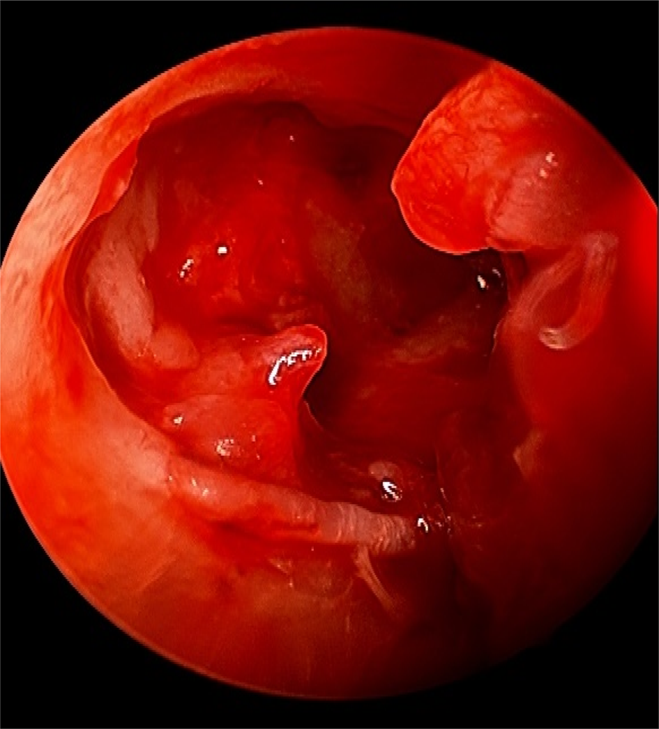
Fig. 5. Middle-ear inspection with a 30○ endoscope, showing granulation tissue and mucosal oedema on the medial wall of the middle ear.
A temporalis fascia graft was placed using the underlay technique. A satisfying air–bone gap closure was achieved (Figure 2b). The patient was given a hearing aid after a brief period, ensuring that the perforation had healed.
Discussion
Aural rehabilitation through a hearing aid is a safe, conservative and non-surgical option preferred by many patients with hearing impairment. Hearing aid fitting is preceded by the creation of an ear mould. However, the process, if not managed carried out with caution, can lead to complications in patients with altered aural anatomy. The silicone impression material used for making an ear mould is viscous, and hence can flow into the mesotympanum, hypotympanum and Eustachian tube, leading to poor middle-ear aeration.Reference Suzuki, Okamura, Yano, Moteki, Kitoh and Takumi2
Persistence of a foreign body in the middle ear can cause complications such as bleeding from the ear, otitis externa, perforation of the tympanic membrane, permanent hearing loss, stapes dislocation, ossicular discontinuity, ossicular erosion, facial nerve palsy, granulation tissue in the middle ear, aural polyps and endolymphatic fistula.Reference Kiskaddon and Sasaki3 Clinical presentations of a foreign body in the middle ear include otalgia, aural fullness, otorrhoea, vertigo, facial nerve palsy and hearing loss acceleration.Reference Jacob, Morris and Welling1,Reference Usmani, Singh, Pandey and Agarwal4 Inflammation of the external auditory canal, debris, granulation tissue and perforation of the tympanic membrane are the usual findings on clinical examination. Imaging studies may be helpful in evaluating the nature, location and extent of the foreign body in the middle ear.
Jacob et al. reported a child with retained ear mould impression material that had flown into the middle ear via a ventilation tube during the preparation of swim moulds.Reference Jacob, Morris and Welling1 Usmani et al. reported a case of a vegetative foreign body in external auditory canal that had migrated to the middle ear after multiple attempts of inadvertent removal by a local physician, causing tympanic membrane perforation and facial nerve palsy.Reference Usmani, Singh, Pandey and Agarwal4 A case of a neglected foreign body in the middle ear that triggered the formation of cholesteatoma and acute mastoiditis was reported by Garag et al.Reference Garag, Rashmi, Shanbag and Arunkumar5 Similarly, a case of a bullet in the external auditory canal was reported by Piromchai et al., where the bullet was pushed through the tympanic membrane into the hypotympanum during an attempt to remove the foreign body. The patient was later referred to an otolaryngologist and the bullet removed by a post-auricular approach.Reference Piromchai, Srirompotong, Lertchanaruengrith and Mills6 Hence, any foreign body in the ear has to be managed carried out with caution by an experienced otolaryngologist, to avoid iatrogenic complications.
The choice of surgical approach for foreign body removal from the middle ear depends on the nature and extent of the foreign body. van den Boer et al. recommended several surgical approaches for foreign body removal from the middle ear.Reference van den Boer, Spronsen, Holland, Ebbens and Waterval7 Forty-nine ears with similar case scenarios were analysed. In their study, in-office removal attempts without high-resolution computed tomography of the temporal bone were described in 12 patients, of which only 1 was successful. A transcanal approach was chosen in 35 patients, a transmastoidal approach with or without posterior tympanotomy was selected in 9 patients, and subtotal petrosectomy was opted for in 1 patient. They also recommended canaloplasty as a useful approach for the removal of a foreign body in the hypotympanum with a prominent anterior canal wall.Reference van den Boer, Spronsen, Holland, Ebbens and Waterval7 Katarkar et al. preferred a post-aural approach, as it provides wide exposure of the medial part of external auditory meatus and middle ear.Reference Katarkar, Katarkar, Jain, Modh and Shah8
• Silicone impression material as a foreign body in the middle ear is a rare complication of an ear moulding procedure
• Symptoms include otalgia, aural fullness, otorrhoea, vertigo, facial palsy and accelerated hearing loss
• Surgical intervention may be needed to remove the foreign body from the middle ear
• An otolaryngologist's opinion is beneficial prior to hearing aid fitting
The hearing aid moulding procedure should be performed by a well-trained hearing aid specialist. Otolaryngologists, audiologists and other hearing instrument dispensers must be aware of the potential hazards associated with the ear moulding procedure. It is highly recommended that the audiologist take a brief otological history, focusing on the patient's history of ear discharge and previous ear surgery. If the anatomy is altered or should the audiologist be in doubt, the patient needs to be referred to an otolaryngologist for further evaluation. For instance, to get an ear mould impression of a large and deep mastoid cavity, the cavity must be packed with otoblocks under microscopic guidance by a trained otolaryngologist.
We recommend a clinical approach algorithm for audiologists, for prior to taking an ear mould impression, in cases when the patient directly consults them without seeing an otolaryngologist (Figure 6). This approach can significantly reduce the risk of complications associated with the ear moulding procedure.

Fig. 6. Clinical approach algorithm for audiologists, for before taking an ear mould impression. TM = tympanic membrane
Conclusion
The ear moulding procedure can be associated with complications. The audiologist or a hearing aid acoustician should perform an otological examination to ensure that the ear canal is clean and the anatomy is optimal for taking an ear mould impression. Ear mould impression material presenting as a foreign body in the middle ear or mastoid may need surgical intervention.
Competing interests
None declared


