


Introduction
Vascular malformations are divided into two main subcategories: ‘low flow’ lesions (e.g. venous and capillary malformations) and ‘high flow’ lesions (e.g. arteriovenous malformations and arteriovenous fistulae).Reference McGill, Mulliken, Cummings, Fredrickson, Harker, Krause and Schuller1
Supratentorial arteriovenous malformations are the most frequently encountered type of intracranial arteriovenous malformation, accounting for approximately 85 per cent.Reference Arnaout, Gross, Eddleman, Bendok, Getch and Batjer2
Infratentorial arteriovenous malformations are less common, and are very rarely seen within the internal auditory meatus. Only two case reports have been previously described.Reference Sundaresan, Eller and Ciric3, Reference Mahran, Samii, Penkert and Ostertag4 These lesions may present with similar symptoms to other, more common pathologies of the internal auditory meatus; however, they are potentially life-threatening and are managed differently.
We present an unusual case of an arteriovenous malformation in the left internal auditory meatus, which caused intermittent vertigo for one year. To our best knowledge, this is the first time that such a lesion has been reported with magnetic resonance imaging and magnetic resonance angiography correlation, and also the first internal auditory meatus arteriovenous malformation to be managed with endovascular embolisation.
Case report
A 50-year-old man presented to the ear, nose and throat clinic following intermittent episodes of vertigo for one year. These episodes had been relatively sparse initially but had increased in frequency until they were occurring daily, lasting for a few seconds at a time. The patient had not developed any auditory symptoms or hearing loss. He had no other significant past medical history.
On examination, both ears appeared normal. There were no demonstrable cranial nerve deficits. In particular, no facial muscle paralysis was present. However, the patient did rotate to the left when performing the Unterberger test.
Magnetic resonance imaging showed a cluster of serpiginous flow voids within the left internal auditory meatus (Figure 1). Further flow voids extended to the left cerebellopontine angle cistern, with an associated prominent draining vein.

Fig. 1 Axial, T2-weighted magnetic resonance imaging scan of the left internal auditory meatus, showing a cluster of serpiginous flow voids within the internal auditory meatus, with some extension to the cerebellopontine angle (white arrow).
Magnetic resonance angiography images confirmed the presence of a cluster of abnormal high flow vessels occupying the left internal auditory meatus and cerebellopontine angle cistern (Figure 2). This area measured 16 × 10 mm in maximum axial diameter. The left anterior inferior cerebellar artery was slightly enlarged and appeared to be supplying the abnormality.
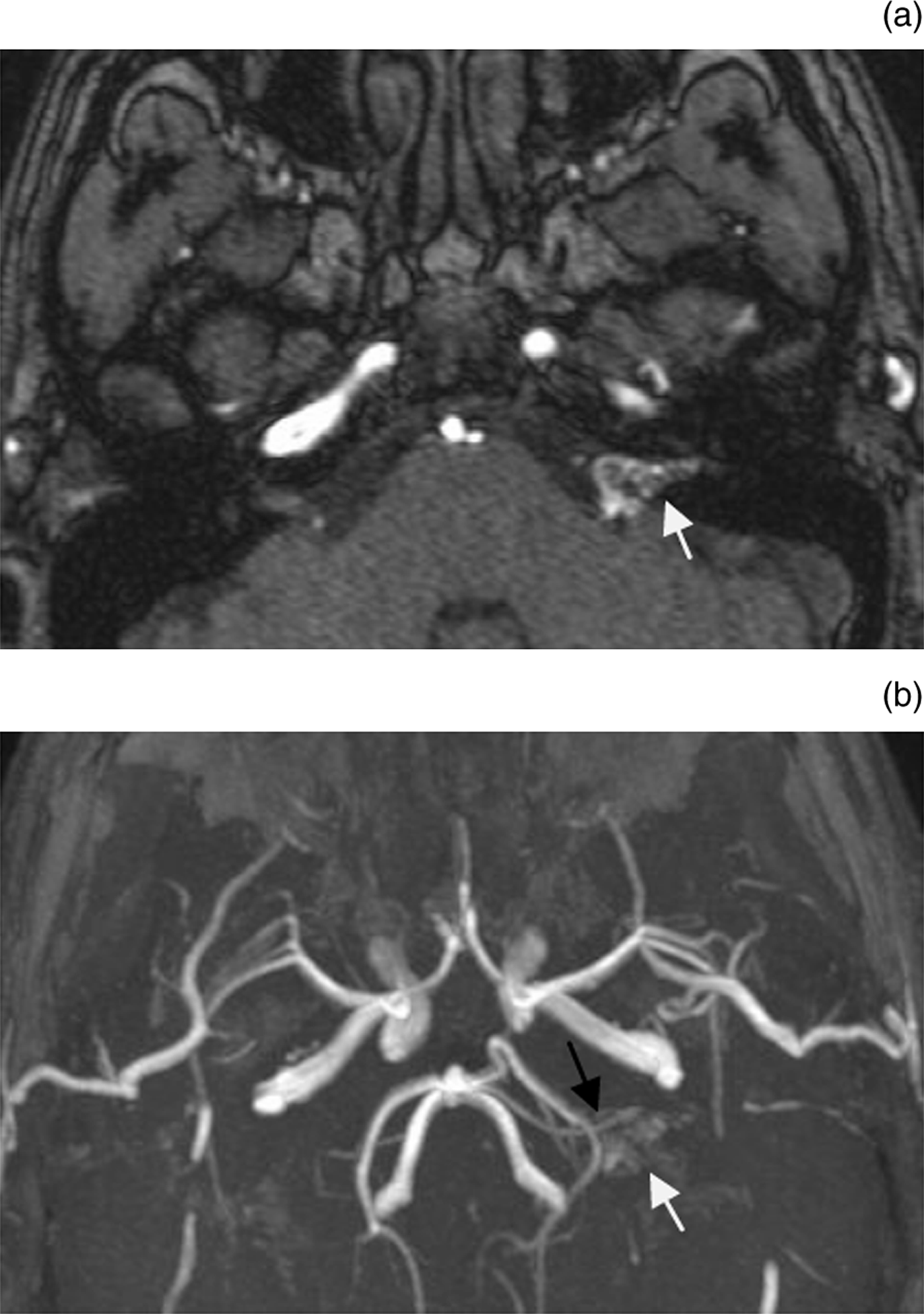
Fig. 2 (a) Axial magnetic resonance angiography source image. (b) Axial maximum intensity angiography image, revealing a cluster of abnormal vessels occupying the left internal auditory meatus and cerebellopontine angle cistern (white arrows). The left anterior inferior cerebellar artery is enlarged and appears to be supplying the abnormality (black arrow).
Six-vessel cerebral angiography was performed. This demonstrated a rapid arteriovenous shunt within the left internal auditory meatus, largely supplied by the labyrinthine artery but with additional transosseous and dural supply (Figure 3). It drained to the superior petrosal sinus, with retrograde flow to the lateral mesencephalic vein.

Fig. 3 Angiography (a) via the right vertebral artery, and (b) with selective microcatheterisation of the anterior inferior cerebellar artery showing a network of abnormal vessels supplied by the left labyrinthine artery (white arrows).
A microcatheter was subsequently positioned within the distal segment of the labyrinthine artery and glue embolisation was performed, achieving occlusion of the labyrinthine artery and much of the lesion.
At follow up, the patient reported experiencing some unsteadiness. This had improved by 10 months post-procedure, at which time there were no residual cerebellar signs. The patient also developed left-sided sensorineural hearing loss and some transient low tone tinnitus, as well as a sensory neuropathy within the mandibular division of the left trigeminal nerve. Facial nerve function was entirely normal.
Repeated magnetic resonance imaging showed an area of infarction in the left middle cerebellar peduncle and lateral pons, consistent with the territory of the anterior inferior cerebellar artery perforating vessels. There were no persistent high flow vessels present within the left internal auditory meatus.
Discussion
Vascular lesions of the internal auditory meatus and cerebellopontine angle are very rare. The majority of lesions in the cerebellopontine angle are vestibular schwannomas; in the largest published surgical series, these represented 61–91 per cent of lesions.Reference Brackmann and Bartels5, Reference Valavanis, Schubiger and Naidich6 Meningiomas, epidermoid cysts and non-vestibular schwannomas account for the majority of the remaining primary lesions of the cerebellopontine angle.Reference Brackmann and Bartels5, Reference Valavanis, Schubiger and Naidich6 Vascular malformations of the cerebellopontine angle, including cavernous haemangiomas (low flow malformations) and arteriovenous malformations, comprise 0.3–1.6 per cent of lesions at this location.Reference Brackmann and Bartels5, Reference Valavanis, Schubiger and Naidich6
Isolated internal auditory meatus lesions comprise mainly vestibular schwannomas (which account for more than 90 per cent). The majority of intracanalicular vascular lesions correspond to cavernous haemangiomas, over 40 cases of which have been reported worldwide.Reference Aquilina, Nanra, Brett, Walsh and Rawluk7, Reference Shim, Song, Lee, Lee, Park and Shin8 These often cause a greater amount of neurological impairment, and are a commoner cause of facial nerve symptoms, than acoustic schwannomas of a similar size.Reference Maya, Lo, Kovanlikaya, Som and Curtin9
An arteriovenous malformation is a high flow vascular malformation with an aberrant nest of vessels (also known as the ‘nidus’) interposed between a feeding artery and a draining vein. Histologically, the arteriovenous malformation may consist of hypertrophied vessels, due to the increased blood flow, and also arteriovenous shunts.Reference Pham, Wong and Allison10
Arteriovenous malformations in the internal auditory meatus or cerebellopontine angle are exceedingly rare. In the English language literature, there have been two previous cases in the internal auditory meatus and four involving the cerebellopontine angle (Table I).
Table I Previously reported internal auditory meatus & cerebellopontine angle avms

AVM = arteriovenous malformations; IAM = internal auditory meatus; HL = hearing loss; HSL = hemisensory loss; HFS = hemifacial spasm; R = right; FW = facial weakness; TN = trigeminal neuralgia; T = tinnitus; V = vertigo; CT = computed tomography; CPA = cerebellopontine angle; NA = not available; MRI = magnetic resonance imaging; AICA = anterior inferior cerebellar artery; n = nerve
Notably, excitatory symptoms due to neurovascular contact (e.g. hemifacial spasm and trigeminal neuralgia) appear to be a common clinical feature of arteriovenous malformations. Arteriovenous malformations in the posterior cranial fossa often present with haemorrhage, more so than supratentorial arteriovenous malformations, which may generate symptoms such as headache and seizures which lead to earlier diagnosis.Reference Khaw, Mohr, Sciacca, Schumacher, Hartmann and Pile-Spellman13, Reference Da Costa, Thines, Dehdashti, Wallace, Willinsky and Tymianski14 Larger arteriovenous malformations are capable of causing ischaemia and hydrocephalus, depending on their location.Reference Arnaout, Gross, Eddleman, Bendok, Getch and Batjer2
Contrast-enhanced computed tomography of arteriovenous malformations shows dilated, opacified vessels. Magnetic resonance imaging can demonstrate serpiginous flow voids related to the high flow vessels, whilst angiography delineates the feeding artery and draining veins.
Despite major advances in the treatment of intracranial vascular lesions, the management of infratentorial arteriovenous malformations remains a significant challenge. Due to the compact nature of the posterior cranial fossa, with a high density of closely packed neurological structures, the treatment of arteriovenous malformations in this site can result in a greater incidence of neurological deficits, compared with other cranial sites.Reference Firsching, Huber and Frowein15
The main goals of the treatment of unruptured arteriovenous malformations are to preclude intracranial haemorrhage and resolve neurological symptoms. The treatment modalities available are endovascular embolisation, radiosurgery and neurosurgical excision. Deciding which treatment mode to employ depends on each individual case, and a combination of modalities may be more appropriate. For example, endovascular embolisation or radiosurgery can be used to reduce the size of arteriovenous malformations prior to surgery.Reference Gobin, Laurent, Merienne, Schlienger, Aymard and Houdart16, Reference Jafar, Davis, Berenstein, Choi and Kupersmith17 There is a dearth of information comparing the clinical outcomes of the various interventional techniques used to treat intracranial arteriovenous malformations. A randomised trial of unruptured brain arteriovenous malformations is currently under way, comparing interventional versus conservative management.Reference Mohr18
• Intracranial arteriovenous malformations are usually supratentorial
• Such lesions in the internal auditory meatus and cerebellopontine angle are rare
• Such a case is presented, the first with magnetic resonance imaging and angiography correlation, and managed with endovascular embolisation
• Excitatory symptoms due to neurovascular contact (e.g. hemifacial spasm, trigeminal neuralgia) are common
• Magnetic resonance angiography can be diagnostic
• Pre-operative diagnosis is imperative, as management differs with aetiology
The most frequently encountered complication following embolisation of arteriovenous malformations is haemorrhage; ischaemia is another, less common complication.Reference Biondi, Le Jean, Capelle, Duffau and Marsault19 The main reason for haemorrhage may be increased pressure in the vessels within or surrounding the nidus, or rupture of an associated aneurysm or pseudoaneurysm. Reflux of embolic material into adjacent veins is thought to increase the risk of haemorrhage.
Following therapeutic embolisation, ischaemia may occur due to: emboli or thrombus developing after vessel catheterisation; anterograde glue injection or retrograde glue reflux into normal pedicles; or thrombus migration into feeding arteries (such as the anterior inferior cerebellar artery, as in our patient).Reference Jahan, Murayama, Gobin, Duckwiler, Vinters and Viñuela20
Conclusion
There have only been two previously documented cases of an arteriovenous malformation involving the internal auditory meatus. Early diagnosis is imperative as management differs from that of other lesions at this location. In the presented case, non-invasive imaging with magnetic resonance angiography confirmed the magnetic resonance imaging detection of a high flow vascular malformation.
There is great debate as to the correct treatment of unruptured arteriovenous malformations, especially those in the posterior cranial fossa, where treatment complications can cause significant morbidity. In the presented case, although the internal auditory meatus arteriovenous malformation was successfully occluded by embolisation, the procedure was complicated by an ischaemic lesion. The natural history of unruptured arteriovenous malformations is not well documented. A randomised trial of unruptured brain arteriovenous malformations would clarify this and facilitate treatment decisions.


