Introduction
Developing foetuses exhibit developmental plasticity and can respond to environmental cues in a manner that produces a phenotype that anticipates the future environment.Reference Godfrey and Barker 1 – Reference Gluckman, Seng, Fukuoka, Beedle and Hanson 3 For example, epidemiological studies in the Netherlands and China have shown that undernutrition during pregnancy at the time of famine induced a ‘thrifty phenotype’ in foetuses; moreover, subsequent mismatch between the predicted environment (i.e. one of hunger) and reality (i.e. sufficient nourishment) may induce metabolic syndrome in foetuses.Reference Painter, de Rooij and Bossuyt 4 – Reference Silveira, Portella, Goldani and Barbieri 7 Animal studies in rats demonstrated that restraint stress (RS) during pregnancy caused high levels of anxiety and depression-like behaviour in the offspring,Reference Darnaudéry and Maccari 8 as well as elevated blood pressure and prolonged changes in heart rate, increased basal plasma corticosterone levels and increased vascular reactivity to neuropeptide Y.Reference Igosheva, Klimova, Anishchenko and Glover 9 , Reference Igosheva, Taylor, Poston and Glover 10 Subsequent studies have also identified long-lasting hyperactivation of the hypothalamic–pituitary–adrenal axis response, an altered circadian rhythm of corticosterone secretion and a depressive state caused by reduced hippocampal corticosteroid receptor density in the offspring of stressed mothers.Reference Viltart, Mairesse and Darnaudéry 11 , Reference Morley-Fletcher, Darnaudéry and Mocaer 12
In humans, the children of mothers who experience severe mental stress, such as bereavement after the death of a close family member, are at increased risk of becoming overweight.Reference Li, Olsen and Vestergaard 13 Thus, a range of stressors may induce physical changes in the foetus that lead to the development of metabolic syndromes.
Glucocorticoids are steroid hormones that control several physiological processes; their expression is regulated by 11β-hydroxysteroid dehydrogenase type 1 (11β-HSD1), an enzyme that catalyses the conversion of inactive cortisone to its active form, cortisol. Overexpression of 11β-HSD1 in transgenic mice results in fatty liver and insulin resistance due to enhanced gluconeogenesis and fatty acid synthesis.Reference Paterson, Morton and Fievet 14 There is also evidence of an association between 11β-HSD1 DNA polymorphisms and metabolic syndromes in humans.Reference Gandhi, Adhikari, Basu and Achappa 15 , Reference Moon, Lee, Kim and Lee 16 Expression of 11β-HSD1 increases in the livers of rat offspring born to diabetic mothers,Reference Fujisawa, Nakagawa, Li, Liu and Ohzeki 17 while undernutrition during pregnancy downregulates the expression of 11β-HSD2, a ‘reverse converter of 11β-HSD1’, in the placentaReference McTernan, Draper and Nicholson 18 and other organs.Reference Takaya, Iharada, Okihana and Kaneko 19 However, 11β-HSD1 null mice are resistant to diabetes and high fat diet-induced obesity.Reference Morton, Holmes and Fiévet 20 , Reference Morton, Paterson and Masuzaki 21 In addition, inhibition of 11β-HSD1 improves metabolic syndromes, such as type 2 diabetes mellitus.Reference Morton, Paterson and Masuzaki 21 , Reference Anderson and Walker 22
These various findings suggest 11β-HSD1 may be associated with the onset of metabolic syndromes. We used 11β-HSD1 expression as a marker to identify effects in the livers of foetuses and pups of pregnant females that had undergone RS. Our results suggest prenatal stress led to fatty liver and upregulated 11β-HSD1 in the offspring.
Methods
All experimental protocols and procedures were approved by the animal care and use committee of the University of Yamanashi.
Animals, experimental protocol and tissue collection
Pregnant female C57BL/6J mice were obtained at embryonic day (E) 6 from SLC Inc. (Shizuoka, Japan) and kept in the laboratory for 2 days. The mice were then assigned to two groups: a non-stress control group (NS group) and a restraint stress group (RS group). For the RS, the mice were placed into 50 ml plastic tubes for 3 h (9.30 am–12.30 pm) each day from E8 to parturition (usually 12 days later). After parturition, the mice were allowed to feed and care for their pups as normal. The pups were weaned at postnatal day 30 (P30). Liver samples were collected for histology and protein and mRNA determinations at E18 (n=21; NS group, n=21; RS group), P15 (n=5; NS group, n=7; RS group), P37 (n=10; NS group, n=11; RS group) and P52 (c). The body weights of the offspring mice were measured every 3 days after birth.
Plasma corticosterone and leptin analyses
Corticosterone in plasma was quantified with a commercial EIA kit (Enzo Life Science Inc., NY, USA), and leptin levels were quantified with a commercial ELISA kit (Morinaga Institute of Biological Science Inc., Yokohama, Japan).
Lipid staining
Liver tissues were fixed in 4% paraformaldehyde at 4°C overnight and then immersed in 30% sucrose in phosphate-buffered saline (PBS) for 2 days. Cryosections (thickness, 12 μm) were prepared and stored at −20°C. For lipid staining, the sections were stained with 0.3% Oil Red O solution/isopropanol for 20 min at 37°C. Nuclei were stained using Mayer’s haematoxylin solution (Wako Pure Chemical Industries Ltd., Osaka, Japan).
Western blotting
Proteins were separated by electrophoresis on 8–10% sodium dodecyl sulphate-polyacrylamide gels and then transferred to polyvinylidene fluoride membranes. The membranes were blocked with 5% skim milk in PBS/0.01% Tween 20 for 1 h at room temperature, and then incubated overnight at 4°C with one of the following antibodies in 5% skim milk in PBS buffer: rabbit polyclonal anti-peroxisome proliferator-activated receptor gamma (PPARγ) (1:500; sc-7273, Santa Cruz, TX, USA), rabbit polyclonal anti-11β-HSD1 (1:500; sc-20175, Santa Cruz), rabbit polyclonal anti-Glucocorticoide receptor (GR) (1:500; sc-1004, Santa Cruz), or mouse monoclonal anti-β-actin (1:1000; Cell Signaling Technology Inc., MA, USA). After three washes in PBS, the membranes were incubated for 1 h at room temperature with horseradish peroxidase-conjugated anti-mouse IgG secondary antibody (1:1000; GE Healthcare, Buckinghamshire, UK) or anti-rabbit IgG secondary antibody (1:1000; GE Healthcare) and washed three times with PBS. Immunoreactivity was visualized with a chemiluminescence detection system (ECL; Nacalai Tesque Inc., Kyoto, Japan) and LAS-4000 imaging analysis software (Fujifilm Inc., Tokyo, Japan). Fluorescence intensities were normalized to β-actin.
RNA extraction and quantitative reverse transcription-polymerase chain reaction
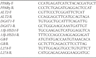
Total RNA was extracted from mouse livers using a RNA TRIzol RT kit (Cosmo Bio Inc., Tokyo, Japan). Each total RNA sample was reverse-transcribed with random primers and a cDNA transcription kit (Applied Biosystems, Life Technologies, Carlsbad, CA, USA) according to manufacturer instructions. One-tenth of the reaction mixture was used for PCR amplification. Gene expression was measured by quantitative RT-PCR (qRT-PCR) with a SYBR Green PCR kit (SYBR Green ER; Invitrogen, Life Technologies, Carlsbad, CA, USA) on an ABI Prism 7500 system. Oligonucleotide primer sequences for PPARγ, alanine aminotransferase 2 (Alt2), diacylglycerol acyl transferase 1 (Dgat1), 11β-hsd1, and Gr are listed in Table 1. Expression was normalized to mouse L27a. All qRT-PCR experiments were performed in duplicate.
Table 1 List of all primer sequences used

11β-HSD1 inhibitor experiments
In some case, we used an 11β-HSD1 antagonist to examine the effect of 11β-HSD1 for lipid accumulation. The pups at postnatal day 20 received daily injections of carbenoxelone (20 mg/kg; Sigma-Aldrich, MA, USA). Liver samples were collected at postnatal day 45 for histology as well as protein and mRNA determinations (n=3; RS+vehicle group, n =4; RS+carbenoxelone group).
Statistical analysis
Data are expressed as the mean±standard error of the mean of n independent observations. Comparisons of multiple groups were made by one-way analysis of variance (ANOVA) followed by Tukey’s post-hoc test or Dunnett’s test when two groups were compared. Differences were considered significant when P values were <0.05.
Results
The effect of maternal stress during pregnancy on the offspring
To evaluate the effect of RS during pregnancy on the metabolic systems of the foetuses, we measured the levels of plasma glucocorticoids in the mothers and their foetuses at E18. Plasma corticosterone was higher in the RS mothers than in the NS mothers (Fig. 1a), while there was no difference between the RS and NS foetuses (Fig. 1b).

Fig. 1 Metabolic changes in the foetuses and offspring of stressed and unstressed mothers. (a) and (b) Plasma corticosterone levels in the pregnant mice and E18 foetuses, respectively. (c) and (d) Histological sections of livers from P37 and P52 mice, respectively, stained with Oil Red O. Red arrows indicate lipid accumulation. (e) and (f) Body weights of the offspring at P1 and P37, respectively. (g) Leptin plasma level at P37 in offspring. RS, restraint stress; NS, no stress.
At P37, cryosections of livers from the offspring of RS mothers showed higher lipid levels than those from NS offspring (Fig. 1c). The difference was especially evident at P52 (Fig. 1d). However, no such increase was observed in the livers of RS offspring at E18 and P15 (data not shown). Body weights did not differ at P1 (Fig. 1e) or P37 (Fig. 1f). Likewise, there was no difference in serum leptin concentration at P37 (Fig. 1g).
Expression of genes associated with lipid accumulation
The lipid accumulation in the livers of RS offspring at P37 led us to examine whether the expression of genes associated with lipid metabolism were altered. We identified significantly elevated expression of three genes in RS offspring: Alt2, a key enzyme of gluconeogenesis (Fig. 2a); Dgat1, a catalyst in triglyceride production (Fig. 2b); and Pparγ, a factor associated with hepatic lipid accumulationReference Harno, Cottrell and Yu 23 (Fig. 2c). PPARγ protein levels were also elevated in the liver of RS offspring (Fig. 2d).

Fig. 2 Expression of genes and proteins associated with lipid accumulation in the livers of stressed and unstressed mothers. Relative mRNA levels at P37 of (a) alanine aminotransferase 2 (Alt2), (b) diacylglycerol acyltransferase 1 (Dgat1), (c) peroxisome proliferator-activated receptor gamma (Pparγ). (d) Level of peroxisome proliferator-activated receptor gamma (PPARγ) protein at P37. All values are expressed as mean±s.e.m., *P<0.05, **P<0.01. NS, no stress; RS, restraint stress.
Expression of upstream regulators of lipid accumulation
11β-HSD1 is a key regulator of the stress response, as it upregulates corticosterone activity without increasing plasma glucocorticoid levels,Reference Matsusue, Haluzik and Lambert 24 which are associated with adipogenesis.Reference Fujisawa, Nakagawa, Li, Liu and Ohzeki 17 , Reference Masuzaki, Paterson and Shinyama 25 , Reference Liu, Park and Pietrusz 26 It has been suggested that 11β-HSD1 regulates the expression of GR, which is associated with the regulation of proteins involved in lipid accumulation, such as PPARγ.Reference Berthiaume, Laplante and Festuccia 27 For these reasons, we investigated the expression of 11β-HSD1 in the offspring of RS mice and found elevated levels of 11β-HSD1 protein at P37 (Fig. 3a), although we found no evidence for the upregulation of gene expression (Fig. 3b). The level of GR protein was also elevated at P37 in the livers of the RS offspring (Fig. 3c). Expression of the Gr gene also increased, although the difference between groups was not significant (Fig. 3d).

Fig. 3 Expression of the upstream regulators of lipid accumulation in the livers of offspring stressed and unstressed mothers. (a) Levels of 11β-hydroxysteroid dehydrogenase type 1 (11β-HSD1) protein at P37. (b) mRNA level of 11β-hsd1 at P37. (c) Level of glucocorticoid receptor protein (GR) at P37. (d) Relative mRNA level of glucocorticoid receptor (Gr), at P37. All values are expressed as mean±s.e.m., *P<0.05, **P<0.01. NS, no stress, RS, restraint stress.
To address the question of when lipid accumulation is initiated, we investigated expression at a late prenatal stage. Expression of 11β-HSD1, Alt2, and Dgat1 was elevated in the livers of RS foetuses at E18 (Figs 4a, 4c and 4d), but expression of Gr and Pparγ did not differ between groups during the foetal period (Fig. 4b, 4e).

Fig. 4 Expression of the upstream regulators of lipid accumulation in the livers in foetuses of stressed and unstressed mothers. Relative mRNA levels at E18 of (a) mRNA 11β-hydroxysteroid dehydrogenase type 1 (11β-hsd1), (b) glucocorticoid receptor (Gr), (c) alanine aminotransferase 2 (Alt2), (d) diacylglycerol acyltransferase 1 (Dgat1), and (e) peroxisome proliferator-activated receptor gamma (Pparγ). All values are expressed as mean±s.e.m., *P<0.05.
Inhibition of 11β-HSD1 and lipid accumulation
Our results showed 11β-HSD1 is a key regulator of lipid accumulation in the stressed liver. Expression increased at E18, perhaps activated by glucocorticoid activity. To verify this, we inhibited 11β-HSD1 with carbenoxeroneReference Rhee, Kim and Park 29 and found that 11β-HSD1 expression was unchanged. However, transcript and protein expression of Gr and Pparγ decreased (Fig. 6a, 6b and 6c). GR expression decreased significantly (P<0.014), and PPARγ decreased between the RS and RS+car groups (Fig. 6d, 6e, 6f and 6g). Histological data suggested that this expression inhibits lipid accumulation in the liver (see Supplementary Fig. 1b). Therefore, lipid accumulation is regulated by 11β-HSD1 activity in our experiment.

Fig. 5 Schematic outlines of the pathways of lipid metabolism in the liver, based on the observations made here on foetuses and the offspring of stressed pregnant mice. 11β-HSD1, 11β-hydroxysteroid dehydrogenase type 1; GR, glucocorticoid receptor; ALT2, alanine aminotransferase 2; DGAT1, diacylglycerol acyltransferase 1; PPARγ, peroxisome proliferator-activated receptor gamma; TG, triglycerides.
Discussion
The underlying premise of the initiative titled ‘Developmental Origins of Health and Disease (DOHaD)’ is that famine in the Netherlands and China caused adulthood diseases in individuals who were affected during gestation.Reference Godfrey and Barker 1 – Reference Gluckman, Seng, Fukuoka, Beedle and Hanson 3 Support for this premise comes from animal studies in which maternal undernutrition caused fatal metabolic disorders in the offspring.Reference Arai, Soga and Ohata 28 , Reference Lillycrop, Phillips and Torrens 31 Famine can have a physical impact through malnutrition, but also imposes mental stress on pregnant women. Therefore, we designed this study to investigate the concept that mental stress during pregnancy might induce metabolic disorders in the offspring.
We observed increased lipid accumulation in the livers of offspring born to mothers that had been subjected to restraint, a form of mental stress, during pregnancy. This increased lipid accumulation occurred without any changes in food intake (data not shown) or weight gain. Expression analysis of genes encoding proteins associated with lipid accumulation (Alt2, Dgat1, Pparγ) or upstream regulators of these proteins (11β-hsd1, Gr) showed that they were upregulated in the livers of the offspring of stressed mice. These effects commenced during foetal development and were maintained postnatally (see scheme Fig. 5).

Fig. 6 Expression of genes and proteins associated with lipid accumulation in the livers of stressed and injected of 11β-HSD1 antagonist. Relative mRNA levels at P45 of mRNA 11β-hydroxysteroid dehydrogenase type 1 (11β-hsd1), (b) glucocorticoid receptor (Gr), (c) peroxisome proliferator-activated receptor gamma (Pparγ). (d), (e), (f), (g) Level of 11β-HSD1, glucocorticoid receptor (GR), peroxisome proliferator-activated receptor gamma (PPARγ) protein at P45. All values are expressed as mean±s.e.m., *P<0.05, **P<0.01. RS+veh, restraint stress+vehicle; RS+car, restraint stress+car.
Although we observed increased levels of 11β-HSD1 and other proteins associated with lipid accumulation, we found no indication of an increase in GR protein during foetal development. One possible interpretation of these results is that the increase in 11β-HSD1 induces conversion of GR from an inactive to an active state, without altering the amount of GR protein.Reference Chapman, Holmes and Seckl 32 The increase in 11β-HSD1 was evident at E18 and P37, suggesting that the change in 11β-HSD1 production was initiated during foetal development, at the time of maternal exposure to stress, and continued into adulthood. This conclusion is consistent with a recent report showing that upregulation of 11β-HSD1 occurs in patients with metabolic syndromes and obesity.Reference Torrecilla, Fernández-Vázquez and Vicent 33
Increased levels of 11β-HSD1 have been found in patients with non-alcoholic fatty liver disease (NAFLD), a disease that presents with alcoholic hepatitis-like pathological changes in the absence of an alcohol habit.Reference Ahmed, Rabbitt and Brady 34 This disease has been associated with the foetal environment,Reference Rueda-Clausen, Dolinsky and Morton 35 , Reference Nobili, Alisi, Panera and Agostoni 36 although its cause remains uncertain. The ‘multiple hits’ hypothesisReference Alisi, Panera, Agostoni and Nobili 37 suggests that ‘primary hits’ such as insulin resistance and fat accumulation may lead to fatty liver and that ‘secondary hits’, including oxidative stress, may induce non-alcoholic steato-hepatitis. Our findings provide a possible explanation for the origin of NAFLD or and ‘primary hits’, suggesting it might be a consequence of trans-placental effects that occur during foetal development. In another report, NAFLD was associated with hepatocellular carcinoma (HCC)-induced obesityReference Nobili, Alisi and Grimaldi 38 in adults, although HCC has also been found in young obese subjects with NAFLD. Our study suggests foetal stress may set the stage for metabolic syndrome or lipid accumulation in the liver at a young age. It is possible that treatment with an 11β-HSD1 inhibitor, a treatment regime that is used to enhance insulin sensitivity and hepatic lipid catabolism in patients with type 2 diabetes and obesity,Reference Andrews, Rooyackers and Walker 39 , Reference Vicker, Su and Ganeshapillai 40 may be effective in patients with a high level of 11β-HSD1 due to maternal stress during pregnancy. Indeed, our results suggest the activity of 11β-HSD1 regulates lipid accumulation in the liver (Fig. 6).
The exposure of pregnant mice to RS led to higher plasma levels of corticosterone, which induced 11β-HSD1 protein expression in the foetuses. A similar effect of RS on plasma corticosterone level in mice was recently reported.Reference Kim, Jung, Kim, Min and Yoon 41 Expression of 11β-HSD2, a ‘glucocorticoid barrier’ enzyme, decreases in the placenta, allowing an increased influx of corticosterone to the foetus;Reference Mairesse, Lesage and Breton 42 indeed, we found that the plasma level of corticosterone increased in the foetuses of RS mice. Therefore, the cause of the increase in 11β-HSD1 remains unclear. Secretion of cortisol is regulated by a hypothalamic-pituitary-adrenal feedback system. Cortisol is inactivated by 11β-HSD2 in the kidney and re-activated by 11β-HSD1 in the liver and adipose tissue.Reference Hollis and Huber 43 In the low-insulin state, glucocorticoids show lipolysis, other hand, it shows lipid accumulation in high-insulin state.Reference Andrews and Walker 44 In this experiment, the expression of 11β-HSD2 was unchanged in liver (data not shown), suggesting that regulation of lipid metabolism in the liver may be associated with 11β-HSD1 regulation. Indeed, 11β-hsd1-overexpressing mice show an increase in visceral fat and insulin resistance, indicating lipid abnormalities, while knockout mice do not exhibit insulin resistance with a high fat diet.Reference Morton, Paterson and Masuzaki 21 , Reference Masuzaki, Paterson and Shinyama 25 Our inhibitor experiments suggest that suppression of 11β-HSD1 limits hepatic lipid accumulation.
Further studies are needed to determine whether high-fat food induces additional lipid accumulation in the liver of RS offspring. It will also be interesting to determine whether 11β-HSD1 is upregulated in adipose tissue, which also contributes to the development of adult disease, since adipose tissue also selectively expresses 11β-HSD1.Reference Lillycrop, Phillips, Jackson, Hanson and Burdge 30 The role of epigenetic gene regulation mechanisms in stress-induced upregulation of 11β-hsd1 should also be examined, as undernutrition during pregnancy induces epigenetically determined upregulation of genes associated with lipid accumulation in the foetal liver.Reference Arai, Soga and Ohata 28 , Reference Rhee, Kim and Park 29
Acknowledgements
The authors thank for the valuable discussion and comment (T. Kubota and K. Moriishi) and technical advice (H. Kasai and K. Mochizuki). They thank for technical support to C. Obata and H. Kasai.
Financial Support
This study was supported by grants for Scientific Research (C) (23591491 to T Hirasawa) and Exploratory Research (25670473 to T Kubota) from JSPS KAKENHI, Japan, and by a grant for Core Research for Evolutional Science and Technology (CREST) from the Japan Science and Technology Agency (JST) (to T Hirasawa).
Conflicts of Interest
None.
Ethical Standards
The authors assert that all procedures contributing to this work comply with the ethical standards of the relevant national guides on the care and use of laboratory animals (University of Yamanashi) and has been approved by the institutional committee (Univesity of Yamanashi).
Supplementary material
To view supplementary material for this article, please visit http://dx.doi.org/10.1017/S2040174415000100