


Introduction
Ovarian follicular development is crucial to female reproductive healthiness and this event depends on several factors such as growth factor and hormone signals. At the beginning, the transition of primordial follicles to secondary stages is controlled by some growth factor as bone morphogenic protein 15 (BMP15), kit ligand, BMP4 and hormones as insulin and growth hormone. All of them are important to maintain oocytes and granulosa cells (GCs) survival through initial follicular development.Reference Palma, Argañaraz and Barrera 1 Physiological levels of androgens have also been linked to a role on modulating the expression and activity of genes related to growth and development of ovarian follicles in mammals.Reference Prizant, Gleicher and Sen 2 , Reference Sen, Prizant and Light 3
In turn, androgen excess may disturb physiological mechanisms of follicular development leading to chronic anovulation, as observed in human clinical diseases. One example is the polycystic ovary syndrome (PCOS), a prevalent condition in which impaired follicular growth and high circulating androgen levels lead to menstrual disturbances and anovulatory infertility.Reference Spritzer 4 Evidence indicates that prenatal or prepubertal androgen excess may be involved with the pathophysiology of PCOS.Reference Franks, McCarthy and Hardy 5 – Reference Sir-Petermann, Codner and Pérez 7 In addition, some animal models of PCOS have been developed by using experimental exposure of high androgen levels.Reference Abbott, Dumesic and Franks 8 – Reference Franks 12 In this sense, the study of ovarian dysfunction promoted by animal models of androgen interventions may provide interesting insights into the initial pathogenic mechanisms of PCOS. Indeed, the pathological ovarian features of PCOS include arrest of follicular development with accumulation of early and mid-antral stage follicles.Reference Franks, Stark and Hardy 10 Abnormalities in cellular investments of the ovarian follicle, such as theca cell (TC), GC and oocyte layers during early follicular development may be a primary cause of ovarian dysfunction leading to reduced responsiveness to follicle-stimulating hormone (FSH) and luteinizing hormone stimulation.Reference Orisaka, Tajima, Tsang and Kotsuji 13 Nevertheless, it is not clear whether reproductive hormone abnormalities are a primary or secondary reflex of follicular disruption, or whether and to what degree androgen overproduction may modulate GCs activity.Reference Gervásio, Bernuci, Silva-de-Sá and Rosa-E-Silva 14
Because of these many uncertainties, animal models are still crucial for the study of PCOS. In fact, the observation that dehydroepiandrosterone (DHEA) levels are increased in women with PCOSReference Loughlin, Cunningham and Moore 15 led to the development of a PCOS animal model using postnatal treatment with DHEA, the first androgen to rise in the early female pubertal period.Reference Apter, Bützow, Laughlin and Yen 16 This postnatal DHEA treatment produced different degrees of ovarian morphological changes, such as cystic ovaries with thin GC layer and consistent with anovulation,Reference Lee, Anderson and Lee 17 – Reference Sander, Luchetti and Solano 20 increased ovarian stroma and increased number of atretic follicles. Animals also exhibit altered steroidogenesis with high serum estrogen and androgen levels.Reference Henmi, Endo and Nagasawa 21 However, these animal models are limited by the need for long experimental periods, precluding the observation of direct androgen effect on early follicular growth and development. Thus, animal models using short periods of follicular stimulation would be useful to investigate this aspect.
A rodent model of acute gonadotropin hyperstimulation and hyperandrogenism has been recently developed.Reference Faut, Elia and Parborell 22 – Reference Velez, Heber and Ferreira 24 In that experimental model, a two-hormone treatment was administered to immature female animals aged 22–25 days: equine chorionic gonadotropin (eCG), an efficient interspecies inductor of follicular recruitment,Reference Parborell, Pecci, Gonzalez, Vitale and Tesone 25 and DHEA, in order to promote a hyperandrogenic condition.Reference Velez, Heber and Ferreira 24 However, that study did not focus on the prepubertal stage, and therefore missed the opportunity to promote a folliculogenesis spurt after the onset of physiological follicular development in the ovaries. In the present study, we aimed to investigate the dynamics of follicular morphology and ovarian function in prepubertal Wistar rats, aged 25–28 days, acutely treated with the same previously validated concentrations of eCG and DHEA.
Materials and methods
All procedures involving animals were conducted in accordance with local regulations (National Council on Research Animal Care, CONCEA, Brazil). The study was approved by the Ethics Committee at Hospital de Clínicas de Porto Alegre (Brazil).
Experimental animal and procedures
In total, 20 prepubertal (25–28 days old) female Wistar rats were divided into three groups, that received one single dose of the following treatments, between 8 and 10 am: intraperitoneal injection of 25 IU of eCG alone (Novormon, Buenos Aires, Argentina) in 0.1 ml of saline solution (eCG group, n=7); 26 subcutaneous injection of 60 mg/kg of DHEA (Sigma–Aldrich, MO, USA) in 0.1 ml of sunflower oil together with 25 IU of eCG (eCG+DHEA group, n=7);Reference Faut, Elia and Parborell 22 and a group with no hormones and subcutaneous injection of vehicle (sunflower oil) (control group, n=6).
Rats were housed under controlled temperature (21°C) and lighting (12 h light/12 h dark cycle; lights from 7 am until 7 pm), with free access to chow (Nuvilab CR-1, PR, Brazil) and water until the experimental day.
Animals were anesthetized with isoflurane and killed by decapitation 8 h after the treatments. Trunk blood was collected and serum was separated by centrifugation at 2500 g for 10 min and stored at −80°C until DHEA, testosterone and estradiol measurements. The left ovarian tissue from each rat was dissected and immediately fixed in 10% buffered formalin for morphological and morphometric studies.
Hormonal assays
Radioimmunoassays (RIA; Immunotech SAS, Marseille, France and Diagnostics Systems Laboratories, TX, USA) were used to measure serum DHEA, testosterone and estradiol, with sensitivity of 0.06 ng/ml, 0.02 ng/ml and 2.20 pg/ml, respectively. Intra- and interassay coefficients of variation were ⩽10%. RIAs were performed according to manufacturer instructions, using control serum samples.
Morphological and morphometric analysis of follicles and oocytes
Formalin-fixed ovaries were dehydrated in ethanol and embedded in paraffin. Serial 5-μm thick sections were prepared on microscope slides and stained with hematoxylin–eosin (Laborclin, PR, Brazil; Vetec, RJ, Brazil).
The identification of follicles was based on strict criteria.Reference Pedersen and Peters 27 Five-μm step sections were mounted at 50 μm intervals onto microscope slides. This interval was chosen to prevent counting the same structure twice. Two trained investigators, who were blinded to treatment group, carried out the morphological analyses (Olympus BX41, Tokyo, Japan).
Follicles were classified as primordial, primary, secondary, antral, atretic and cystic.Reference Cruz, Barra, González, Sotomayor-Zárate and Lara 28 Primordial follicles were those with one oocyte surrounded by flattened GCs. Primary follicles presented a single layer of cuboidal GCs, whereas secondary follicles had two or more GC layers. Antral follicles were recognized by the presence of both visible one single vacuole and oocyte nuclei. Atretic follicles were defined as follicles with >5% of the GCs having pyknotic nuclei. Cystic follicles were characterized as those with a large antral cavity, thickened theca interna layer and attenuated GC layer.
For morphometric evaluation, 15 structures of each follicle stage were chosen in each group for analysis. Follicular diameters were determined as the average of two measurements at a right angle across the midpoints. In both primordial and primary follicles, the diameter was measured from the outer layer of GCs. For secondary and antral stages, the diameter was measured from the outer wall of the thecal layer. All diameters were measured using the Image-Pro Plus 4.5 Software (Media Cybernetics, MD, USA), after calibration with a stage micrometer (Media Cybernetics, MD, USA).
Statistical analysis
Statistical analyses were carried out using GraphPAD PRISM 5 Software (San Diego, CA, USA). Data were tested for normal distribution using the Shapiro–Wilk test and described as median and interquartile range (25–75%). Non-parametric analysis was carried out with Kruskal–Wallis test for comparing the control, eCG and eCG+DHEA groups. Dunn’s post-hoc test was used as the adjustment procedure for multiple comparisons. Data were considered statistically significant at P<0.05. The P for trend was estimated for the comparisons among the groups.
Results
Effect of treatments on DHEA, testosterone and estradiol levels
Animals treated with eCG+DHEA presented higher serum estradiol [61 (38–596) pg/ml] and testosterone [4.2 (2.3–17.0) ng/ml] levels in comparison with the control group [8.0 (5.6–10.0) pg/ml and 0.09 (0.09–0.11) ng/ml]. Animals under eCG stimulation showed intermediate levels of these steroid hormones [10.0 (8.5–15.0) pg/ml and 0.23 (0.18–0.25) ng/ml] (Fig. 1a and 1b). Serum DHEA levels were elevated in eCG+DHEA [23.00 (21.00–23.00) ng/ml] in relation to the other two groups [0.69 (0.40–2.40) and 0.60 (0.56–0.72) ng/ml, respectively, for control and eCG groups) (Fig. 1c).

Fig. 1 Serum estradiol (a), testosterone (b) and dehydroepiandrosterone (DHEA) (c) levels from control (n=6), equine chorionic gonadotropin (eCG) (n=7) and eCG+DHEA (n=7) groups. Values are expressed as median and 25–75% interquartile range (lower and upper limit of the box); maximum and minimum values are shown by the limits of vertical lines. P<0.01 by Kruskal–Wallis test. Different letters indicate significant differences (P<0.05).
Morphological analysis of ovarian follicles
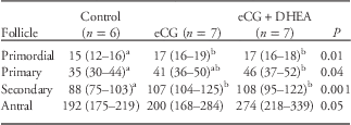
Table 1 shows the number of follicles according to follicular stage and treatment. The total number of primary and secondary follicles was higher in the eCG+DHEA group than in the control group (Table 1; Fig. 2). The eCG group did not differ from either the control or the eCG+DHEA group regarding the number of primary and secondary follicles. The total number of antral follicles was also higher in the eCG and eCG+DHEA groups compared with controls. Primordial, atretic and cystic follicles were similar in all groups (Table 1).

Fig. 2 Representative ovarian micrographs from control (a), equine chorionic gonadotropin (eCG) (b) and eCG+dehydroepiandrosterone (DHEA) (c) groups. PF, primary follicle; SF, secondary follicle. Scale bar is 100 μm.
Table 1 Number of follicles according to follicular stage and treatment

eCG, equine chorionic gonadotropin; DHEA, dehydroepiandrosterone.
Values are expressed as median and interquartile range (25–75%) and percentage of total number of follicles for each follicle stage and per group (Kruskal–Wallis test).
a,bDifferent superscript letters indicate significant differences (P<0.05).
Morphometric analysis of ovarian follicles
Primordial follicle diameter was larger in the eCG and eCG+DHEA groups compared with controls (Table 2). The size of primary and secondary follicles was greater in eCG+DHEA animals compared with controls, whereas the diameter of primary follicles was similar in the eCG group in comparison with both controls and eCG+DHEA animals (Table 2; Fig. 2). Antral follicular diameter was similar in all groups.
Table 2 Follicular diameter in μm according to follicular stage and treatment
eCG, equine chorionic gonadotropin; DHEA, dehydroepiandrosterone.
Values are expressed as median and interquartile range (25–75%) of follicular diameter for each follicle stage and per group (Kruskal–Wallis test).
a,bDifferent superscript letters indicate significant differences (P<0.05).
Discussion
In the present study, acute administration of eCG plus androgen affected initial follicular recruitment, causing an increase in the number of primary, secondary and antral follicles in ovaries of prepubertal rats. A similar animal model has been previously described in immature Sprague Dawley rats (22–25 days of age), showing early apoptosis of antral follicles.Reference Faut, Elia and Parborell 22 The present study is the first to employ both morphological and morphometric analysis in order to measure follicular growth in different stages of follicular development after acute stimulation with eCG plus DHEA in Wistar rats. The decision to use this rat strain is warranted by the fact that this is the experimental strain for which the largest amount of reproductive physiology data are available.
Moreover, our study was carried out specifically with prepubertal animals (25–28 days of age). In prepubertal 25–30-day-old animals, the hypothalamus–pituitary–ovary axis is undergoing maturation; follicular development has initiated, and both antral and atretic follicles are already present. Therefore, eCG stimulation may be very useful to promote follicular development in terms of speed and quantity.Reference Gal and Orly 23 Actually, the study of Gal et al.,Reference Gal and Orly 23 using eCG stimulation followed by human chorionic gonadotropin administration, resulted in a growth spurt of peri-ovulatory follicles. Interestingly, in the present study, stimulation with eCG only produced a slightly and non-significant increase in the proportion of small follicles. This probably occurred because of the short-term duration of the experimental model, with no sufficient time to allow growing follicles could secrete androgens. Nevertheless, eCG stimulation served as priming for the stimulation with DHEA,Reference Elia, Sander and Luchetti 29 , Reference Misugi, Ozaki and El Beltagy 30 generating an androgen excess state and promoting greater ovarian morphological changes. In turn, although we did not tested the effect of isolated DHEA treatment, it is important to point out that prepubertal animals present only an early slight follicular growth. Based on evidence from previous studies using the same treatments,Reference Faut, Elia and Parborell 22 – Reference Velez, Heber and Ferreira 24 we hypothesize that a priming effect of eCG would be needed to detect a DHEA effect on ovarian changes. Further studies are needed to confirm this hypothesis.
Previous in vitro studies suggest that high doses of androgens could impair early follicular development.Reference Okutsu, Itoh, Takahashi and Ishizuka 31 – Reference Walters, Allan and Handelsman 33 Indeed, experimental animal models using prenatal exposure to androgens have resulted in reproductive disorders with increased preantral follicles and elevated estradiol and testosterone levels in adult age, resembling PCOS.Reference Walters, Allan and Handelsman 33 , Reference Wu, Li and Wu 34 Taken together, these studies and our present results support the hypothesis that prenatal exposure to androgens serves to ‘program’ both ovaries and adrenals of prepubertal animals to secrete high levels of androgens that interfere with ovarian function.Reference Abbott, Dumesic and Franks 8 , Reference Franks 12
We found significant increases in estradiol, testosterone and DHEA levels in animals treated with eCG plus DHEA in comparison with controls, indicating the consistency of our experimental model. These results are in agreement with those of a previous studyReference Velez, Heber and Ferreira 24 with a different female rat strain. Moreover, these findings suggest that follicles remain healthy, with functional cells, following acute exposure to eCG. This was confirmed by our morphological findings and visualization of healthy follicles. Indeed, it is known that biosynthesis of estradiol-like hormones is a basic function of GCs,Reference Palma, Argañaraz and Barrera 1 initiated early in primary follicles. In turn, androgens could be derived from TC secretion from small follicles and adrenal gland. In fact, a recent studyReference Zhou, Kang, Chen, Han and Ma 35 has demonstrated significantly increased expression of aromatase in rat ovaries and increased 3β-hydroxysteroid dehydrogenase (3β-HSD), 17β-HSD and aromatase messenger RNA (mRNA) levels in adrenal gland. Therefore, DHEA could also be stimulating adrenal cells to synthetize androgens to act as substrate to aromatase enzymes in follicular GCs.
Interestingly, we found an increased proportion of primary, secondary and antral follicles following acute hormonal stimulus with eCG and DHEA. Our results differ slightly from those of a previous study,Reference Velez, Heber and Ferreira 24 which reported greater proportion of small follicles. Although Velez et al.Reference Velez, Heber and Ferreira 24 studied Sprague Dawley rats in the late postnatal age (22–25 days old), in our study Wistar rats were studied at the late prepubertal age (25–28 days old), and presented both small and antral follicles. The presence of higher numbers of primary, secondary and antral follicles in our animals underscores the role of DHEA added to eCG in initial follicular recruitment, despite the acute nature of the intervention. DHEA possibly interacts with androgen receptors expressed in GCs of primary and early antral follicles. Similarly, a previous studyReference Lenie and Smitz 36 observed higher expression of androgen receptors in small follicles. Indeed, acute DHEA treatment seems to enhance intracellular factors to ensure primary to secondary follicular transition.Reference Collado-Fernandez, Picton and Dumollard 37 In addition, several studies have shown that androgens mediate local amplification of both insulin-like growth factor-1 (IGF-1) and FSH actions, inducing an increase in IGF-1, IGF-1 receptor and FSH receptor mRNA in the ovaries of experimental animals.Reference Vendola, Zhou, Wang and Bondy 38 – Reference Weil, Vendola, Zhou and Bondy 40 These findings show that androgens play an important role in modulating growth factors responsible for early folliculogenesis.
An increased diameter of primordial, primary and secondary follicles was also found in our study. We hypothesize that exogenous DHEA amplifies the responsiveness of IGF-1 and perhaps other growth factors by acting on androgen receptors,Reference Lebbe and Woodruff 41 and that it stimulates the activity and proliferation of GCs in these small follicles. Amplified proliferation of GCs would be maintained through follicular transitions from primary to secondary follicles.
Limitations of the present study are the absence of molecular analyses such as gene and protein expression in ovarian tissue, which could have provided further mechanistic insight. However, to the best of our knowledge, this is the first study to assess morphological aspects of early follicular development in late prepubertal female Wistar rats acutely stimulated with eCG plus DHEA. The histological analyses were able to demonstrate an initial change that may potentially impair normal follicular development during the ulterior reproductive cycles. Therefore, further studies with this experimental model and longer periods of observation are needed in order to detect whether the observed increased growth of small and early antral follicles is associated with later arrest of preantral follicles throughout the reproductive life course.
Conclusion
In conclusion, the present experimental model using an acute intervention with eCG plus DHEA triggered an increase in the growth and number of small and early antral follicles and could be a reliable tool to study the mechanisms of detrimental follicular development with possible reproductive consequences later in life.
Acknowledgments
None.
Financial Support
This work was supported by grants from Conselho Nacional de Desenvolvimento Científico e Tecnológico (CNPq INCT 573747/2008-3), Research Incentive Fund of Hospital de Clínicas de Porto Alegre (FIPE-HCPA 11-0607), Brazil, Agencia Nacional de Promoción Científica y Tecnológica (PICT 71/2010, PICT 577/2012, PICT 689/2013) and Consejo Nacional de Investigaciones Científicas y Técnicas (CONICET) PIP 185, Argentina.
Conflicts of Interest
None.
Ethical Standards
The authors assert that all procedures contributing to this work comply with the ethical standards of the relevant national guides on the care and use of laboratory animals (Wistar rats) and have been approved by the institutional committee [Ethics Committee of the Hospital de Clínicas de Porto Alegre, Brazil (GPPG 14-0125)].


