


Staphylococcus aureus is one of the most prevalent major mastitis pathogens in dairy herds worldwide (Tenhagen et al. Reference Tenhagen, Köster, Wallmann and Heuwieser2006; Zecconi et al. Reference Zecconi, Calvinho and Fox2006; Persson et al. Reference Persson, Nyman and Grönlund-Andersson2011; Dieser et al. Reference Dieser, Vissio, Lasagno, Bogni, Larriestra and Odierno2014). Most authors agree that, albeit with limitations to predict the outcome of therapy, susceptibility testing should precede antibiotic treatment, mainly in case of subclinical mastitis (Haveri et al. Reference Haveri, Suominen, Rantala, Honkanen-Buzalski and Pyörälä2005; Barkema et al. Reference Barkema, Schukken and Zadoks2006). Penicillin G has been considered the drug of choice for treating Staph. aureus IMI; however, resistant isolates were early detected and its prevalence reported to vary widely between countries from 1·8 to 100% (Aarestrup & Jensen, Reference Aarestrup and Jensen1998; De Oliveira et al. Reference De Oliveira, Watts, Salmon and Aarestrup2000; Russi et al. Reference Russi, Bantar and Calvinho2008; Persson et al. Reference Persson, Nyman and Grönlund-Andersson2011). Production of β-lactamase is considered the most frequent mechanism of penicillin resistance among Staph. aureus isolated from bovine IMI (Watts & Salmon, Reference Watts and Salmon1997; Haveri et al. Reference Haveri, Suominen, Rantala, Honkanen-Buzalski and Pyörälä2005, Olsen et al. Reference Olsen, Christensen and Aarestrup2006). Mastitis diagnostic laboratories usually perform standard disk diffusion (DD) or dilution tests to determine Staph. aureus susceptibility to penicillin. However, β-lactamase producing Staph. aureus isolated from bovine IMI had penicillin minimum inhibitory concentrations (MIC) near or below the breakpoint currently recommended by CLSI (2013); suggesting that this breakpoint could be too high to detect penicillin resistance, mainly for those isolates yielding test results close to the detection limit (Watts & Salmon, Reference Watts and Salmon1997; Haveri et al. Reference Haveri, Suominen, Rantala, Honkanen-Buzalski and Pyörälä2005; Klement et al. Reference Klement, Chaffer, Leitner, Shwimmer, Friedman, Saran and Shipgel2005; Russi et al. Reference Russi, Bantar and Calvinho2008). In such cases, additional testing should be required to correctly identify β-lactamase producing isolates. Detection of the blaZ gene by PCR is considered the reference method, since it correlates well with β lactamase production (Olsen et al. Reference Olsen, Christensen and Aarestrup2006). Few published studies describe the relationship between genotypic and phenotypic methods for detecting penicillin resistance in Staph. aureus isolated from bovine IMI (Haveri et al. Reference Haveri, Suominen, Rantala, Honkanen-Buzalski and Pyörälä2005; Pitkälä et al. Reference Pitkälä, Salmikivi, Bredbacka, Myllyniemi and Koskinen2007). However, there is no information about comparison of DD test, the most widely used in routine laboratories, with presence of blaZ gene. In addition, the clover leaf is an easy, cost-effective and sensitive method for detection of β-lactamase production in staphylococci (Jarløv & Rosdahl, Reference Jarløv and Rosdahl1986); however, there are only few reports about its use in Staph. aureus isolated from bovine IMI (Gianneechini et al. Reference Gianneechini, Concha and Franklin2002; Persson et al. Reference Persson, Nyman and Grönlund-Andersson2011) and only one compares its performance with presence of blaZ gene (Pitkälä et al. Reference Pitkälä, Salmikivi, Bredbacka, Myllyniemi and Koskinen2007). The aim of this study was to compare phenotypic methods for penicillinase detection currently used by laboratories that carry out routine mastitis diagnosis with detection of blaZ.
Materials and methods
Bacterial isolates
One hundred and fifty Staph. aureus isolates were obtained from either quarter or composite milk samples from lactating cows; ninety-nine were from subclinical cases and fifty-one from clinical cases. Isolates belonged to 95 dairy farms located in 5 Argentinian provinces that concentrate more than 90% of the country dairy production: Santa Fe (n = 48), Buenos Aires (n = 40), Córdoba (n = 55), Entre Ríos (n = 3) and La Pampa (n = 4). A maximum number of 3 isolates from the same dairy herd were included. Samples were collected and cultured according to standard methodology (Oliver et al. Reference Oliver, Gonzalez, Hogan, Jayarao and Owens2004). Isolates were tentatively identified as Staph. aureus on the basis of conventional biochemical reactions. Briefly, colonies were tested for cell morphology after Gram staining, catalase production, clumping factor, coagulase production using rabbit plasma, acetoin production and selective growth on P Agar added with 7 μg/ml acriflavin (Roberson et al. Reference Roberson, Fox, Hancock and Besser1992). Isolates characterised as Staph. aureus by biochemical reactions were further identified by PCR amplification of a specific genomic DNA fragment as previously described (Martineau et al. Reference Martineau, Picard, Roy, Ouellette and Bergeron1998). Following identification, isolates were kept as frozen stocks in BHI/glycerol 15% at −70 °C.
Determination of MIC and agar diffusion test
Minimal inhibitory concentration (MIC) of penicillin was determined by an agar dilution procedure according to CLSI (2008) recommendations, using Staph. aureus ATCC 29213 as control strain. Prior to susceptibility testing, bacteria were activated from frozen stocks by overnight culture at 35 °C on Columbia agar base (Laboratorios Britania, Buenos Aires) supplemented with 5% bovine defibrinated blood. Muller-Hinton agar (Merck & Co., Inc. Whitehouse Station NJ, USA) plates with different penicillin concentrations (dilution range 0·015–2 μg/ml) were inoculated with bacterial suspensions by a multipoint inoculator. The breakpoint to consider isolates as resistant was ≥0·25 μg/ml (CLSI, 2008). The agar disk diffusion method was performed according to CLSI (2008) guidelines using the following disks: penicillin (10 U) and oxacillin (1 μg) (Laboratorios Britannia). Interpretive criteria for penicillin and oxacillin were those adopted by the CLSI (2008). Oxacillin disks were used to rule out presence of methicillin-resistant Staph. aureus.
Penicillinase detection
The following tests were performed: (i) chromogenic cephalosporin (nitrocefin) disk method (Cefinase™ Paper Disc BBL™, Sparks MD, USA) carried out according to the manufacturers’ directions; (ii) acidimetric method (Diatabs™ beta-lactamase diagnostic tablet, Rosco Diagnostica, Taastrup, Denmark) performed and interpreted according to the manufacturer's directions; (iii) iodometric method, a penicillin starch paper strip, performed and interpreted according to previous descriptions (Oberhofer & Towle, Reference Oberhofer and Towle1982); (iv) clover leaf test was performed as described by Bergan et al. (Reference Bergan, Bruun, Digranes, Lingaas, Melby and Sander1997) using Staph. aureus Oxford strain (ATCC 9144), kindly provided by Dr R.E. Gianneechini, as an indicator on Mueller-Hinton agar (Merck & Co.). The test was interpreted as positive if the indicator strain grew with the test isolate into the inhibition zone and as negative if the inhibition zone was circular with no shape of a cloverleaf. For every test, Staph. aureus ATCC 29213 and 25923 were included as positive and negative controls for β-lactamase production, respectively.
Detection of blaZ gene
Whole genomic DNA was isolated as described by Pospiech & Neumann (Reference Pospiech and Neumann1995). PCR amplification of the internal region of the blaZ gene was carried out using primers designed by Vesterholm-Nielsen et al. (Reference Vesterholm-Nielsen, Olhom Larsen, Elmerdahl Olsen and Aarestrup1999) with the following sequences: blaZ primer1: AAG AGA TTT GCC TAT GCT TC and blaZ primer2: GCT TGA CCA CTT TTA TCA GC. The PCR reaction mixture (25 μl) contained 1-μm of primer 1 and 2, 0·2-mKm of dNTP, 0·25 μl of Taq buffer 10x, 0·25 U of Taq polymerase (Invitrogen CA, USA) and 25 ng of DNA template. Amplification was carried out on thermal cycler Techne TC 3000 G (Techne Inc. NJ, USA) using a program as follows: an initial 5-min denaturation step at 94 °C, followed by 35 cycles of 30 s of denaturation at 94 °C, 30 s of annealing at 55 °C, and 1 min of extension at 72 °C; with a final extension step at 72 °C for 10 min (Haveri et al. Reference Haveri, Suominen, Rantala, Honkanen-Buzalski and Pyörälä2005). PCR products were analysed by electrophoresis on ethidium bromide-stained 2% agarose gels (Biodynamics, Buenos Aires, Argentina). A positive (Staph. aureus ATCC 29213) and a negative control (without DNA template) were included in each PCR run.
Statistical analysis
Kappa coefficient was used to determine rate of agreement of qualitative tests with detection of blaZ gene by PCR. In the case of MIC and DD methods the above mentioned breakpoints were used to define positive and negative results. In addition, for these later tests, analysis of the receiver operating characteristic curve (ROC) was performed to define the relationship between sensitivity and specificity of the test based on the cut-off value used to define a result as positive or negative. Area under the curve (AUC) values near to 1 indicate a higher discriminative power of the test and can estimate the best cutoff point for a given test. A Student's t test was carried out to determine variation of MIC and DD test values among isolates with respect to presence or absence of blaZ gene. Linear correlation between MIC and DD test was evaluated using Spearman's correlation coefficient and the relationship between MIC and size of inhibition zone determined by the DD test was determined by an error-rate bounded classification scheme using one MIC breakpoint (Metzler & DeHaan, Reference Metzler and DeHaan1974).
Results
All isolates were confirmed as being Staph. aureus based on genotypic typing. No oxacillin-resistant isolates were detected. Performance of the six phenotypic methods compared with detection of blaZ gene by PCR is shown in Table 1.
Table 1. Performance of seven phenotypic tests for detection of penicillin resistance or production of β-lactamase compared with presence of blaZ gene detected by PCR

† Presence of blaZ gene
‡ Absence of blaZ gene
Most isolates with MICs ≥0·25 μg/ml and inhibition zone diameters ≤28 mm (CLSI breakpoints) carried the blaZ gene, although two PCR-negative isolates with MICs ≥0·25 μg/ml and inhibition zone diameters ≤28 mm were observed (2/67). Phenotypic expression of β-lactamase was not detected for these isolates, except for one isolate that yielded a positive result by the iodometric method. Conversely, 8 isolates that carried the blaZ gene showed MICs in the range 0·12–0·03 μg/ml; six of those isolates showed inhibition zone diameters ≥29 mm, while the remainder showed inhibition zone diameters of 26 and 27. Three of those 8 isolates produced β-lactamase detected by Diatabs™ and Cefinase™ methods. Combining simultaneously DD with Diatabs™ or Cefinase™ and considering as positive the result of either test, sensitivity increased to 99%, reducing false negative results.
MIC50 and MIC90 were 0·12 and 1 μg/ml, respectively. MICS and inhibition zone diameters distributions of isolates according to the presence or absence of blaZ gene are shown in Figs. 1 & 2.

Fig. 1. Distribution of minimum inhibitory concentrations (MIC) of penicillin G for 150 Staphylococcus aureus isolates from bovine intramammary infections. Arrow indicates MIC breakpoint. Bars depict presence (positive) or absence (negative) of blaZ gene.
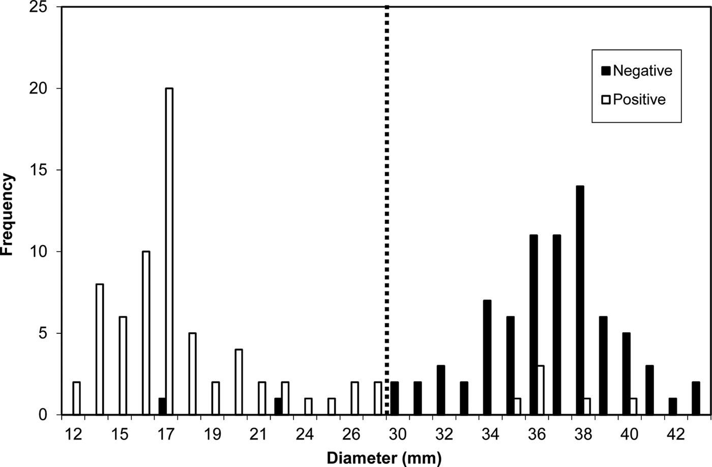
Fig. 2. Distribution of inhibition zone diameters of penicillin G disks for 150 isolates from bovine intramammary infections. Dotted line indicates breakpoint. Bars depict presence (positive) or absence (negative) of blaZgene.
For those tests yielding quantitative results (MIC and DD test) a ROC curve analysis was performed to define the relationship between sensitivity and specificity of each test according to the cut-off point used to define the test as positive or negative. For MIC the calculated AUC was 0·969 and the cutoff value ≥0·185 μg/ml; while for DD test AUC was 0·961 and the cut-off value was 28·5 mm. Correlation coefficient between the two tests was 0·87. Two major errors (false resistant) and no very major errors (false susceptible) were detected. One isolate could not be evaluated by the clover leaf method since an inhibition zone against the indicator strain precluded test interpretation.
Discussion
In the present study, the highest sensitivity was observed for DD (93%) while the highest specificity for Diatabs™ (98·7%). Among the colour-based tests, sensitivity and specificity for Cefinase™ was similar to that obtained by Haveri et al. (Reference Haveri, Suominen, Rantala, Honkanen-Buzalski and Pyörälä2005) using liquid nitrocefin (92·3 and 96·5%, respectively). In addition Pitkälä et al. (Reference Pitkälä, Salmikivi, Bredbacka, Myllyniemi and Koskinen2007) observed sensitivities ranging from 70 to 100% using Cefinase and nitrocefin disks, respectively. The proportion of false negative and false positive results for iodometric method in the present study was high. False positive results observed for this test have been attributed to nonspecific reactions of iodine with bacterial proteins (Livermore, Reference Livermore1995). Only one previous report evaluated detection of penicillin resistance by clover leaf method compared with presence of blaZ gene (Pitkälä et al. Reference Pitkälä, Salmikivi, Bredbacka, Myllyniemi and Koskinen2007). Proportion of false positive results obtained in the present study was similar to that reported by Pitkälä et al. (Reference Pitkälä, Salmikivi, Bredbacka, Myllyniemi and Koskinen2007); however a high percentage of false negative results (14/71) was observed which disagrees with previous findings (Pitkälä et al. Reference Pitkälä, Salmikivi, Bredbacka, Myllyniemi and Koskinen2007).
A good correlation between MIC determination and DD test was found in the present investigation which agrees with results from previous studies (Gianneechini et al. Reference Gianneechini, Concha and Franklin2002; Pengov & Ceru, Reference Pengov and Ceru2003). Conversely, Schlegelova et al. (Reference Schlegelova, Rysánek, Sedivá and Babák2001) reported an accordance of 84·11% between standard broth microdilution and DD methods and Klement et al. (Reference Klement, Chaffer, Leitner, Shwimmer, Friedman, Saran and Shipgel2005) reported a substantially lower susceptibility to penicillin G by DD test (42·1%) than by MIC (59·5%) with a correlation between methods of 0·7; indicating that discrepancies in the classification of isolates as susceptible or resistant were mainly related to inadequacy of interpretive criteria. In an early study, 23·4% of Staph. aureus isolates with MICs of 0·06–0·125 μg/ml were shown to possess the blaZ gene and most of them produced β-lactamase when tested by nitrocefin (Haveri et al. Reference Haveri, Suominen, Rantala, Honkanen-Buzalski and Pyörälä2005). In addition, in a previous study we have shown that two (4%) Staph. aureus isolates with MICs of 0·19 μg/ml produced β-lactamase when tested by nitrocefin (Russi et al. Reference Russi, Bantar and Calvinho2008). Taken together, these data support the evidence that CLSI proposed breakpoint to identify penicillin-resistant isolates may be too high. In this regard, Klement et al. (Reference Klement, Chaffer, Leitner, Shwimmer, Friedman, Saran and Shipgel2005) obtained an estimated cut-off point of 21 mm for Staph. aureus susceptibility to penicillin G instead of 29 mm recommended by CLSI for DD test. The nature of the differences found between Klement et al. (Reference Klement, Chaffer, Leitner, Shwimmer, Friedman, Saran and Shipgel2005) study and the present study can be explained not only by the higher correlation between MIC and DD test observed in our study leading to a lower percentage of false resistant (major errors) and lack of false susceptible (very major errors), but also by the use of blaZ detection as a reference method. The ≥0·185 μg/ml and 28·5 mm resistant cut-off points estimated in the present study can be considered close to the ones indicated by CLSI (2013). However, it has to be taken in account that, albeit low, a number of false negative isolates potentially capable of producing β-lactamase with 35–40 mm inhibition diameters and a MICs 0·03–0·12 μg/ml were detected and less than half of them were positive to β-lactamase by Cefinase™ or Diatabs™. Current CLSI recommendations to perform β-lactamase testing when diameter zones are >29 mm or MIC < 0·12 μg/ml can certainly minimise these potential false negative outcomes.
In conclusion, the DD method used for routine clinical practice in veterinary laboratories yielded the least number of false negative results compared with genotypic detection of blaZ gene. Considering that from a clinical standpoint a false negative result is the most unfavourable situation, for routine laboratories a combination of standard DD test with Diatabs™ or Cefinase™ should be advisable to minimise false negative results.
This research was supported by funds of Asociación Cooperadora de la E.E.A. Rafaela INTA, INTA AESA 52:203992 and grants from SECyT – FONCYT (PICTO N° 30368/05).


