Sudden cardiac arrest (SCA) is a leading cause of death, with approximately 350,000–700,000 individuals per year affected in Europe (Reference Koster, Baubin and Bossaert1). Defibrillation is a key link in the “chain of survival” and chances of survival are closely related to the time that elapses from collapse to the delivery of an effective shock (Reference Deakin, Nolan, Sunde and Koster2). Cardiopulmonary resuscitation (CPR) plus defibrillation within 3 to 5 minutes of collapse can result in survival rates of 49–75 percent; while CPR alone cannot convert ventricular fibrillation (VF) to a normal sinus rhythm, and each minute of delay in defibrillation reduces the chance of survival to discharge by 10–12 percent (Reference Koster, Baubin and Bossaert1;Reference Caffrey3).
Improvements in the survival rate were obtained in prehospital and in-hospital settings where an organization for emergency SCA support, including early defibrillation, was implemented (Reference Destro, Marzaloni, Sermasi and Rossi4–Reference Zafari, Zarter and Heggen6). Incidence of SCAs in hospitals is high, due to the medical conditions of patients and a large number of staff and visitors. A common result of the epidemiological studies concerning in-hospital SCAs is that the rate of survival to hospital discharge is low for patients who had an arrest in general wards, where more time may be needed for rescuers equipped with a defibrillator to reach the victim (Reference Chan, Krumholz, Nichol and Nallamothu7–Reference Tunstall-Pedoe, Bailey and Chamberlain9). To facilitate in-hospital early defibrillation (IHED), and improve this outcome, a defibrillator should be immediately available to the first-responders to a SCA (Reference Destro, Marzaloni, Sermasi and Rossi4;Reference Bossaert, Callanan and Cummins10;Reference Sharieff and Kaulback11).
Improvements in the design of defibrillators have led to the availability of fully automated and semiautomated external defibrillators (often collectively referred to as AEDs) (Reference Varon, Sternbach, Marik and Fromm12). These devices allow trained nonprofessionals to respond to emergencies effectively (Reference Koster, Baubin and Bossaert1;Reference Caffrey3;Reference Domanovits, Meron and Sterz13;Reference Kette, Boni and Liberti14). AEDs should be considered as a means to facilitate early defibrillation within hospitals, especially in areas where advanced life support (ALS) rescuers are not readily available (Reference Deakin, Nolan, Sunde and Koster2;Reference Cusnir, Tongia and Sheka15;Reference Link, Atkins and Passman16). While considering the use of AEDs within a hospital, it should be taken into account that manual defibrillators are most likely more appropriate than AEDs when rescuers with ALS experience are supposed to be present. Manual units allow cardiac rhythm to be analyzed more quickly, with shorter interruptions of CPR chest compressions (Reference Domanovits, Meron and Sterz13;Reference Chan, Krumholz and Spertus17). Furthermore, they allow physicians to diagnose and treat a wider range of arrhythmias. However, some AEDs include manual capabilities and may be used for immediate rescue by first-responding staff, and manually operated when the ALS team reaches the victim.
Our aim was to develop a checklist and a measurement protocol to evaluate AEDs, by taking into account the features that could affect the response to SCAs occurring in areas of a hospital where ALS trained personnel may not be immediately available. This approach was used to evaluate a representative set of AEDs from a clinical/ergonomic and technical point of view. However, it was beyond the scope of the present study to address all of the demands for an effective IHED plan.
MATERIALS AND STUDY DESIGN
Literature Review
PubMed and ISI Web of Knowledge databases were searched for pertinent articles by using different combination of the following keywords: automatic/automated; characteristics; comparison; defibrillator/defibrillation; evaluation; external; guideline; in-hospital. For instance, the following search string can be used: (((external[Title/Abstract]) AND defibrillat*[Title/Abstract]) AND automat*[Title/Abstract]) AND (in-hospital[Title/Abstract] OR comparison[Title/Abstract] OR evaluation[Title/Abstract] OR guideline[Title/Abstract] OR characteristics). Approximately 250 abstracts were analyzed, leading to a deeper, full text, analysis of approximately 110 articles. Eventually, almost 70 articles have been taken into account in the development of the present study.
Semiautomated External Defibrillators
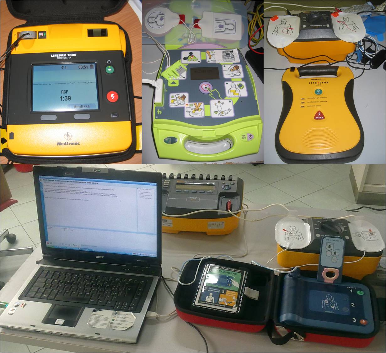
An AED is a device that, once activated by the operator, uses voice-synthesized instructions and/or video prompts to: guide the rescuer, analyze the electrocardiogram (ECG) obtained from disposable adhesive electrodes placed on the patient's chest surface, notify the rescuer whether a shockable cardiac rhythm is detected, and charge to treat the patient. Semiautomated units require an operator to activate the discharge. Six models of semiautomated defibrillators were tested at Bambino Gesù Children's Hospital. Informed consent to the publication of test results was gathered from all of the suppliers.
Analysis of Technical Specifications
Specifications were gathered from the technical datasheets. AEDs have a variety of capabilities and features, which were grouped into six categories: general features, electrodes, power supply, display, recording capabilities, and instructions/messages/alarms (Table 1).
Table 1. Technical specifications.

(1) Configurable 3-shocks sequences
(2) Configurable number of shocks
(3) Configurable by the manufacturer
(4) Configurable
(5) 1 with rechargeable battery
(6) Only adult electrodes
(7) Paediatric adapter required
(8) Connectors only
(9) Depending on model
(10) Depending on model
(11) AP = audio prompt or alarm, AV = audio vocal message, V = visual message or alarm
(12) Bar graph for compression depth
Assessment of Ergonomics and Appropriateness
Ergonomics and appropriateness were evaluated by a resuscitation team composed by an anesthesiologist (certified PALS provider) and by four nurses (certified BLS providers). A general questionnaire, which focused on features that should be included in AEDs for IHED, was submitted to the team members at the beginning of the study (Supplementary Table 1, which can be viewed online at http://dx.doi.org/10.1017/S0266462313000652). Ergonomics was assessed by using both user questionnaires and usability tests (Reference Wiklund18). The team members were preliminary trained in the use of each AED model by a product specialist; afterward, they answered the ergonomics/usability questionnaire on model-specific characteristics (Supplementary Table 2, which can be viewed online at http://dx.doi.org/10.1017/S0266462313000652); finally, a formal usability test was performed. The assessment referred to IHED in nonintensive care areas of Bambino Gesù Children's Hospital.
Measurement Protocol
To test performances of the AEDs, a measurement protocol, based on current technical standards and guidelines, was implemented (19–21). The measurements are aimed at assessing the ability of defibrillators to correctly recognize shockable and nonshockable rhythms, the accuracy of delivered energy, and the charging time (Supplementary Table 3, which can be viewed online at http://dx.doi.org/10.1017/S0266462313000652). The testing equipment consisted of calibrated Impulse 7000DP Defibrillator Analyzer (firmware version 2.03) and Impulse 7010 Selectable Defibrillator Load (Fluke Biomedical, Cleveland, OH). As both measurement instruments were calibrated, measurements were not repeated, thus no statistical analysis of data was performed. Tests on AEDs were repeated only when they yielded out of range results or were failed. Time measurements were taken with a stopwatch, and were performed independently by two operators. The protocol was implemented in the Fluke's Ansur Software (version 2.8.3) to automate measurements. The experimental setup is shown in Supplementary Figure 1, which can be viewed online at http://dx.doi.org/10.1017/S0266462313000652.
Tests for Rhythm Recognition
AEDs use an algorithm to interpret cardiac electrical signals and determine whether the patient is experiencing a life-threatening arrhythmia or not. Devices were tested against their ability to recognize 1 mV ECG rhythms simulated by the defibrillator analyzer. The following nonshockable rhythms were tested: normal sinus, pair of premature ventricular complexes, coarse and fine atrial fibrillation, atrial flutter, atrial tachycardia, supraventricular tachycardia, asystole, first-degree, second-degree (type I and type II), and third-degree blocks. The shockable arrhythmia waveforms tested were: coarse (CVF) and fine (FVF) ventricular fibrillation, and five types of polymorphic ventricular tachycardia (PVT). Input waveforms in the range of 120–300 bpm (with 5 bpm steps) were used to test the minimum defibrillation frequency for monomorphic ventricular tachycardia (MVT). The sensitivity of rhythm recognition with respect to the amplitude of ECG was tested by varying the amplitude of CVF and FVF input signals (with 0.05 mV steps in the range 0.05–0.45 mV and 0.5 mV steps in the range 0.5–5.0 mV), and measuring the minimum shocked amplitude.
Tests for Accuracy of Delivered Energy
AEDs were tested on impedance loads in the 25–175 Ω range (with 25-Ω steps) to ensure that the correct amount of current was delivered when the impedance between electrodes was varied (Reference Link, Atkins and Passman16). For each combination of energy level and impedance, accuracy was evaluated by comparing the measured delivered energy with the corresponding rated value, that is, the value specified by the manufacturer for that specific energy-load combination (where rated values were not provided by the manufacturers, the selected energy level was considered as the reference value). To detect the initial deliverable energy, the “shock” button was pressed as soon as indicated by the device. Tests were repeated with a 50-Ω load, pressing the “shock” button within 2 seconds before the automatic disarming to measure the residual deliverable energy (22). The most common energy levels for pediatric and adult defibrillation were chosen for these latter tests (50 J and 150 J, respectively).
Tests for Charging Time
A set of characteristic times were measured. The start time (ST) was measured as the time interval from power-on (with electrodes connected both to the defibrillator and to the load) to notification of the start of rhythm analysis. The analysis time (AT) was defined as the time interval from notification of the start of rhythm analysis to indication of rhythm recognition (or equivalent message). Measurements with shockable and nonshockable 1 mV input rhythms were averaged to test the AT. The charge time (CT) was defined as the time interval from the indication of rhythm identification to the “ready for discharge” condition. CT was measured for every energy level of each AED. The time from the initial power switch-on to the “ready for discharge” condition (SRT) was therefore determined as ST+AT+CT. Similarly, the time from the notification of rhythm analysis start to the “ready for discharge” condition (ART) was calculated as AT+CT.
RESULTS
Analysis of Technical Specifications
Features and technical parameters of AEDs are shown in Table 1 (note that other releases of the models tested, with slightly different specifications, may exist). The devices tested in this study were satisfactorily light (1.5–3.2 kg); however, differences in size were more relevant, with ZOLL AED Plus (>9,000 cm3) being the largest model and Philips HeartStart FRx (<2,400 cm3) the smallest model. All of the AEDs featured an impedance compensated biphasic waveform, which was rectilinear for AED Plus, pulsed truncated exponential for SCHILLER Fred easy, and truncated exponential for the remaining devices. Energy delivery sequences for adults and pediatric patients were fixed-level for most defibrillators (HeartStart FRx, Fred easy, Defibtech Lifeline AED, and CU Medical Systems/Progetti Rescue 1), escalating for AED Plus, and configurable as fixed-level or escalating for Physio-Control/Medtronic LIFEPAK 1000. The initiation of rhythm analysis was automatic in all devices except for LIFEPAK 1000, which also allowed the rescuer to start the analysis at any time by pressing a button, and for SCHILLER Fred easy, which can be configured by the manufacturer for automatic or manual initiation. Fred easy and LIFEPAK 1000 were also the only two models that, optionally, allowed for manual mode. However, in the SCHILLER defibrillator, the manual mode could only be activated during certain phases of rescue and, to prevent inadvertent activation, two buttons (including the “shut-down” button) must be simultaneously pressed twice. Daily self-tests can be performed by all the devices. All AEDs provided electrodes for adults and pediatric patients, except for Philips HeartStart FRx, which requires a pediatric adapter to be connected in cases of pediatric patient defibrillation. ZOLL AED Plus and Defibtech Lifeline AED allowed the preconnection of both types of electrodes to the AED, while the other models could only be prearmed with electrodes for adults, excluding Rescue 1 which does not allow preconnection of any electrode. The shelf-life of electrodes for devices are 2 years; with the exception of Rescue 1 (shelf-life of 18 months), and the electrodes for adults of AED Plus (shelf-life of 5 years). Most AEDs were equipped with a single disposable internal battery; however different solutions have been implemented for power supply. AED Plus was powered by ten disposable batteries, while Rescue 1 was equipped with a rechargeable battery and Lifeline AED had a secondary battery dedicated to the device status indicator. The shelf life of batteries ranged from 1 to 7 years. Philips HeartStart FRx and Defibtech Lifeline AED did not feature any display, while the remaining models could display ECG. This latter feature is usually optional, while it is standard in Rescue 1 (together with values of heart rate and delivered energy). Finally, all devices allowed for ECG recording, but there were differences in the support for data storage (internal and/or removable memory), availability of audio recording (only in AED Plus, Fred easy, Lifeline AED, and Rescue 1) and length of recording both for ECG and audio.
Assessment of Ergonomics and Appropriateness
The emergency team members expressed their subjective judgments in a general questionnaire on features that should be included in AEDs for IHED (Supplementary Table 1) and in model-specific questionnaires (Supplementary Table 2). With regard to the assessment of features for IHED, manual mode defibrillation and manual start of rhythm analysis were considered not advisable. Similarly, the team members answered that the presence of a monitor was not required, and displaying ECG was not advisable. They preferred to have different electrodes for defibrillation of adult and pediatric patients rather than connecting an adapter when the pediatric mode is required. Finally, availability of instructions for defibrillation and instructions, and feedback, for CPR were regarded as highly relevant features that should be provided both in audible and visual format.
Regarding the assessment of model-specific characteristics, waveforms and sequences of energy levels were judged acceptable for all devices. According to the results of questionnaires and usability tests, ease of use and handiness were also evaluated positively for all models. The overall suitability of ZOLL AED Plus, SCHILLER Fred easy, and Physio-Control LIFEPAK 1000 were rated positively for all types of rescuers. HeartStart FRx and Lifeline AED were rated acceptable for their suitability for members of a resuscitation team and positively for other rescuers. Rescue 1 was rated acceptable with regard to its suitability to all types of rescuers. However, specific ergonomic aspects affected single units. Rescuers reported that in AED Plus, the identification of the “shock” button by rescuers that have not been previously adequately trained may not be immediate, due to the presence of icons and LEDs representing the phases of the rescue chain. When the Fred easy defibrillator was turned on, a few seconds were required before the responder could be reassured about the correct switch-on. Moreover, if this model is operated in manual mode (which requires a complex maneuver, as described above), the “shut-down” button must be pushed to start the rhythm analysis. According to the rescuers that participated in this study, these aspects may result in a delayed defibrillation in cases where the AED is operated by inexperienced or inadequately trained first-responders. Electrodes were judged positively, except for HeartStart FRx and Rescue 1 that were rated only as acceptable because they require rescuers to mount an adapter for pediatric defibrillation (a solution considered inappropriate in a children's hospital). Models that allow the operator to preconnect electrodes for both adults and pediatric patients were preferred. For AED models featuring a display, rescuers judged the display size and the visibility of waveforms, parameters, and messages positively. All models can provide both audio and visual prompts and alarms (Table 1; devices without a display feature LEDs and/or have labels placed on their case). In cases where an AED is turned on with electrodes disconnected, the device prompts for electrode connection. Nevertheless, only three models (ZOLL AED Plus, Philips HeartStart Frx, and SCHILLER Fred easy) inform the operator about the type of electrodes connected. For the other devices, the responder can recognize the type of electrodes by the color code of the cable (Lifeline AED), or of the cable connector. Finally, CPR instructions are provided by all AEDs. Three models (HeartStart FRx, Rescue 1 and, optionally, Fred easy) feature a self-paced metronome for CPR rate, while two other models (Lifeline AED and LIFEPAK 1000) have a CPR countdown. ZOLL AED Plus is the only device featuring a CPR feedback, with a metronome for CPR rate and a bar graph for CPR compression feedback (only available with electrodes for adult patients). Rescuers judged Philips HeartStart FRx as the AED with the most detailed CPR instructions. Depending on the model, additional information is given by the AED, such as the number of delivered shocks, time elapsed since power-on, and battery status. Ultimately, additional messages are given during the phases of rescue (rhythm analysis phase, end/results of rhythm analysis, ready for discharge condition, confirmation of shock delivery).
Measurement Protocol
Rhythm Recognition
Overall, the AEDs performed well when the selected shockable and nonshockable waveforms were given as input signals. However, only ZOLL AED Plus, SCHILLER Fred easy and Progetti Rescue 1 correctly recognized all of the five PVT waveforms as shockable rhythms. Furthermore, when the frequency of 1 mV MVT input waveforms was varied, differences between models were observed in the minimum frequency at which the rhythm was identified as shockable. Lifeline AED and LIFEPAK 1000 discharged at frequencies lower than 150 bpm, while HeartStart FRx and Fred easy needed frequencies higher than 200 bpm to deliver a shock. Regarding the recognition of shockable rhythms (CVF and FVF) while varying the amplitude of input signals, large differences were found in the minimum shocked amplitude, with Physio-Control LIFEPAK 1000 (that can recognize 0.05 mV CVFs and 0.15 mV FVF as shockable) being the most sensitive device and HeartStart FRx the least sensitive (0.5 mV and 1.0 mV, respectively). Finally, all units were able to defibrillate when the maximum amplitude (5 mV) input signals were used.
Accuracy of Delivered Energy
To assess accuracy, delivered energy was measured for each combination of impedance and available energy levels. The difference between measured energy and the corresponding rated value should not exceed ±15 percent (20;21). The ZOLL AED Plus defibrillator fell in this range in 41 of 42 measurements performed, with 18 percent difference at 200 J/125 Ω. Both HeartStart FRx and Fred easy performed within the ±15 percent range in all twelve measurements; however, these models could not deliver shocks, both in adults and in pediatric configuration, when they were tested with 25-Ω loads. In fourteen measurements, the Lifeline AED defibrillator consistently fell within the ±15 percent limit, except for a 34 percent difference at 50 J/25 Ω. Physio-Control LIFEPAK 1000, in 126 measurements, consistently fell within the range. In ten measurements performed, Rescue 1 failed to fall in the ±15 percent interval when the 50 J/25 Ω combination was set; when the 50 J energy level was selected, this model failed to deliver shocks with loads ≥125 Ω, while it did not discharge with a 175 Ω load when the 150 J level was chosen. Figure 1 shows measurement results for 50 J (A) and 150 J (B) energy levels. Finally, delivered energy before automatic disarming was measured with a 50-Ω load at the 50 J and 150 J energy levels. All devices fulfilled the criterion that a fully charged unit should not lose more than 15 percent of the initial deliverable energy before automatically disarming (Figure 1C) (22).

Figure 1. Accuracy of delivered energy for 50 J (A) and 150 J (B) energy levels, while varying the load in the range 25–175 Ω (with 25-Ω steps). For each combination of energy level and impedance, accuracy was evaluated by comparing the measured delivered energy with the corresponding rated value. To detect the initial deliverable energy, the “shock” button was pressed as soon as indicated by the device. (C) Difference between energy delivered to a 50-Ω load before automatic disarming and rated energy value at the 50 J and 150 J energy levels.
Charging Time
Remarkable differences were found between the tested AEDs when timing was analyzed. ST varied from a minimum of 4.1 seconds for Defibtech Lifeline AED, to a maximum of 11.7 seconds for AED Plus (Figures 2A and 2B). Regarding the AT, Physio-Control LIFEPAK 1000 (with an average of 6.7 s) was faster than other defibrillators in recognizing shockable rhythms. For half of the AEDs, no noteworthy difference was found between AT when a shockable rhythm was given as an input rather than when a nonshockable input was presented (Figure 2C). However, in cases of nonshockable rhythms, longer ATs were recorded for Lifeline AED (+3.4 s) and Fred easy (+4.8 s). Conversely, longer ATs were required for shockable rhythms with the HeartStart FRx model (+6.1 s), because the charging phase could not be distinguished from the rhythm analysis phase. Figures 2A and 2B show CTs for 50 J and 150 J, which were chosen as reference energy levels to compare CTs of different devices for pediatric and adult defibrillation, respectively. For the Philips AED, CT measured the time interval from the notification of the start of rhythm analysis to the “ready for discharge” condition. For both energy levels, the longest CTs were recorded with AED Plus, while SCHILLER Fred easy performed best at 50 J and Physio-Control LIFEPAK 1000 at 150 J. LIFEPAK 1000, differently from all other devices, had shorter CT at the 150 J energy level than at 50 J. Notably, relevant differences were found for SRT (= ST+AT+CT), with the shortest values obtained with the Philips HeartStart FRx AED (16.6 and 17.8 s for 50 J and 150 J, respectively) and the longest SRTs recorded with AED Plus (36.7 and 39.4 s). Similar results were found in regard to ART (= AT+CT).

Figure 2. Start Time (ST), analysis time (AT), and charge time (CT) for 50 J (A) and 150 J (B) energy levels: the time from the initial power switch-on to the “ready for discharge” condition (SRT) is ST+AT+CT, while the time from the notification of rhythm analysis start to the “ready for discharge” condition (ART) is AT+CT. Average analysis time for 1 mV shockable and non-shockable rhythms (C). *For the HeartStart FRx, the charging phase could not be distinguished from the rhythm analysis phase. Therefore, when shockable rhythms were presented, AT and CT were measured together as the time interval from the notification of the start of rhythm analysis to the “ready for discharge” condition.
DISCUSSION
Epidemiological studies have shown that the rate of survival to hospital discharge for patients who suffered SCA in general wards is low (Reference Chan, Krumholz, Nichol and Nallamothu7–Reference Tunstall-Pedoe, Bailey and Chamberlain9). Programs based on CPR plus early defibrillation with AEDs can improve the survival rate (Reference Sanna, La Torre and de Waure23) and AEDs should be considered as a means to facilitate IHED in areas where ALS rescuers are not immediately available (Reference Deakin, Nolan, Sunde and Koster2;Reference Link, Atkins and Passman16). A previous study (Reference Fleischhackl, Losert and Haugk24) measured and compared the time to first shock and the influence on basic life support of voice prompts given by commercially available AEDs. Other authors compared waveforms, energy and current delivery of both manual and automated defibrillators (Reference Walker, Melnick and Chapman25–Reference Zelinka, Buić and Zelinka28). Our study provides a clinical and technological comparison of six AEDs, based on the analysis of technical specifications and the assessment of ergonomics and appropriateness for IHED, and on performance tests. The tools used in this research, summarized in Table 2 and provided as supplementary tables, might be used as a checklist to evaluate AEDs.
Table 2. Summary of checklists for evaluating AEDs.

Analysis of the datasheets revealed several differences between devices. The emergency team members recruited for this study expressed no preference for any of the energy delivery protocols. The optimal biphasic waveform, energy levels, and shock sequence are unknown; the recommendations are based on a consensus, while there is no evidence to support either a fixed or escalating energy protocol (Reference Deakin, Nolan, Sunde and Koster2). Designs differ as to whether manual defibrillation capabilities are included or not. Although AEDs with these functionalities are often used in noncritical areas within hospitals instead of the more expensive manual defibrillators, the resuscitation team members judged manual mode defibrillation and manual start of rhythm analysis as not indicated. Indeed, AEDs with manual mode should provide an easy switch between modalities, clearly inform the operator which mode is active, and protect against inadvertent activation of the manual mode. Otherwise, this feature could impair activation of the analysis and delivery of a shock, which should be easy when using an AED (Reference Fleischhackl, Losert and Haugk24). Furthermore, a potential drawback of units providing manual start of rhythm analysis is that shock delivery can be delayed if the operator forgets to press the “analyze” button promptly. The rescuers of Bambino Gesù Children's Hospital preferred units with electrodes for both adult and pediatric patients, rather than with an adapter for the pediatric mode. Moreover, they appreciated defibrillators that allow prearming in both adult and pediatric modalities, because configuration of AEDs for adults is suitable for use in children older than 8 years, while for children between 1 and 8 years the pediatric mode should be used to limit the delivered energy (Reference Koster, Baubin and Bossaert1;Reference Link, Atkins and Passman16). The operators also considered the presence of a monitor not required, and the display of ECG waveforms as not advisable. This judgment was based on two reasons: first, most first-responders would not be trained in rhythm recognition and qualified to interpret ECG; and second, the monitor could distract the operators and may cause them to question the analysis program, thus hesitating to deliver a shock. AEDs should allow for recording data and events from the time the unit is switched on until it is turned off. However, differences were also found in the recording capabilities of AEDs, and some models do not allow for audio recording. Modality of presentation of instructions for defibrillation, and availability of instructions and feedback for CPR are highly relevant features for improving the response to SCA. From this point of view, instructions placarded on the units, voice prompting, indicators, and text messages differed markedly between defibrillators. Finally, all models can provide daily self-checks of their operating condition, thus limiting inspections to a visual check of the AED status indicator. In conclusion, although all units were sufficiently easy to operate, the ergonomics of most models could be further improved; specific aspects affecting some of the tested devices may delay defibrillation in cases where the AED is operated by inexperienced bystanders. However, the results of both the general questionnaire and the model-specific questionnaire for assessment of ergonomics and appropriateness might highly depend on factors such as the training and skills of first-responders, procedures, distribution of manual defibrillators within the healthcare facility, and categories of potential victims of SCAs. All of these aspects may influence the choice of the most appropriate AED. Besides the technical features analyzed above, other elements should be evaluated before purchasing the devices. An important factor is standardization. Indeed, standardization of AEDs within a hospital can facilitate operator training and ALS trained emergency team members who may have to follow different protocols depending on which AED is in use. Finally, the shelf-life of batteries and electrodes can remarkably influence the total cost of ownership, and this should be taken into account when comparing the purchase cost of AEDs.
The measures included in the laboratory protocol allowed the comparison of additional performance parameters. AEDs have already been tested against libraries of recorded cardiac rhythms in trials in both adults and children (Reference Atkinson, Mikysa and Conway29;Reference Kerber, Becker and Bourland30). In the present study, differences between defibrillators in recognizing shockable and nonshockable rhythms emerged when PVT waveforms were given as input by a defibrillator tester. Furthermore, performance differences were found when the frequency of MVT input waveforms and the amplitude of CVF and FVF input signals were varied. Transthoracic impedance can vary considerably, and all of the defibrillators tested measured impedance between the electrodes and adjusted energy delivery accordingly. To test their accuracy, delivered energy was measured for all combinations of impedance and energy levels. These tests revealed that most units have poor performance at low and high impedance levels (Reference Deakin, Nolan, Sunde and Koster2;Reference Walker, Melnick and Chapman25). Finally, when timing was tested, remarkable differences were found between the AEDs, regarding, more specifically, the time for initial autotest, analysis time, and charge time. Importantly, differences greater than 20 seconds were found for the time from the initial power switch-on to the “ready for discharge” condition. These results suggest that, to minimize interruptions to chest compressions during rhythm analysis, and given the correlation of outcome with the time interval between collapse and defibrillation, timing should be further improved in most AEDs (Reference Deakin, Nolan, Sunde and Koster2).
One of the limitations of our study was that no simulation on manikins was performed by the operators (Reference Fleischhackl, Losert and Haugk24). Moreover, because the evaluation of appropriateness was specifically restricted to Bambino Gesù Children's Hospital, results cannot be immediately extended to other healthcare facilities. In addition, further performance parameters, including peak current values, could be part of an assessment protocol for AEDs. Finally, connectivity of AEDs to the hospital LAN/WLAN, and its effects on IHED and the “chain of survival,” were outside of the scope of this study.
CONCLUSIONS
The goal of this research was to develop an assessment protocol for AEDs used in areas of a hospital where ALS trained personnel may not be available. We also aimed at performing an evaluation of commercially available AEDs from a clinical/ergonomic point of view and by measuring their technical performances. A checklist for evaluating devices was provided (Table 2). Results of this assessment show that either ergonomics and/or performances (particularly timing and accuracy) of each defibrillator model may be improved, while the choice of the most appropriate defibrillator should depend on the organizational context in which it is intended to be used. A comprehensive Health Technology Assessment report on this issue is needed, including ethical, legal and organizational implications.
SUPPLEMENTARY MATERIAL
Supplementary Table 1–3 and Supplementary Figure 1 can be found at: http://dx.doi.org/10.1017/S0266462313000652
CONTACT INFORMATION
Federico Nocchi, PhD (federico.nocchi@opbg.net), Clinical Technology Innovation Research Area and Clinical Engineering Department, Bambino Gesù Children's Hospital, IRCCS, Roma, Italy
Gerardina Masucci, Ingegneria Biomedica Santa Lucia S.p.A., Piacenza, Italy; Clinical Engineering Department, Bambino Gesù Children's Hospital, IRCCS, Roma, Italy
Carlo Capussotto, Clinical Engineering Department, Bambino Gesù Children's Hospital, IRCCS, Roma, Italy
Corrado Cecchetti, MD, Emergency Department, Bambino Gesù Children's Hospital, IRCCS, Roma, Italy
Matteo Ritrovato, PhD, Clinical Technology Innovation Research Area, Bambino Gesù Children's Hospital, IRCCS, Roma, Italy
Pietro Derrico, Head of Clinical Technology Innovation Research Area and Clinical Engineering Department, Bambino Gesù Children's Hospital, IRCCS, Roma, Italy
CONFLICTS OF INTEREST
The authors have no financial arrangements with companies whose products are discussed in the study or their competitors. All authors report they have no potential conflicts of interest.