


1. Introduction
The upper Elliot Formation of southern Africa is well known by vertebrate palaeontologists as one of the most important Lower Jurassic terrestrial tetrapod-bearing assemblages (Knoll, Reference Knoll2005; Irmis & Knoll, Reference Irmis and Knoll2008). Nevertheless, complete or subcomplete specimens are generally rare, as are articulated remains.
In his revision of basal sauropodomorph systematics, Galton (Reference Galton, Weishampel, Dodson and Osmólska1990) only recognized one valid species, based on several dozen partial skeletons, in the upper Elliot Formation: Massospondylus carinatus Owen, Reference Owen1854. Galton & Upchurch (Reference Galton, Upchurch, Weishampel, Dodson and Osmólska2004) added Melanorosaurus thabanensis Gauffre, Reference Gauffre1993. Nevertheless, the holotype and unique specimen of the latter taxon (a femur) comes from the lower Elliot Formation (Knoll, Reference Knoll2005). Vasconcelos & Yates (Reference Vasconcelos, Yates and Ashwal2004) revived Gryponyx africanus Broom, Reference Broom1911 as a basal massospondylid, while Yates, Hancox & Rubidge (Reference Yates, Hancox and Rubidge2004) reported the presence of an upper Elliot Formation sauropod probably related to Vulcanodon karibaensis Raath, Reference Raath1972. At the same time, Barrett (Reference Barrett2004) documented the existence of an upper Elliot Formation basal sauropodomoph that he recently described in details and named Massospondylus kaalae Barrett, Reference Barrett2009. Preliminary results also point to a fairly diverse upper Elliot Formation sauropodomorph fauna (Yates, Bonnan & Neveling, Reference Yates, Bonnan and Neveling2007).
Knoll (F. Knoll, unpub. Ph.D. dissertation, MNHN, 2002) referred to the existence of a skeleton of a young ‘prosauropod’ individual from the upper Elliot Formation of Lesotho and figured the right ilium (F. Knoll, unpub. Ph.D. dissertation, MNHN, 2002, pl. 10). The aim of this paper is to fully describe, compare and discuss this specimen.
Institutional abbreviations. BM – Lesotho National Museum, Maseru, Lesotho; BP – Bernard Price Institute for Palaeontological Research, Johannesburg, South Africa; MB – Museum für Naturkunde, Berlin, Germany; MNCN – Museo Nacional de Ciencias Naturales, Madrid, Spain; MNHN – Muséum National d'Histoire Naturelle, Paris, France; NM – Nasionale Museum, Bloemfontein, South Africa; QG – Queen Victoria Museum, Harare, Zimbabwe; SAM – Iziko South African Museum, Cape Town, South Africa; SMNS – Staatliches Museum für Naturkunde, Stuttgart, Germany; UCR – University of Zimbabwe, Harare, Zimbabwe.
2. Systematic palaeontology
Dinosauria Owen, Reference Owen1842
Saurischia Seeley, Reference Seeley1888
Sauropodomorpha von Huene, Reference Huene1932
Ignavusaurus genus novum
Etymology. From Latin ignavus, coward, and ancient Greek σαύρος (masc.), a lizard, because the type locality, Ha Ralekoala, literally means ‘The place of the father of the coward’.
Diagnosis. As for type and only known species.
Ignavusaurus rachelis species nova
Figures 2–14
Etymology. In honour of the paleontologist Raquel López-Antoñanzas from the National Museum of Natural Sciences in Madrid (CSIC). The name Raquel comes from the Hebrew רָחֵל (ewe); its most common Latin form (Rachel) is a noun of the third declension, hence the genitive ending in ‘-is’.
Holotype. BM HR 20, a partial, articulated skeleton. The specimen is provisionally housed in the National Museum of Natural History in Paris. It will eventually return to Lesotho as soon as the National Museum is built in Maseru.
Type locality. The specimen was found at a remote site in southern Lesotho, whose coordinates are S: 30°04′; E: 28°23′ (Fig. 1a, b). It lay close to the place named Ha Ralekoala (Qacha's Nek district), not far from Sekake, at an elevation of approximately 1725 m.

Figure 1. Discovery site of BM HR 20: Ignavusaurus rachelis gen. et sp. nov. (a) Situation in Africa; (b) aerial photograph of the area, scale bar equals 50 m; (c) excavation point (rock pick to right of photograph centre is about 33 cm long).
Type horizon and age. The skeleton, which is largely articulated, was isolated in well-indurated reddish siltstone (Fig. 1c) of the upper Elliot Formation. A palynological study of the matrix has been carried out by R. Rauscher (Université de Strasbourg 1, Strasbourg) and E. Masure (Université Paris VI, Paris). It proved unsuccessful, but the upper Elliot Formation is most often thought to be Hettangian in age (Knoll, Reference Knoll2005 and references therein).
Diagnosis. A non-sauropod sauropodomorph dinosaur with the following unique combination of character-states: transverse width of the ventral ramus of the postorbital greater than its rostrocaudal width at midshaft; height of the postorbital rim of the orbit raised so that it projects laterally to the caudal dorsal process; first dentary tooth adjacent to symphysis; teeth linearly placed within the jaws; orientation of the dentary tooth crowns slightly procumbent; distribution of the serrations along the mesial and distal carinae of the tooth restricted to the apical half of the crown; 14 vertebrae between cervicodorsal transition and primordial sacral vertebrae; transverse processes of the dorsal vertebrae dorsally directed; caudal margin of middle dorsal neural spines concave in lateral view with a projecting caudodorsal corner; first caudal centrum shorter than high; shape of the metacarpal V about as wide as it is long with a strongly convex proximal articulation surface; caudal margin of the postacetabular process of the ilium bluntly pointed; lateral margins of the pubic apron concave in dorsal view; no longitudinal dorsolateral sulcus on proximal ischium; roughly hemispherical femoral head with no sharp medial distal corner; subrectangular astragalus in proximal view; pyramidal process on the craniolateral corner of the proximal surface of the astragalus; transverse width of the proximal end of the fifth metatarsal 50 % of the length of this bone. Although none of these features are proper to Ignavusaurus rachelis, they do not occur in this combination in any other taxon.
3. Description and comparisons
Numerous teeth (some of them associated) and many skull fragments, including the quadrate and the postorbital, have been retrieved. Much of the vertebral column is preserved, including most of the dorsal series and the proximal third or so of the tail (Fig. 2). The pectoral girdle is reduced to a fragment of scapula, whereas the pelvis is fairly complete. A substantial part of the left forelimb is present as well as a large portion of the right leg, a fragmentary left femur and phalanges of the left foot. Measurements are provided in Table 1.
Table 1. Measurements of Ignavusaurus rachelae gen. et sp. nov.


Figure 2. BM HR 20: Ignavusaurus rachelis gen. et sp. nov. Part of the specimen as exposed in the course of preparation. Scale bar equals 12 cm.
Ignavusaurus rachelis was compared with the wealth of material of the Elliot Formation housed in the MNHN at first hand. Among these specimens, those studied by Gauffre (F.-X. Gauffre, unpub. Ph.D. dissertation, MNHN, 1996) are determined here as Melanorosaurus readi Haughton, Reference Haughton1924. All the material appears indeed to belong to a single species, whose femur is similar to that of Melanorosaurus readi (see Galton, Van Heerden & Yates, Reference Galton, Van Heerden, Yates, Tidwell and Carpenter2005), with a subrectangular, very tall, fairly vertical and straight fourth trochanter that is located near the medial border of the shaft . . . Dental comparison with this taxon was made on the basis of the observation of the skull of NM QR3314 and its published description (Yates, Reference Yates2007b). Most of the remaining specimens kept in the MNHN were studied by Costedoat (D. Costedoat, unpub. D.E.S. dissertation, Univ. Paris, 1962) and are identified here as Massospondylus cf. carinatus. These latter observations were complemented by comparisons with the description of this taxa provided by Cooper (Reference Cooper1981), as well as by observations of additional specimens of M. carinatus housed in the SAM. Comparisons with other taxa were essentially made through comprehensive material in the SMNS and MB (plateosaurids), a cast in the MNCN (Mussaurus patagonicus) and literature.
3.a. Skull and teeth
Unfortunately, the skull is fragmented in more than a hundred and twenty small bits, which limits comparisons. Among the largest fragments, the right quadrate and the left postorbital (Fig. 3) can be recognized. The quadrate is elongate with a strongly concave caudal margin in lateral and medial views. This latter makes an angle of about 80° with the condyle, which is simple and longer than wide. Although the rostrodorsal and caudal branches have been broken, the postorbital no doubt displayed a typical Y-shape. Its latero-medial width decreases sharply caudally. In lateral view, the medial axes of the three branches cross over in a flat zone of the bone, which contrasts with the condition in Plateosaurus longiceps (MB.R.1937) in which the intersection of these axes takes place on a lateral eminence.

Figure 3. BM HR 20: Ignavusaurus rachelis gen. et sp. nov. Left postorbital: (a) lateral view; (b) medial view; (c) caudal view. Scale bar equals 1 cm.
Some thirty complete or fragmentary teeth are present (Fig. 4). The height of most of the crowns does not reach 4 mm. All the teeth are narrow, labio-lingually compressed, and carinated. However, some are pointed and devoid of denticulation, whereas others are more spatulate, with a rather flat lingual surface and a convex labial one, and with a slight neck and up to five, relatively salient, apically directed denticles on each carina. Intermediate morphologies also occur, some teeth having in particular very attenuated denticles. The heterodonty of Ignavusaurus rachelis is fairly remarkable since a dentition made of similar high-crowned teeth is the prevalent condition in basal sauropodomorphs. The single dentigerous bone found (a fragment of the left dentary; Fig. 4e) suggests that tapering teeth were rostrally positioned in the jaws and slightly procumbent. The preservation of a series of nine teeth in their original respective position (Fig. 4d) suggests that the overlap was weak or non-existent. All in all, the dentition of I. rachelis suggests a more opportunistic diet than the possibly fairly herbivorous one that evolved in less basal sauropodomorph taxa such as Plateosaurus (see e.g. Barrett, Reference Barrett and Sues2000 and Barrett & Upchurch, Reference Barrett and Upchurch2007).

Figure 4. BM HR 20: Ignavusaurus rachelis gen. et sp. nov. Teeth and dentary: (a) labial view of an isolated tooth; (b) lingual view of the same tooth; (c) distal view of the same tooth; (d) labial view of a series of associated teeth; (e) labial view of left dentary with teeth. Scale bar equals 2 mm (a–c) and 5 mm (d, e).
The skull and dentition of Massospondylus carinatus are relatively well known (see e.g. Gow, Kitching & Raath, Reference Gow, Kitching and Raath1990; Sues et al. Reference Sues, Reisz, Hinic and Raath2004; Barrett & Yates, Reference Barrett and Yates2006). Without even invoking the difference in size, the teeth of M. carinatus differ from those of Ignavusaurus rachelis in their more marked neck and the shape and distribution of their denticles. The denticles of most of the teeth of I. rachelis are indeed very sharp and are distributed on the apical half of the crown, whereas, in general, they are quite rounded and present essentially on the apical third of the crown in M. carinatus.
The teeth of Melanorosaurus readi (Yates, Reference Yates2007b; pers. obs.) are larger and relatively wider (mesiodistally) than those of Ignavusaurus rachelis. In addition, the denticles are serration-like in M. readi; they are more numerous and rather rounded than salient, in contrast with the condition in I. rachelis.
3.b. Vertebrae, ribs and hemal arches
Except for the most cranial preserved vertebra (namely the 2nd dorsal, which is reduced to the caudal half of the centrum) the following 29 vertebrae are fairly complete (Fig. 2). They constitute two articulated series disjointed in the sacral region. Based on the configuration (position of the parapophyses. . .) in a number of species belonging to the genera Plateosaurus, Lufengosaurus, Yunnanosaurus and Riojasaurus (Galton, Reference Galton, Weishampel, Dodson and Osmólska1990, Reference Galton1999; pers. obs.), the series of 31 vertebrae of BM HR 20 comprises the last 13 dorsals (from the fragmentary 2nd to the 14th), 1 dorsosacral, 2 sacrals and the 15 first caudals. Breaks in several of the centra have made it possible to see that the cortical bone is very thin (less than 1 mm) and that it covered a very poorly ossified spongiosa.
The dorsal centra (Fig. 5) are slightly amphicoelous. Their ventral border becomes progressively more rounded caudally. They are more than twice as long as wide. In lateral view, the ventral borders appear concave as well as the lateral faces in ventral view. Numerous ‘pleats’ of the bone surface are present near the articular extremities. The neural arches are not fused to the centra. They are about twice as tall as the centra and their base is well developed, being about as high as the neural spine. They are relatively simply organized. The cranial centrodiapophyseal lamina is similar in shape (thickness, roundness and straightness) and length to the caudal centrodiapophyseal lamina. These laminae form a constant angle of 75° along the entire length of the dorsal series. A discrete migration of the parapophysis from the base of the neural arch toward the diapophysis can be observed in the series from the most cranial dorsal vertebra caudally. In the 3rd and 4th dorsals and possibly in the 5th also, the parapophysis involves a small part of the centrum. In all well-preserved dorsal vertebrae (namely all but the 2nd), the parapophysis is associated with the cranial centrodiapophyseal lamina, which is therefore actually a paradiapophyseal lamina. The transverse processes of the dorsal vertebrae are dorsally directed, being at an angle of about 80° from one another. Although accounted for in part by preservational artifact (lateral crushing), this is likely a genuine character-state to some extent because this angle increases all along the vertebral series with the result that the last preserved caudals have almost horizontal transverse processes. The caudal margin of the middle dorsal neural spines is concave in lateral view with a projecting caudodorsal corner. The dorsal portion of the vertebral series was associated with numerous ribs, which are fragmented and crushed along their length, but appear to be of the general basal sauropodomorphan gross morphology (see Galton & Upchurch, Reference Galton, Upchurch, Weishampel, Dodson and Osmólska2004).
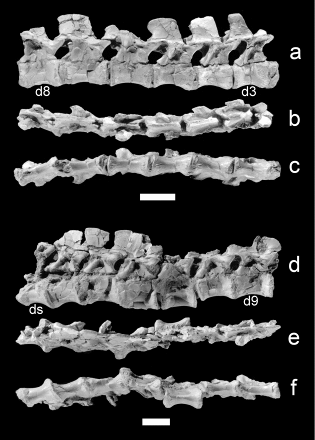
Figure 5. BM HR 20: Ignavusaurus rachelis gen. et sp. nov. Articulated dorsal vertebrae, plus dorsosacral (slightly reconstructed): (a) right lateral view of second to eighth dorsal vertebrae; (b) dorsal view of second to eighth dorsal vertebrae; (c) ventral view of second to eighth dorsal vertebrae; (d) right lateral view of ninth to fourteenth dorsal vertebrae, plus dorsosacral; (e) dorsal view of ninth to fourteenth dorsal vertebrae, plus dorsosacral; (f) ventral view of ninth to fourteenth dorsal vertebrae, plus dorsosacral. d3 – third dorsal vertebra; d8 – eighth dorsal vertebra; d9 – ninth dorsal vertebra; ds – dorsosacral vertebra. Scale bar equals 2 cm.
There is no coosification in the sacral region: there was no fusion between the sacrals and the sacral ribs and between these and the blade of the ilium, nor between the centra and the neural arches. The configuration of this zone is a bit uncertain due to imperfect preservation, but one dorsosacral and two sacrals appear to be present (in Plateosaurus longiceps and related species, there are generally two sacrals and a caudosacral; Galton & Upchurch, Reference Galton, Upchurch, Weishampel, Dodson and Osmólska2004). The first sacral is shorter than the dorsosacral and the second sacral, but otherwise all the three are rather similar. The facet for the articulation of the sacral rib is located cranially on the centrum (due to deficient preservation, no detail can be provided on the shape and position of the sacral ribs attachment on the ilium). The centrum is rounded ventrally. The centrum of the dorsosacral and of the first sacral shows a bilateral depression ventral to the pedicel of the neural arch.
The height/length ratio of the caudal centra (Fig. 2) decreases along the series: from the seventh caudal vertebra caudally, the centra are longer than high. The articular faces are ovular and the ventral faces of the centra are unobtrusively ‘carinated’. The poor ossification of the caudal vertebrae is visible in the absence of fusion of the neural arches with the centra and between the neural arches and the transverse processes. The latter are situated very low. The caudals bear a hemal arch from the second vertebra caudally. The length of the hemal arches is about a third greater than the height of the vertebra.
With respect to the dorsal vertebrae, differences with Massospondylus carinatus are found mostly in the neural arches. For instance, the relative height from the neurocentral suture to the level of zygapophyseal facets is much higher in Ignavusaurus rachelis. In the 13th dorsal vertebra of I. rachelis this height is more than 80 % the height of the centrum, whereas it is about 60 % in M. carinatus (Cooper, Reference Cooper1981, fig. 10). Important differences with Melanorosaurus readi are also present. In this taxon, the dorsal centra are more derived in being always proportionally shorter. The neural arch height/centrum height ratio is higher in I. rachelis than in M. readi and the plateosaurids. In the latter, the base of the neural arch is flat, the cranial centrodiapophyseal lamina tends to be thinner and shorter than the caudal one (especially in caudal dorsals) and they orientate differently than in I. rachelis.
The caudals of Ignavusaurus rachelis are very different from those of Massospondylus carinatus as figured by Cooper (Reference Cooper1981, figs 15, 16c). For instance, in M. carinatus the articular surfaces of the midcaudals are not ovular, but rather heart-shaped. In contrast with the condition in I. rachelis, the caudal articular surfaces appear somewhat convex in lateral view. In M. carinatus, the ventral border of the caudals appears much more concave in lateral view than in I. rachelis. Moreover, the neural arch of the caudal vertebrae of M. carinatus covers only the cranial-central portion of the centrum versus almost all the dorsal surface in I. rachelis. In comparison with MNHN MAF 977 (a middle caudal vertebra of Melanorosaurus readi), some differences are also obvious. For instance, the articular surfaces are more rounded than ovular, the caudal articular apophyses are less elevated and, above all, the transverse processes are both situated and directed more ventrally. In plateosaurids, the length of the hemal arch is about equal to the height of the vertebra that bears it.
3.c. Gastralia
The gastralia are long ‘spaghetti-like’ rods that are ovoid in cross-section, instead of being rather cylindrical as in Massospondylus carinatus (Cooper, Reference Cooper1981).
3.d. Scapula
Unfortunately, only the distal extremity of the left scapula is preserved (Fig. 6a, b). It is gently, proximo-distally arched. The distal expansion is narrower than in Massospondylus carinatus (Cooper, Reference Cooper1981, fig. 23a, b) and might have been partly cartilaginous.

Figure 6. BM HR 20: Ignavusaurus rachelis gen. et sp. nov. Distal extremity of left scapula and distal end of left humerus: (a) lateral view of scapula; (b) medial view of scapula; (c) cranial view of humerus; (d) caudal view of humerus; (e) lateral view of humerus; (f) medial view of humerus. Scale bar equals 1 cm.
3.e. Humerus
Only about the distal third of the left humerus is present (Fig. 6c–f). In cranial view, both the longitudinal bulges are strongly convex. The intercondylar surface is roughly triangular. In caudal view, the concave surface (olecranon fossa) is limited laterally by a proximo-distally lengthened swelling. The medial condyle extends slightly more distally than the lateral one.
In cranial view, the distal condyles of the humerus of Massospondylus carinatus are separated by a rather deep semicircular fossa (Cooper, Reference Cooper1981, figs 26, 27), which is lacking in Ignavusaurus rachelis. In Melanorosaurus readi (MNHN MAF 981), the medial bulge of the cranial side is much flatter than in I. rachelis, approaching the condition in eusauropods.
3.f. Ulna
Only the left, quite well-preserved, ulna is present (Fig. 7a–d). The proximal articular surface is roughly triangular in proximal view. The olecranon process is moderately developed, but the coronoid process is prominent. The shaft has an elliptical cross-section. The distal epiphysis is relatively weakly transversally expanded and only slightly convex.

Figure 7. BM HR 20: Ignavusaurus rachelis gen. et sp. nov. Left ulna and left radius: (a) lateral view of ulna; (b) medial view of ulna; (c) cranial view of ulna; (d) caudal view of ulna; (e) lateral view of radius; (f) medial view of radius; (g) cranial view of radius; (h) caudal view of radius. Scale bar equals 1 cm.
The extreme weakness of the intercotylar crest makes the ulna of Ignavusaurus rachelis strikingly different from that of a number of taxa such as Plateosaurus longiceps, Massospondylus carinatus and Melanorosaurus readi (MNHN MAF 765). In addition, the sigmoid notch is much more pronounced in I. rachelis than in M. carinatus, so that both the olecranon and coronoid process are more conspicuous in the former. In I. rachelis, the extremities are disposed at around 40° from one another. This is close to the condition in Melanorosaurus readi, but clearly lower than in M. carinatus, in which the twisting is about 60° (Cooper, Reference Cooper1981). In comparisons with M. carinatus (Cooper, Reference Cooper1981, p. 735, fig. 31), I. rachelis is also distinguished by the absence of a distal tubercle for the attachment of the radio-ulnar ligament. On the whole, the ulna of BM HR 20 clearly differs from the ulna of M. carinatus, as well as from that of larger Triassic forms.
3.g. Radius
Only the left radius is present (Fig. 7e–h). In the matrix, it was positioned inside out at about 130° from the humerus and 135° from the ulna. It is a lightly built bone in which, as usual, the proximal extremity is concave and cranio-caudally expanded. The distal one, which is incomplete, is rather flat. The diaphysis is straight; the distal and proximal extremities are disposed at nearly 30° from one another.
The twisting of the diaphysis of the radius in Ignavusaurus rachelis is not far from that in Plateosauravus cullingworthi (van Heerden, Reference Van Heerden1979, pls 27, 28). However, it is much less than that in Massospondylus carinatus (about 45° according to Cooper, Reference Cooper1981; a difference whose amplitude can hardly be accounted for by crushing). Moreover, in comparisons with the radius of M. carinatus and P. cullingworthi, that of I. rachelis appears less broad at the level of the extremities, the articular surfaces of which are also different. This is especially true for M. carinatus, for which the distal condyle is said to be strongly convex (Cooper, Reference Cooper1981, fig. 29) and the proximal articular surface is also rounded.
3.h. Carpus
No element of the carpus can be determined with certainty. However, close to the distal extremity of the ulna (but possibly a little displaced), a small (diameter of almost 5 mm), somewhat lentil-like bone was uncovered. The identification of this bone is difficult. Its shape is different from the carpal bones of Massospondylus carinatus as figured by Cooper (Reference Cooper1981, figs 32–36), but it recalls the possible remnant of radiale described by Broom (Reference Broom1911, p. 295) in Gryponyx africanus.
3.i. Metacarpal III and phalanges of manual digit III
Metacarpal III and the phalanges of manual digit III are well preserved (Fig. 8). Metacarpal III has strongly concave lateral margins. The ventral face is much flatter than the dorsal one. The proximal articular surface is flat, whereas the distal one is convex. The first phalanx is short, wider than deep in proximal view. The second phalanx is also short, but it is deeper. The third phalanx has a more elongate morphology. The ungual is weakly hooked, much deeper than wide in proximal view.

Figure 8. BM HR 20: Ignavusaurus rachelis gen. et sp. nov. Left metacarpal III and phalanges of manual digit III: (a) dorsal view; (b) right lateral view. Scale bar equals 1 cm.
When compared with metacarpal III of Massospondylus carinatus (QG1282; Cooper, Reference Cooper1981, fig. 41), that of Ignavusaurus rachelis appears especially distinct in caudal view because of the strong concavity of the ventral side in the former. The phalanges of manual digit III of I. rachelis show some morphological differences with the equivalents in the manus of M. carinatus (Cooper, Reference Cooper1981, figs 43, 45, 46), but they are unobtrusive. The ungual phalanx of digit III of I. rachelis appears more ovular than that of M. carinatus (QG1160; Cooper, Reference Cooper1981, fig. 43) in proximal view.
The collection of the MNHN contains several dissociated metacarpals and phalanges that are difficult to identify. Gauffre (F.-X. Gauffre, unpub. Ph.D. dissertation, MNHN, 1996, fig. 32B) only described as metacarpal III MNHN MAF 728, which shows a morphology that is on the whole similar to that of Ignavusaurus rachelis. However, some differences are noticeable. For instance, the ventral border of the distal articular surface is convex in metacarpal III of Melanorosaurus readi, whereas it is flat in that of I. rachelis. Moreover, the proximal articular surface makes a sharper angle with the longitudinal axis of the bone in M. readi than in I. rachelis.
3.j. Metacarpal IV and phalanges of manual digit IV
The metacarpal IV is similar to the metacarpal III, but it is only about 80 % as large. The first phalanx is short and wide. The second phalanx has a close morphology, but it is much smaller. The third phalanx is a minuscule, short, slightly hooked bone.
In comparison with Massospondylus carinatus (Cooper, Reference Cooper1981, fig. 40h, i), the metacarpal IV of Ignavusaurus rachelis is wider at the extremities. The proximal articular surface of the two first phalanges appears more quadrangular in I. rachelis than in M. carinatus (Cooper, Reference Cooper1981, fig. 43). The third phalanx of the manual digit III is not hooked at all in M. carinatus (Cooper, Reference Cooper1981, fig. 45).
3.k. Metacarpal V and phalanx of manual digit V
The metacarpal V is a distinctive, short bone, wider proximally than distally. Both articular ends are convex. The medial, lateral, dorsal and ventral sides are more or less concave. The phalanx is small, wider than deep, with a simple proximal concavity.
The metacarpal V of Ignavusaurus rachelis is distinct from that of Massospondylus carinatus (Cooper, Reference Cooper1981, fig. 40a–d) in that its proximal extremity is much deeper than the distal one, instead of being about equally deep. The first phalanx of the manual digit V is remarkably less massive in I. rachelis than in M. carinatus as figured by Cooper (Reference Cooper1981, fig. 45).
3.l. Ilium
Both ilia are preserved (Fig. 9). They are both incomplete but, fortunately, the unbroken parts of each are different so that one can have an accurate idea of the complete ilium. It is typical of the brachyiliac condition (Colbert, Reference Colbert1964), which is characteristic of ‘prosauropods’. The blade is short and deep. Its dorsal margin is only weakly, but evenly, convex. The preacetabular process is pointed, about as long as deep. It does not project further cranially than the pubic peduncle and its depth is much less than that of the blade dorsal to the acetabulum. The postacetabular process is bluntly pointed. Its length is nearly equivalent to the distance separating the pubic and ischial peduncles. The pubic peduncle is elongated (about as long as the iliac blade is deep in its central part). The ischial peduncle does not bear any caudally projecting ‘heel’ at its articular end. The acetabulum is fully open.

Figure 9. BM HR 20: Ignavusaurus rachelis gen. et sp. nov. Ilia (partly reconstructed): (a) right ilium, lateral view; (b) left ilium, lateral view. Scale bar equals 2 cm.
The differences between the ilium of Ignavusaurus rachelis and that of Massospondylus carinatus (Cooper, Reference Cooper1981, figs 39, 51, 52) are numerous. Thus, the preacetabular process forms a much more open angle with the pubic peduncle in I. rachelis than in M. carinatus. There is generally a ridge ventral to the swollen surface of the postacetabular process that borders a rather deep depression for the origin of the M. caudifemoralis brevis in M. carinatus (Cooper, Reference Cooper1981, fig. 85). In I. rachelis, the brevis shelf is not so distinct. The pubic peduncle of the ilium of I. rachelis is straight, not curved in its midlength, and the ischiatic peduncle is more ventrally directed than in M. carinatus. The supra-acetabular buttress forms a more largely opened collar to the acetabulum on the ilium of I. rachelis than in M. carinatus.
The ilium of Ignavusaurus rachelis is distinguished from that of Massospondylus carinatus and Melanorosaurus readi by the dorsal border of the blade, from the preacetabular process to the postacetabular one, which generally does not form a regular curve. The shape of the postacetabular process of the ilium of M. readi is in between the thin and (moderately) pointed one of I. rachelis and the thick and blunt one of plateosaurids.
3.m. Pubis
A large part of the left pubis (proximal portion damaged), as well as fragments of the right, are preserved (Fig. 10a–d). This is a thin, relatively narrow bone. The whole of the dorsal surface is fairly flat, but there is a central longitudinal eminence on the ventral face in the proximal half of the pubic blade. The lateral edge is bowed toward the midline symphysis. The medial margin is marked by a small, badly preserved expansion proximally. The obturator notch is completely visible in dorsal view. Lateral to the obturator foramen, there is a shallow concavity on the ventral surface.

Figure 10. BM HR 20: Ignavusaurus rachelis gen. et sp. nov. Left pubis (partly reconstructed) and articulated ischia: (a) dorsal view of pubis; (b) ventral view of pubis; (c) lateral view of pubis; (d) medial view of pubis; (e) dorsal view of ischia; (f) ventral view of ischia. Scale bar equals 2 cm.
In comparison with Massospondylus carinatus (Cooper, Reference Cooper1981, figs 57, 58), the lateral border of the pubis of Ignavusaurus rachelis is clearly more curved. From this point of view, the pubis of M. carinatus appears intermediate between that of I. rachelis and that of Melanorosaurus readi (F.-X. Gauffre, unpub. Ph.D. dissertation, MNHN, 1996, fig. 36, pl. 17; Galton, Van Heerden & Yates, Reference Galton, Van Heerden, Yates, Tidwell and Carpenter2005, fig. 1.10c, e), which approaches the condition in plateosaurids (wide pubic blade with a rectilinear lateral border). Also, the ischium of M. carinatus bears a completely closed obturator foramen, whereas that of I. rachelis appears largely open as in ‘Anchisaurus’ (Galton, Reference Galton1976; Galton & Upchurch, Reference Galton, Upchurch, Weishampel, Dodson and Osmólska2004), but admittedly preservation of this zone is frequently imperfect (see e.g. Sereno, Reference Sereno2007).
3.n. Ischium
The ischia are articulated and rather well preserved, although a bit deformed and incomplete (Fig. 10e, f). In overall morphology, the ischium shows two regions: a plate-like obturator section proximally and a rod-like distal shaft. The passage from the obturator plate to the shaft is very gradual. The ischial component of the acetabular rim is large and rectilinear. There is no longitudinal dorsal sulcus (for the origin of the ischio-femoralis), but rather a barely noticeable groove. The distal extremity of the shafts, which was possibly cartilaginous, is lacking. In transverse section, the shaft is about as deep as wide and triangular in shape. No elongate interischial fenestra is present.
The ischium of Ignavusaurus rachelis differs in several respects from that of Massospondylus carinatus (Cooper, Reference Cooper1981, fig. 54). Thus, the participation of the ischial proximal plate to the acetabulum is well marked in M. carinatus by a widely open notch, which is absent in I. rachelis. The proximal shaft of the ischium of M. carinatus is somewhat heart-like in cross-section (Cooper, Reference Cooper1981, fig. 55) due to a deep dorsal groove, which is lacking in I. rachelis. The transverse width of the conjoined distal ischial expansions is much less in M. carinatus than in I. rachelis.
The morphological differences with the ischia of Plateosauravus cullingworthi (Van Heerden, Reference Van Heerden1979, fig. 14, pls 34, 35) are even more important. As a matter of fact, the lateral margin of the ischial shaft is continuously curved (concave) in this taxon, whereas the two margins are much more parallel in Ignavusaurus rachelis. Other differences include the more pronounced robustness of the area of articulation with the ilium in P. cullingworthi.
3.o. Femur
Both femora are present, but fractured. The left femur is much damaged proximally, the distal extremity is lacking and a histological examination revealed that the spongiosa is badly preserved as well. The right femur (Fig. 11) is in a much better state, even if both extremities are incompletely preserved. In medial and lateral views, the longitudinal axis of the femur was strongly bent and the angle between the long axis of the femoral head and the transverse axis of the distal condyles is also pronounced (at least 30°). The femoral head is dorsomedially projected, not offset from the shaft on a neck and appears roughly hemispherical, but this condition might be due to the state of preservation of this zone. The cranial (= ‘lesser’) trochanter is positioned near the centre of the cranial face, so that it is not visible in caudal view of the femur. It is shaped like a proximo-distally lengthened raised process. The proximal tip of the cranial trochanter reaches about the level of the femoral head. There is no transverse ridge extending laterally from it. The fourth trochanter is positioned in the proximal half of the femur in a central location along the mediolateral axis. It is not perfectly preserved but, as far as can be determined, it is a low rugose ridge, whose length is about 1/5 the total length of the femur, and it is subtrapezoidal in profile, with a slightly steeper distal angle than the proximal slope. No extensor depression is well discernible on the cranial surface of the distal end of the femur.

Figure 11. BM HR 20: Ignavusaurus rachelis gen. et sp. nov. Right femur: (a) cranial view; (b) caudal view; (c) lateral view; (d) medial view. ct – cranial trochanter; ft – fourth trochanter. Scale bar equals 1 cm.
The breakages, a small amount of deformation and the imperfect state of preservation of the femoral head limit the comparisons between the femur of Ignavusaurus rachelis and that of other taxa. The femur of Massospondylus carinatus has been well illustrated by Cooper (Reference Cooper1981, figs 59, 60). It shows many differences with that of I. rachelis. In caudal view, the internal outline of the femur of M. carinatus (Cooper, Reference Cooper1981, fig. 60) shows a swelling at the level of the fourth trochanter. In this view, the internal outline of the femur is straight all along its length in Ignavusaurus rachelis. Moreover, the fourth trochanter of the femur of I. rachelis is slightly curved in caudal view, not sigmoidal like in M. carinatus. In cranial view, the cranial trochanter appears in a more central position in I. rachelis than in M. carinatus, although this difference could be in part due to deformation. The femoral shaft of M. carinatus is clearly more slender in its middle part, whereas the width of the shaft is nearly constant all along its length (in cranial and caudal views) in I. rachelis. With respect to the distal extremity, the differences are significant. For instance, it is not as caudally expanded in I. rachelis as it is in M. carinatus. In medial view, the distal articular surface in I. rachelis is not flat as in M. carinatus, but rather gently curved.
The femur of Melanorosaurus readi has been illustrated in particular by Gauffre (F.-X. Gauffre, unpub. Ph.D. dissertation, MNHN, 1996, pls 3–5; see also Galton, Van Heerden & Yates, Reference Galton, Van Heerden, Yates, Tidwell and Carpenter2005, figs 1.11e–h, 1.12a–d, 1.13a–c, g–i). The most obvious difference concerns the relative distance separating the femoral head from the fourth trochanter. Whereas the fourth trochanter is close to the femoral head in Ignavusaurus rachelis (a primitive character-state for sauropodomorphs), a deep neck makes the high fourth trochanter of M. readi extend onto the distal half of the bone. Moreover, in M. readi the fourth trochanter has a relatively shorter base than in I. rachelis. In cranial view, the cranial trochanter is rather lateral in M. readi, whereas it is central in I. rachelis. In medial and lateral views, the condyles of the femur of M. readi have a rounded caudal outline, whereas they become angular near the junction with body of the bone in I. rachelis.
3.p. Tibia
Only the right tibia is present in BM HR 20 (Fig. 12). Unfortunately, a great part of the cranio-proximal extremity is missing (the cnemial crest is not preserved) and the external region of the distal extremity is a bit damaged. The bone has an elongate shape, but it is still slightly shorter than the femur (tibia/femur length ratio ≈ 0.95). The preserved caudo-proximal articular surface bears a characteristic groove, in continuity with the shaft of the bone, for part of the powerful caudal cruciate ligament. There is neither cranio-caudal nor significant transverse widening at the distal extremity, which is longer (cranio-caudally) than wide (latero-medially). The caudo-lateral process does not flare laterally. The distal articular surface is subrectangular.

Figure 12. BM HR 20: Ignavusaurus rachelis gen. et sp. nov. Right tibia: (a) cranial view; (b) caudal view; (c) lateral view; (d) medial view. Arrow points to posterior intercondyloid groove. Scale bar equals 1 cm.
In Massospondylus carinatus (Cooper, Reference Cooper1981, figs 65, 66), there is no groove on the proximal articular surface. Moreover, the malleoli show a greater difference (in lateral view) in their respective distal extension in M. carinatus than in Ignavusaurus rachelis. In medial view, the distal region widens more in M. carinatus than in I. rachelis.
A groove similar to that on the tibia of Ignavusaurus rachelis is not present on the tibiae of Melanorosaurus readi kept in the MNHN (MAF 927, 987, 1039, II LC, IV LC). Moreover, the distal extremity shows a straight border in I. rachelis in medial view, not a convex one as in MNHN MAF 987. Contrary to the condition in M. readi, the cranial malleolus was not clearly more expanded medio-laterally than the caudal malleolus in I. rachelis. In addition, the distal articular region in I. rachelis is not strongly convex (but rather flat), the cranial outline is convex (not concave) and the internal outline is rather concave (not convex). Noticeable differences also occur between the tibia of I. rachelis and that of Plateosauravus cullingworthi as illustrated by van Heerden (Reference Van Heerden1979, fig. 17, pls 42–44), such as the widening of the distal extremity.
3.q. Astragalus
The astragalus of the right pes has been preserved almost completely (Fig. 13a, b). It is an angular, subrectangular bone. The distal side is weakly convex. The proximal one (about half of which is occupied by the medial cotylus) is concave, except for the ascending process. The latter provides an articular surface for the cranial malleolus of the tibia, whereas the caudal malleolus fitted in the small medial cotylus.

Figure 13. BM HR 20: Ignavusaurus rachelis gen. et sp. nov. Right astragalus, right distal tarsal III and right distal tarsal IV: (a) proximal view of astragalus; (b) distal view of astragalus; (c) proximal view of distal tarsal III; (d) distal view of distal tarsal III; (e) proximal view of distal tarsal IV; (f) distal view of distal tarsal IV. Scale bar equals 1 cm.
In general shape, the astragalus of Ignavusaurus rachelis, with its ‘interlocking’ tibial–astragalar articulation, looks like that of other basal sauropodomorphs such as Saturnalia tupiniquim (Langer, Reference Langer2003, fig. 6a–f). Therefore, in comparison with the astragalus of Massospondylus (Cooper, Reference Cooper1981, figs 70a–f, 71a–d; MNHN unnumbered specimen from Ha Noosi, Lesotho), there are unmistakable resemblances. However, in proximal and distal views, the caudomedial margin of the astragalus of Ignavusaurus rachelis form a well-marked corner (approximately 105°), whereas this edge is more evenly rounded in Massospondylus carinatus.
The astragalus of Melanorosaurus readi (MNHN MAF1038; F.-X. Gauffre, unpub. Ph.D. dissertation, MNHN, 1996, pl. 11.1) is easily distinguished from that of Ignavusaurus rachelis. In particular, it has a more latero-medially lengthened shape.
3.r. Distal tarsal III
Distal tarsal III is a small rounded bone that shows few characters (Fig. 13c, d). Fortunately, as it was found in association with distal tarsal IV and metatarsals II to IV, the orientation of this bone is known in Ignavusaurus rachelis. The lateral and dorsal margins are slightly convex, whereas the medial one is rather straight with a little depression at mid-length. The ventral corner is blunt. The proximal articular surface is much flatter than the distal one.
The distal tarsal III of Ignavusaurus rachelis is different from that of Massospondylus carinatus figured by Cooper (Reference Cooper1981, fig. 70o–r) in a number of respects. In particular, it is fairly triangular in proximal and distal views, whereas that of M. carinatus is rather ovoid.
3.s. Distal tarsal IV
Distal tarsal IV is a subtrigonal bone (Fig. 13e, f). It is reminiscent of distal tarsal III, except that it is larger and more elongated medio-laterally.
Distal tarsal IV of Ignavusaurus rachelis is distinct from that of Massospondylus carinatus (Cooper, Reference Cooper1981, fig. 70k–n) in that its medial side is not concave. Also, the medio ventral corner extends much more ventrally in the latter than in the former.
3.t. Metatarsals II to IV
The proximal extremities of the right metatarsals II to IV were preserved together with distal tarsal III and IV, metatarsal V and the first phalanx of this digit (Fig. 14a, b). The proximal articular surface of metatarsal II is hourglass-shaped, that of metatarsal III looks like an inverted isosceles triangle with a narrow base whose opposed angle is rounded and that of metatarsal IV is a very shallow triangle.

Figure 14. BM HR 20: Ignavusaurus rachelis gen. et sp. nov. Right metatarsals II to V (a) dorsal view of metatarsals II to V; (b) proximal view of metatarsals II to V; (c) dorsal view of metatarsal V; (d) ventral view of metatarsal V. II – metatarsal II; V – metatarsal V. Scale bar equals 1 cm.
The proximal articular surface of the metatarsal II of Massospondylus carinatus (Cooper, Reference Cooper1981, fig. 72j) is clearly broader than that of Ignavusaurus rachelis. In addition, that of the metatarsal III is asymmetric (Cooper, Reference Cooper1981, fig. 72j). The articular surface of metatarsal III is also very different in M. carinatus due to the fact that its ventral side is strongly concave (Cooper, Reference Cooper1981, fig. 72j), instead of flat as in I. rachelis.
The proximal view of metatarsal III of Melanorosaurus readi (MNHN MAF 1037; F.-X. Gauffre, unpub. Ph.D. dissertation, MNHN, 1996, fig. 33) is roughly wedge-shaped like that of Ignavusaurus rachelis. The metatarsal IV of M. readi (MNHN MAF 1219) is similar to that of I. rachelis in proximal articular view.
3.u. Metatarsal V and first phalanx of pedal digit V
The metatarsal V of Ignavusaurus rachelis has a typical Eiffel-tower shape in dorsal and ventral views (Fig. 14a–d) with concave medial and lateral margins and a proximal half much wider than the distal one. In contrast with the ventral side, the dorsal side is slightly and evenly convex. The phalanx is a small, narrow bone. Its ventral border is slightly concave, whereas the dorsal one is strongly convex.
The metatarsal V of Ignavusaurus rachelis shows differences with that of Massospondylus carinatus (Cooper, Reference Cooper1981, figs 16b, 77). For instance, the medial border in I. rachelis is straighter than in M. carinatus, in which the proximal extremity is also wider. Moreover, the lateral border shows, in ventral view, a crest that continues up to the distal articular surface in I. rachelis, but not in M. carinatus. With respect to the first phalanx of the digit V, the differences between I. rachelis and M. carinatus (Cooper, Reference Cooper1981, figs 72e–g, 79–82) are insignificant.
The metatarsal V of Melanorosaurus readi (MNHN MAF 1178; F.-X. Gauffre, unpub. Ph.D. dissertation, MNHN, 1996, fig. 30) differs from that of Ignavusaurus rachelis in particular in that the lateral border is less convex, whereas the medial one is much more concave. Further comparisons based on the available material are difficult.
3.v. Second and ungual phalanges of pedal digit II
The second phalanx of pedal digit II is well developed. Its proximal articular surface is biconcave. In dorsal view, the caudal border shows a central caudal extension, whereas in ventral view it is much more evenly convex. The ungual is dorsoventrally deep and longer than the previous phalanx. Its proximal articular surface is biconcave and ovoid in shape, slightly deeper than wide.
3.w. Second, third and ungual phalanges of pedal digit III
Only a fragment of the distal extremity of the first phalanx of pedal digit III has been preserved, still in articulation with the second phalanx. The latter is much stronger than the third phalanx, which is also much shorter than the ungual. The ungual is deeper than wide. No particular feature distinguishes these phalanges from those of other basal sauropodomorphs.
3.x. First phalanx of pedal digit IV
Only a fragmentary proximal extremity of this bone has been recovered. Its shows a simple concavity, which is convex in outline except for the ventral side, which is flat.
4. Discussion
4.a. Age determination
The holotype of Ignavusaurus rachelis probably had a body length of about 1.50 m. With an estimated humeral circumference of 31 mm and a femoral circumference of 69 mm, its weight would have been about 22.5 kg, according to the appropriate formula of Anderson, Hall-Martin & Russell (Reference Anderson, Hall-Martin and Russell1985). This size, although moderate, suggests that BM HR 20 was not recently hatched (the hatchlings seem to have been very small in basal sauropodomorphs: Galton & Upchurch, Reference Galton, Upchurch, Weishampel, Dodson and Osmólska2004).
The morpho-anatomical examination of the specimen does not permit confident age estimation. No character positively suggests a non-adult state. For instance, the lack of fusion of the neural arch-centrum articulation can occur in large ‘prosauropod’ specimens (Moser, Reference Moser2003; see also Irmis, Reference Irmis2007). Moreover, the femur of Ignavusaurus rachelis plots as from a fully grown individual in the graph of Callison & Quimby (Reference Callison and Quimby1984, fig. 7; coordinates: x = 1.19; y = 6.00).
With the purpose of resolving the issue of the ontogenetic age of the holotype of Ignavusaurus rachelis, thin histological sections of the humerus and femur were made and examined (Fig. 15). The cortex shows a densely vascularized laminar to subplexiform fibrolamellar bone complex. There are no lines of arrested growth and no haversian reworking. There is generally active perimedullar erosion without remarkable endosteal bone deposition, indicating diameter growth of the medullar cavity, and no sign of decrease of the subperiosteal growth. These features indicate that this specimen is a young, fast-growing individual, maybe less than a year old. The pattern appears close to that of Thecodontosaurus (C. Cherry, unpub. Masters thesis, Univ. Bristol, 2002; Ricqlès et al. Reference Ricqlès, Padian, Knoll and Horner2008). A femur of Massospondylus (BP/1/4267a) studied by Chinsamy (Reference Chinsamy1993) with a diameter similar to that of BM HR 20 had five growth rings. Likewise, a femur of this taxon with the same length as that of the holotype of Ignavusaurus rachelis would have about three growth rings according to Chinsamy (Reference Chinsamy1993, fig. 7; see also Chinsamy-Turan, Reference Chinsamy-Turan2005, fig. 5.4). Interestingly enough, the postcranial skeleton of Massospondylus mainly changed in its proportion during ontogenetic development (Reisz et al. Reference Reisz, Scott, Sues, Evans and Raath2005).

Figure 15. BM HR 20: Ignavusaurus rachelis gen. et sp. nov. Transverse section in the mid-shaft of the humerus showing highly vascularized, fibrolamellar bone without any growth interruption visible (azonal bone). The vascular canals are predominantly laminar and circular. Scale bar equals 500 μm.
4.b. Phylogenetic position
Ignavusaurus shows some derived traits, but overall a large number of primitive character-states. For instance, a distal tibia not wider (latero-medially) than long (cranio-caudally) is an uncommon condition in non-sauropod sauropodomorphs, recalling only the taxa more plesiomorphic than Efraasia (see e.g. Upchurch, Barrett & Galton, Reference Upchurch, Barrett and Galton2007; Yates, Reference Yates2007b). Likewise, Ignavusaurus lacks a number of character-states that are generally present in the sauropodomorphs more derived than Efraasia, such as a distally placed fourth trochanter (see e.g. Langer et al. Reference Langer, Ezcurra, Bittencourt and Novas2009).
In order to assess with more accuracy the relationships of Ignavusaurus, a cladistic analysis was conducted. Recently, some very thorough phylogenetic studies of basal sauropodomorphs have been made available, such as those of Barrett et al. (Reference Barrett, Upchurch, Zhou and Wang2007), Kutty et al. (Reference Kutty, Chatterjee, Galton and Upchurch2007), Upchurch, Barrett & Galton (Reference Upchurch, Barrett and Galton2007), Upchurch et al. (Reference Upchurch, Barrett, Zhao and Xu2007) and Yates (Reference Yates2007a,Reference Yatesb). Although all have considerable merit, those of Yates (Reference Yates2007a,Reference Yatesb) bear a special interest in the context of the present study because they include both the basal sauropodomorphs Thecodontosaurus and Pantydraco (Yates, Reference Yates2003; Galton, Yates & Kermack, Reference Galton, Yates and Kermack2007). The most recent of these two analyses (Yates, Reference Yates2007b), with the moderate amendment and addition of Smith & Pol (Reference Smith and Pol2007), was therefore chosen for the phylogenetic analysis of Ignavusaurus. The matrix (Table 2) was processed using PAUP* ver. 4.0b10 (Swofford, Reference Swofford2001) using the PaupUp ver. 1.0.3.1. graphical interface (Calendini & Martin, Reference Calendini and Martin2005). The relatively high number of terminal taxa and characters precluded exact tree building, so a heuristic search by stepwise addition (‘closest’ option) was performed.
Table 2. Full coding for Ignavusaurus rachelis gen. et sp. nov.

Sixty most parsimonious trees (1128 steps long) have resulted. Their characteristics are: C.I. = 0.370 and R.I. = 0.693. The insertion of Ignavusaurus in the matrix used by Smith & Pol (Reference Smith and Pol2007) did not perturb the topology of the strict consensus tree obtained by those authors, with polytomies mostly present in the relationships between taxa more derived than Vulcanodon (Eusauropoda). Ignavusaurus is positioned between Thecodontosaurus–Pantydraco and Efraasia (Fig. 16). As already suggested by the comparisons above, it does not appear especially closely related to the other taxa known in the Elliot Formation, such as the coeval Massospondylus. However, it should be noted that only one step is required to collapse Thecodontosaurus, Pantydraco, Ignavusaurus, Efraasia and a branch leading to the more derived taxa to the same node.

Figure 16. Phylogenetic relationships of Ignavusaurus rachelis gen. et sp. nov. within Sauropodomorpha. Strict consensus tree of 60 most parsimonious trees (L = 1128, C.I. = 0.370, R.I. = 0.693).
With respect to Thecodontosaurus–Pantydraco, Ignavusaurus and more derived taxa share the following unambiguous synapomorphies: (1) one dorsosacral vertebra, (2) length of midcaudal centra less than twice the height of their cranial faces, (3) lack of deep distal extensor pits on the second and third metacarpals, (4) phalangeal formula of manual digits four and five greater than 2–0, respectively, (5) fully open acetabulum and (6) presence of a phalanx in pedal digit five. On the other hand, Efraasia and more derived taxa display the following unambiguous synapomorphies with respect to Ignavusaurus: (1) longest chevron less than twice the length of the preceding centrum, (2) entepicondyle of the distal humerus with a flat distomedially facing surface bounded by a sharp proximal margin, (3) transverse width of the distal tibia greater than its craniocaudal length and (4) femoral length of at least 400 mm. Ignavusaurus allows a reassessment of some character-states of the sauropodomorph systematics. For instance, a proximal metatarsal IV expanded transversely was considered a potential synapomorphy of a fairly diverse monophyletic Prosauropoda by Upchurch, Barrett & Galton (Reference Upchurch, Barrett and Galton2007). However, this could not be coded for Efraasia and more basal taxa. Its presence in Ignavusaurus suggests that it is a basal synapomorphy of a more inclusive group than Prosauropoda sensu Sereno (Reference Sereno1998).
If Ignavusaurus is one of the most primitive sauropodomorphs, being only more derived than the recently described Panphagia protos (Carnian, Argentina; Martínez & Alcober, Reference Martinez and Alcober2009) and, as detailed above, Saturnalia tupiniquim (Carnian, Brasil), Thecodontosaurus antiquus (Rhaetian, United Kingdom) and Pantydraco caducus (?Rhaetian, United Kingdom), then it has to be compared with two recently described sauropodomorphs of possible similar phylogenetic position: Lamplughsaura dharmaramensis and Pradhania gracilis (Sinemurian, India; Kutty et al. Reference Kutty, Chatterjee, Galton and Upchurch2007). Whereas a basal nesting within Sauropoda is favoured for Lamplughsaura (Kutty et al. Reference Kutty, Chatterjee, Galton and Upchurch2007, p. 1234), as supported by a number of characters such as the textured surface of tooth enamel (cf. Wilson & Sereno, Reference Wilson and Sereno1998), the generally incomplete nature of the type specimen of Pradhania prevents the phylogenetic position of this taxon from being decided. In any case, Pradhania is clearly different from Ignavusaurus. For instance, whereas the mandibular symphysis of the latter tapers (Fig. 4e), that of Pradhania is enlarged dorso-ventrally (Kutty et al. Reference Kutty, Chatterjee, Galton and Upchurch2007, fig. 17.5–6), probably much as in Riojasaurus incertus (Bonaparte & Pumares, Reference Bonaparte and Pumares1995, fig. 3) and even more derived sauropodomorphs. This suggests that Ignavusaurus may be more primitive than Pradhania. The same difference occurs between Ignavusaurus and Mussaurus patagonicus (Bonaparte & Vince, Reference Bonaparte and Vince1979; Pol & Powell, Reference Pol and Powell2007; pers. obs. of cast of holotype). A cladistic analysis involving the extremely young material on which this species is based suggested a phylogenetic position between Thecodontosaurus and Efraasia (Upchurch, Barrett & Galton, Reference Upchurch, Barrett and Galton2007), as for Ignavusaurus. However, a parallel examination based on subadult individuals suggested a nesting among derived non-sauropod sauropodomorphs (Pol & Powell, Reference Pol, Powell, Kellner, Henriques and Rodrigues2005; see also Pol & Powell, Reference Pol and Powell2007), as actually suspected by Upchurch, Barrett & Galton (Reference Upchurch, Barrett and Galton2007).
At this point, it should be emphasized that the phylogenetic position of Ignavusaurus rachelis has to be taken with some reservation because of the ontogenetic stage of the holotype. Indeed, as for other tetrapods, juvenile basal sauropodomorphs may show more plesiomorphic characters than adults (Upchurch, Reference Upchurch, Currie and Padian1997). However, the issue is compounded by the increasing evidence suggesting that major morphological features of the Sauropoda may have originated through paedomorphic processes from more basal sauropodomorphs (Bonaparte & Vince, Reference Bonaparte and Vince1979; Reisz et al. Reference Reisz, Scott, Sues, Evans and Raath2005; Pol & Powell, Reference Pol and Powell2007).
5. Conclusions
Ignavusaurus rachelis is a non-graviportal (subcursorial), rather primitive, non-sauropod sauropodomorph that contributes to our knowledge of basal dinosaurs and their early diversification in Gondwana. It appears to be preceded in the basal sauropodomorph phylogenetic series only by Panphagia, Saturnalia and Thecodontosaurus–Pantydraco. If the latter two taxa are indeed Rhaetian in age (see Whiteside & Marshall, Reference Whiteside and Marshall2008), then their temporal separation with Ignavusaurus is possibly a gap of only a few million years.
This new species underlines how much the upper Elliot Formation, together with a few other strata such as those of the Lufeng Formation in China, constitutes a remarkable window into the early evolution and diversity of sauropodomorph saurischians and thereby into the origin of sauropods. At the time of formation of the upper Elliot Formation, southern Africa was inhabited minimally by one very primitive sauropodomorph (Ignavusaurus), some plateosaurians (Massospondylus, Gryponyx) and two sauropods (unnamed taxa) (Barrett, Reference Barrett2004; Vasconcelos & Yates, Reference Vasconcelos, Yates and Ashwal2004; Yates, Hancox & Rubidge, Reference Yates, Hancox and Rubidge2004; Yates, Bonnan & Neveling, Reference Yates, Bonnan and Neveling2007). The discovery of Ignavusaurus confirms Barrett's (2009) suspicion of an overlooked upper Elliot Formation sauropodomorph diversity.
Acknowledgements
I acknowledge B. Battail (MNHN) for having entrusted BM HR 20 to me, originally as part of my thesis material. I also thank him for his company in the field in Lesotho, where prospecting has been made possible thanks to the permits kindly issued by the relevant authorities. D. Schwarz-Wings (MB), R. Smith and S. Kaal (SAM), and J. Welman (then NM) kindly welcomed me in their respective institutions. A. de Ricqlès (Collège de France, Paris) carried out preliminary histological analyses of the specimen. Thanks are due to A. Heckert (Appalachian State University, Boone, NC), M. Moser (Bayerische Staatssammlung für Paläontologie und Geologie, München) and an anonymous referee for their comments. D. Pol (Museo Paleontológico Egidio Feruglio, Trelew) kindly sent the matrix from Smith & Pol (Reference Smith and Pol2007). R. Vacant (MNHN) skillfully prepared most of the specimen. J. Olivier (Sasolburg Hoërskool, Sasolburg) shed light on the meaning of the name Ha Ralekoala. K. Cowan (Publishing Consultancy, Bozouls) kindly improved the linguistic aspect of the manuscript. The author holds a ‘Ramón y Cajal’ research contract and is supported by the research project CGL2009–12143 from the Ministerio de Ciencia e Innovación (Madrid). Trips in southern Africa were funded by the Chancellerie des Universités de Paris (Paris) and the Jurassic Foundation (Chicago), whereas work in the SMNS and MB was supported by the A. von Humboldt Foundation (Bonn) through a research fellowship and a sponsorship of renewed research stay in Germany, respectively.


