Background
In recent years several warnings have been issued by regulatory authorities on the risk of electrocardiogram (ECG) abnormalities among individuals exposed to psychotropic drugs (Meyer-Massetti et al. Reference Meyer-Massetti, Cheng, Sharpe, Meier and Guglielmo2010; Kogut et al. Reference Kogut, Crouse, Vieweg, Hasnain, Baranchuk, Digby, Koneru, Fernandez, Deshmukh, Hancox and Pandurangi2013). In 2007 an alert was disseminated by the US Food and Drug Administration (FDA) on a risk of torsades de pointes and QT prolongation, that could lead to sudden unexplained death (Beach et al. Reference Beach, Celano, Noseworthy, Januzzi and Huffman2013), in patients receiving haloperidol, especially when the drug was administered intravenously or in doses higher than recommended (Meyer-Massetti et al. Reference Meyer-Massetti, Cheng, Sharpe, Meier and Guglielmo2010). Soon after the release of this alert, several national medicines agencies recommended ECG monitoring before and during treatment with haloperidol, and suggested similar vigilance when prescribing other antipsychotic (AP) drugs (Meyer-Massetti et al. Reference Meyer-Massetti, Cheng, Sharpe, Meier and Guglielmo2010). In August 2011, the FDA announced that the antidepressant (AD) citalopram had been associated with QT prolongation at high doses, informing clinicians that ‘citalopram causes dose-dependent QT interval prolongation’, on the basis of the results of a study undertaken in healthy volunteers given daily doses of 20 and 60 mg citalopram (Nose & Barbui, Reference Nose and Barbui2014). In Europe, the European Medicines Agency issued a similar warning on citalopram first, and on the antidepressant escitalopram thereafter (Nose & Barbui, Reference Nose and Barbui2014). In 2012 the FDA pointed out that although citalopram use should be avoided in patients with certain conditions because of the risk of QT prolongation, ECG monitoring should be performed if citalopram is used in such patients (Nose & Barbui, Reference Nose and Barbui2014).
As a consequence of these warnings, monitoring of the QT interval corrected for heart rate (QTc) has become increasingly common, although the possibility of identifying a pathological QTc value depends on the considered cut-off, which is not clearly defined, and on gender, as women are expected to have higher values. Under everyday circumstances, therefore, the increased risk of QTc prolongation associated with psychotropic drug exposure might be counterbalanced by increased awareness and routine ECG monitoring.
This study was therefore conducted to measure the frequency of QTc prolongation in unselected psychiatric patients, and to document the associated factors using a cross-sectional approach.
Methods
Participants
The study was carried out in 35 Italian psychiatric services that are part of the STAR (Servizi Territoriali Associati per la Ricerca) Network, a research group established to produce scientific knowledge by collecting data under ordinary circumstances of clinical practice. In Italy psychiatric services typically include acute inpatient wards and networks of community outpatient facilities, providing mental health care to all residents in a well-defined catchment area (Conti et al. Reference Conti, Lora, Cipriani, Fortino, Merlino and Barbui2012). During a 3-month recruitment period, a consecutive unselected series of both in- and out- patients were invited to participate. Inpatients aged 18 or above were included if they gave informed written consent, performed an ECG during hospital stay, and were receiving pharmacological treatment with psychotropic drugs on the day of ECG recording. For inpatients with more than one ECG during hospital stay, the first was considered. Outpatients aged 18 or above were included if they gave informed written consent, underwent ECG examination during the recruitment period, and were receiving pharmacological treatment with psychotropic drugs on the day of ECG performance. For outpatients with more than one ECG during the recruitment period, the first was considered. A specific psychiatric diagnosis was not a requirement for inclusion in the study. The study received ethical approval in each participating site, and all participants gave their informed written consent.
Data collection and management
Sociodemographic and clinical characteristics were collected from medical records, including ICD-10 psychiatric diagnosis, alcohol/substance use, recruitment setting, being admitted for drug overdose, heart rate, blood pressure, cardiovascular disorders, cardiovascular and psychotropic drug treatment, with information on dose regimens. Psychotropic drugs were classified following the Anatomical Therapeutic Chemical Classification (ATC) system. Antidepressants were defined as medicines in the N06A ATC group; antipsychotics in the N05A ATC group (excluding N05AN, lithium); and mood stabilisers in the N03AF, N03AG, N03AX and N05AN ATC groups. The QTc interval estimation was obtained automatically in each participating site from standard 12-lead ECG. The most common way for interpreting QTc is to divide its value by the square root of the RR interval expressed in seconds, namely, using Bazett's formula for correction. The QTc was determined by examining lead II with automatic data acquisition and was confirmed by a cardiologist who was blind to the patient's clinical condition. For descriptive purposes, based on current recommendations, the following thresholds of QTc lengthening were calculated: QTc > 450 ms; QTc > 460 ms; QTc > 480 ms; QTc > 500 ms (Taylor, Reference Taylor2003; Vieweg et al. Reference Vieweg, Hasnain, Howland, Hettema, Kogut, Wood and Pandurangi2012). However, considering that in men and women no accepted threshold for QTc lengthening has been established, and that the cut-off in women and men is different, in multivariate analyses QTc was considered as a continuous variable.
Psychotropic drug doses were converted into multiples of the defined daily dose (DDD) for each drug by dividing the prescribed daily dose (PDD) by the DDD (PDD/DDD) (Nose & Barbui, Reference Nose and Barbui2008). The DDD is the international unit of drug utilisation approved by the World Health Organisation for drug use studies. It is a theoretical unit of measurement defined as the assumed average maintenance daily dose for a drug, used for its main indication in adults. Expression of drug use in terms of multiples of DDDs allows calculating, for each patient, a cumulative measure of drug consumption taking into account the concurrent use of more than one agent. A PDD/DDD ratio of one indicates that the dose prescribed is equal to the DDD of that drug; a ratio greater than one indicates a dosage higher than the DDD of that drug, while a ratio lower than one means a dose lower than the DDD of that drug (Nose et al. Reference Nose, Tansella, Thornicroft, Schene, Becker, Veronese, Leese, Koeter, Angermeyer and Barbui2008).
Data analysis
We first tested QTc as a continuous measure for evidence of association with socio-demographic information, clinical data and drug use. Spearman's rank correlation coefficients were calculated for pairs of continuous variables, and Mann–Whitney statistics were used to analyse QTc as a continuous measure by dichotomous variables.
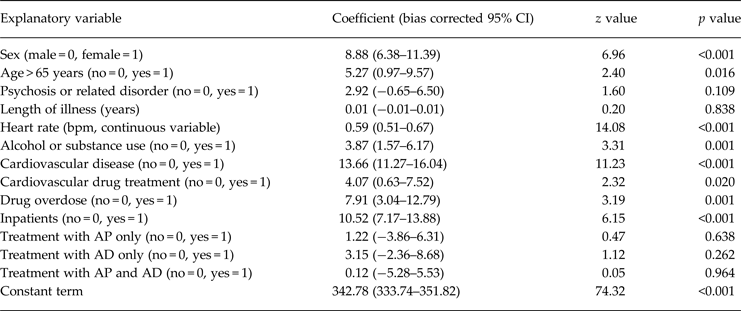
In the whole sample of participants exposed to psychotropic drugs (model 1), linear regression analysis was run to assess the association between QTc interval and the following independent variables: sex (female = 1, male = 0), age (years, continuous variable), psychosis or related disorder (no = 0, yes = 1), length of illness (years, continuous variable), heart rate (beats per minute (bpm), continuous variable), alcohol or substance use (no = 0, yes = 1), cardiovascular disease (no = 0, yes = 1), cardiovascular drug treatment (no = 0, yes = 1), drug overdose (no = 0, yes = 1), inpatients (no = 0, yes = 1), treatment with AP only (no = 0, yes = 1), treatment with AD only (no = 0, yes = 1), and treatment with AP and AD (no = 0, yes = 1).
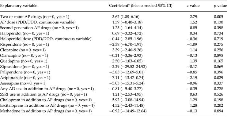
In the sample of participants exposed to AP drugs (model 2), linear regression analyses were run to assess the association between QTc interval and each of the following drug treatment factors: two or more AP drugs (no = 0, yes = 1), AP dose (PDD/DDD, continuous variable), second-generation AP drugs (no = 0, yes = 1), haloperidol (no = 0, yes = 1), haloperidol dose (PDD/DDD, continuous variable), risperidone (no = 0, yes = 1), clozapine (no = 0, yes = 1), olanzapine (no = 0, yes = 1), quetiapine (no = 0, yes = 1), ziprasidone (no = 0, yes = 1), paliperidone, (no = 0, yes = 1), aripiprazole (no = 0, yes = 1), asenapine (no = 0, yes = 1), any AD use in addition to AP drugs (no = 0, yes = 1), selective serotonin reuptake inhibitors (SSRI) use in addition to AP drugs (no = 0, yes = 1), citalopram in addition to AP drugs (no = 0, yes = 1), escitalopram in addition to AP drugs (no = 0, yes = 1), methadone in addition to AP drugs (no = 0, yes = 1). In order to adjust for potential confounding effects of sociodemographic and clinical variables, each of the above-reported drug-treatment factors was analysed together with the covariates from model 1.
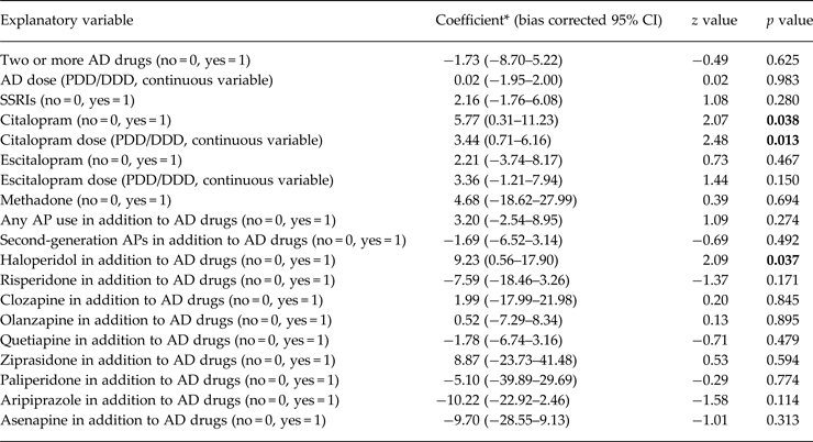
Finally, in the sample of participants exposed to AD drugs (model 3), linear regression analyses were run to assess the association between QTc interval and the following drug treatment factors: two or more AD drugs (no = 0, yes = 1), AD dose (PDD/DDD, continuous variable), SSRI use (no = 0, yes = 1), citalopram (no = 0, yes = 1), citalopram dose (PDD/DDD, continuous variable), escitalopram (no = 0, yes = 1), escitalopram dose (PDD/DDD, continuous variable), methadone (no = 0, yes = 1), any AP use in addition to AD drugs (no = 0, yes = 1), second-generation AP in addition to AD drugs (no = 0, yes = 1), haloperidol in addition to AD drugs (no = 0, yes = 1), risperidone in addition to AD drugs (no = 0, yes = 1), clozapine in addition to AD drugs (no = 0, yes = 1), olanzapine in addition to AD drugs (no = 0, yes = 1), quetiapine in addition to AD drugs (no = 0, yes = 1), ziprasidone in addition to AD drugs (no = 0, yes = 1), paliperidone in addition to AD drugs (no = 0, yes = 1), aripiprazole in addition to AD drugs (no = 0, yes = 1) and asenapine in addition to AD drugs (no = 0, yes = 1). In order to adjust for potential confounding effects of sociodemographic and clinical variables, each of the above-reported drug-treatment factors was analysed together with the covariates from model 1.
A non-parametric bootstrap method of statistical accuracy was used, assuming that the observed distribution of the present sample was a good estimate of the true population distribution (Efron B & Tibshirani R, Reference Efron and Tibshirani1986).
Results
Patient characteristics
During the recruitment period a total of 2411 patients were identified, agreed to participate and were included in the study. No patients refused to participate. The main sociodemographic and clinical characteristics are presented in Table 1. The mean age was approximately 50 years, with an equal distribution between men and women; on average, length of illness was 12 years, and more than one-third had a diagnosis of psychotic disorder. More than two-thirds were recruited as inpatients. The majority was receiving treatment with AP drugs, less than half with AD drugs. On the average, both AP and AD drugs were given at doses very close to the DDD (Table 1).
Table 1. Demographic and clinical characteristics of patients exposed to psychotropic drugs (N = 2411)

AP, antipsychotic drugs; AD, antidepressant drugs; PDD, prescribed daily dose; DDD, defined daily dose.
Prevalence of QTc prolongation
Table 2 reports the prevalence of QTc prolongation in men and women. According to the threshold used, it ranged from slightly less than 20% for the cut-off of 450 milliseconds (ms) to around 1% for the cut-off of 500 ms. As expected, the prevalence was higher in women.
Table 2. Prevalence of QTc prolongation in patients exposed to psychotropic drugs (n = 2411)

ms, milliseconds; CI, confidence interval.
Factors associated with QTc prolongation – univariate analyses
The relationship between the sociodemographic and clinical variables reported in Table 1 and QTc prolongation was initially tested in univariate association analyses. As reported in Table 1, female sex, age, length of illness, heart rate, cardiovascular diseases, drug overdose, inpatients status, treatment with AD, treatment with AP only and AP dose, were all significantly associated with QTc prolongation.
Factors associated with QTc prolongation – multivariate analyses
In the first multivariate model conducted in the whole sample of patients exposed to psychotropic drugs (Table 3), female sex, age, heart rate, alcohol and/or substance abuse, cardiovascular diseases and cardiovascular drug treatment, drug overdose and being inpatients were significantly associated with QTc prolongation (model 1).
Table 3. Factors associated with QTc prolongation in patients exposed to psychotropic drugs (model 1): linear regression analysis (bootstrapped 95% CIs)

CI, confidence interval; bpm, beats per minute; AP, antipsychotic drugs; AD, antidepressant drugs.
After adjustment for the variables included in model 1, we investigated drug treatment factors associated with QTc prolongation (Table 4) in the sample of patients exposed to AP drugs (model 2). Use of two or more AP drugs was positively associated with QTc prolongation, while use of aripiprazole decreased the risk. Use of haloperidol, as well as use of citalopram or escitalopram in addition to AP drugs, was not associated with QTc prolongation.
Table 4. Drug treatment factors associated with QTc prolongation in patients exposed to antipsychotic drugs (model 2): linear regression analysis (bootstrapped 95% CIs)

CI, confidence interval; AP, antipsychotic drugs; AD, antidepressant drugs; SSRI, selective serotonin-reuptake inhibitors.
* Adjusted for the following variables: sex, age, psychosis or related disorder, heart rate, alcohol or substance use, cardiovascular disease, cardiovascular drug treatment, drug overdose, inpatients.
Similarly, after adjustment for the variables included in model 1, we investigated drug treatment factors associated with QTc prolongation (Table 5) in the sample of patients exposed to AD drugs (model 3). Use of citalopram, and citalopram dose, were positively associated with QTc prolongation, as was the case of use of haloperidol in addition to AD drugs.
Table 5. Drug treatment factors associated with QTc prolongation in patients exposed to antidepressant drugs (model 3): linear regression analysis (bootstrapped 95% CIs)

CI, confidence interval; AP, antipsychotic drugs; AD, antidepressant drugs; SSRI, selective serotonin-reuptake inhibitors.
* Adjusted for the following variables: sex, age, psychosis or related disorder, heart rate, alcohol or substance use, cardiovascular disease, cardiovascular drug treatment, drug overdose, inpatients.
Discussion
In a large unselected sample of ordinary practice in- and out-patients, the prevalence of individuals with a QTc above 500 ms was around 1%, while slightly more than 3% had a QTc above 480 ms. These findings are in agreement with recent data collected in adult psychiatric inpatients by Girardin et al., who found that among 6790 psychiatric inpatients 0.9% qualified as long QT case subjects (QTc > 500 ms) (Girardin et al. Reference Girardin, Gex-Fabry, Berney, Shah, Gaspoz and Dayer2013). Other previous surveys reported prevalence rates between 1.2 and 2.6% (Sadanaga et al. Reference Sadanaga, Sadanaga, Yao and Fujishima2004; Ramos-Rios et al. Reference Ramos-Rios, Rrojo-Romero, Paz-Silva, Carballal-Calvo, Bouzon-Barreiro, Seoane-Prado, Codesido-Barcala, Crespi-Armenteros, Fernandez-Perez, Lopez-Morinigo, Tortajada-Bonaselt, Diaz and De2010; Pasquier et al. Reference Pasquier, Pantet, Hugli, Pruvot, Buclin, Waeber and Aujesky2012).
Strengths of this pharmacoepidemiological survey include recruitment of ordinary practice patients employing very wide entry criteria with almost no exclusion criteria, and the multicentre approach which most likely increased generalisability of study findings. The identification of female sex, older age, increased heart rate, alcohol and/or substance use disorders and cardiovascular disorders as correlates of prolonged QTc further corroborates generalisability of study findings, as these are variables known to be associated with ECG abnormalities, including prolonged QTc (Beach et al. Reference Beach, Celano, Noseworthy, Januzzi and Huffman2013).
However, there are limitations that should be considered. First, the cross-sectional design that was employed does not allow generating information about the temporal relationship between drug exposure and QTc prolongation, and therefore no causality assessment can be undertaken. Second, no outcome data were collected, which means that the clinical relevance of QTc prolongation, in terms of probability of torsade de pointes and sudden death, was not assessed. In the study carried out by Girardin et al., for example, it was possible to establish that sudden cardiac death was recorded in five patients, and torsade de pointes in seven other patients (Girardin et al. Reference Girardin, Gex-Fabry, Berney, Shah, Gaspoz and Dayer2013). Although a cut-off at 500 ms is usually accepted as indicative of a high risk for arrhythmic events, the lack of outcome data leaves uncertainty on which QTc threshold is clinically meaningful in terms of risk of ECG abnormalities. We consequently employed different thresholds for descriptive purposes, and QTc values were used as continuous variable in multivariate analyses. Third, the small numbers of patients taking some individual drugs resulted in limited statistical power to detect their possible associations with QTc prolongation, as reflected by wide confidence intervals. Fourth, we did not enrol a control group of individuals who were not receiving psychotropic drugs. Although this would have been useful to have a risk in a comparison population, we argued that in- and out-patients who do not receive any drug treatment would have been too different from our population to act as a reliable comparison group. Last, the choice of using ECG data collected for clinical purposes, which may have introduced some heterogeneity in terms of different centres measuring the QT interval in slightly different ways, was motivated by an attempt to resemble clinical practice as much as possible.
Overall, the relatively low prevalence of QTc prolongation may be interpreted in two different directions. One possibility is that the true risk of ECG abnormalities associated with psychotropic drugs has been over estimated. For example, the citalopram study that prompted safety warnings by regulatory authorities did not measure the proportion of subjects with QTc above a certain threshold, but only reported a mean change from baseline to follow-up in QTc of 7.5 ms with citalopram 20 mg/day and 16.7 ms with citalopram 60 mg/day. Similarly, for escitalopram the change from baseline in QTc was 4.3 ms with 10 mg/day and 10.7 ms with 30 mg/day. Clearly, the clinical relevance of increases of 10–15 ms is questionable, and may also have different meanings depending on baseline values. For antipsychotics, including haloperidol, most data suggested a dose-dependent risk increase, with low doses associated with almost no increase (Reilly et al. Reference Reilly, Ayis, Ferrier, Jones and Thomas2000; Hasnain & Vieweg, Reference Hasnain and Vieweg2014). Considering that in our sample of ordinary practice patients AP doses were on average very close to the DDD of each agent, very little impact on QTc values may be expected.
A second possible interpretation is that the relatively low prevalence of QTc prolongation may be the result of safety warnings. Safety warnings have indubitably increased attention on ECG abnormalities in individuals treated with psychotropic drugs, and therefore those who receive treatment might be those who have been selected for having no ECG abnormalities. In other clinical populations prevalence rates are much higher. In medical inpatients, for example, the prevalence of QTc prolongation may be as high as 22% (Pasquier et al. Reference Pasquier, Pantet, Hugli, Pruvot, Buclin, Waeber and Aujesky2012), whereas in surgical patients QTc prolongation was shown to affect 6% of a sample of elderly surgical patients (van Haelst et al. Reference van Haelst, van Klei, Doodeman, Warnier, De Bruin, Kalkman and Egberts2014). It is also interesting to note that the safety warning for haloperidol, issued in 2007, might have been better integrated into routine clinical practice in comparison with that on citalopram and escitalopram, which was issued more recently, in 2011. This might explain the finding that while use of haloperidol, and haloperidol dose, was not associated with QTc prolongation (which may be the result of careful ECG monitoring before drug use), use of citalopram, and citalopram dose, was found to be significantly associated with QTc prolongation.
In terms of AP-related factors, use of two or more AP drugs significantly increased the risk. This is a very relevant finding as the number of AP drugs is a proxy of total AP dose, which is an established risk factor of QTc prolongation (Sala et al. Reference Sala, Vicentini, Brambilla, Montomoli, Jogia, Caverzasi, Bonzano, Piccinelli, Barale and De Ferrari2005). Another interesting finding on AP drugs refers to aripiprazole, which resulted, in comparison with all other AP drugs, associated with a reduced risk of QTc prolongation. This epidemiological finding is highly consistent with experimental data from placebo and head-to-head comparisons between different AP drugs, recently reviewed by Leucht et al. (Reference Leucht, Cipriani, Spineli, Mavridis, Orey, Richter, Samara, Barbui, Engel, Geddes, Kissling, Stapf, Lassig, Salanti and Davis2013). They found that aripiprazole was not associated with significant QTc prolongation compared with placebo, and that it was the second best choice in terms of risk of ECG abnormalities after lurasidone (not licensed in Italy).
In terms of AD-related factors, we could detect noteworthy differences for citalopram but not for escitalopram. This is unlikely to be a reflection of different safety warning modalities, which were very similar, and should not reflect a power problem, as can be inferred by the confidence intervals around the coefficient estimates. However, it might be possible that the two AD drugs actually bear a different risk of QTc prolongation. This possibility has recently been suggested by Beach et al., who carried out a systematic review of prospective studies investigating the association between SSRI use and QTc values. The review found that the association between citalopram and QTc prolongation was stronger than for most other SSRIs (Beach et al. Reference Beach, Kostis, Celano, Januzzi, Ruskin, Noseworthy and Huffman2014). Castro et al., by contrast, who carried out an analysis of electronic health records, suggested a modest QTc prolongation for both citalopram and escitalopram (Castro et al. Reference Castro, Clements, Murphy, Gainer, Fava, Weilburg, Erb, Churchill, Kohane, Iosifescu, Smoller and Perlis2013).
The confirmation of a link between AP polypharmacy and QTc prolongation supports current guidelines that recommend avoiding the concurrent use of two or more AP drugs, and the confirmation of a link between citalopram and QTc prolongation supports the need for routine QTc monitoring. The relatively low proportion of patients with QTc prolongation suggests compliance with current safety warnings issued by regulatory authorities, but also casts some doubts on the clinical relevance of QTc prolongation related to psychotropic drugs. What still remains to be formally assessed is the cost-utility of such monitoring programmes.
Acknowledgements
The authors wish to thank the STAR network for continuous administrative and scientific support.
Financial support
None.
Conflict of Interest
None.
Ethical Standard
The authors assert that all procedures contributing to this work comply with the ethical standards of the relevant national and institutional committees on human experimentation and with the Helsinki Declaration of 1975, as revised in 2008.
Appendix 1 LIST OF STAR NETWORK GROUP INVESTIGATORS
T. Acciavatti2, A. Adamo3, A. Aguglia4, C. Albanese5, S. Baccaglini6, C. Barbui1, F. Bardicchia7, R. Barone8, Y. Barone9, F. Bartoli10, C. Bergamini11, F. Bertolini1, I. Bighelli1, S. Bolognesi5, A. Bordone12, P. Bortolaso13, M. Bugliani14, C. Calandra12, S. Calò15, G. Cardamone7, M. Caroleo16, E. Carra17, G. Carrà10, D. Carretta10, M. Castellazzi1, L. Chiocchi5, M. Clerici10, M. Corbo2, E. Corsi5, R. Costanzo18, G. Costoloni5, F. D'Arienzo19, S. Debolini5, A. De Capua5, W.A. Di Napoli20, M. Dinelli21, E. Facchi7, F. Fargnoli5, F. Fiori2, A. Franchi5, F. Gardellin22, E. Gazzoletti17, L. Ghio14, M. Giacomin21, M. Gregis23, N. Iovieno24, D. Koukouna5, A. Lax10, C. Lintas23, A. Luca25, M. Luca12, C. Lucii5, M. Lussetti7, M. Madrucci7, N. Magnani7, L. Magni26, E. Manca7, G. Martinotti2 ,C. Martorelli5, R. Mattafirri7, M. Nosè1,G. Ostuzzi1, M. Percudani8, G. Perini28, P. Petrosemolo28, M. Pezzullo7, S. Piantanida13, F. Pinna29, K. Prato8, D. Prestia14, D. Quattrone30, C. Reggianini17, F. Restaino8, M. Ribolsi9, G. Rinosi14, C. Rizzo31, R. Rizzo32, M. Roggi5, G. Rossi26, S. Rossi5, S. Ruberto16, M. Santi6, R. Santoro2, M.S. Signorelli33, F. Soscia7, F. Sozzi1, P. Staffa16, M. Stilo16, S. Strizzolo34, F. Suraniti33, N. Tavian21, L. Tortelli7, F. Tosoni8, M. Valdagno5, V. Zanobini5, and C. Barbui1
1 WHO Collaborating Centre for Research and Training in Mental Health and Service Evaluation, Section of Psychiatry, Department of Public Health and Community Medicine, University of Verona, Verona, Italy (Coordinating site)
2 Department of Neuroscience, Imaging and Clinical Sciences, University of Chieti, Italy
3 University of Palermo, Italy
4 Department of Neuroscience, University of Turin, Italy
5 Department of Mental Health, Siena, Italy
6 Ospedale Villa S. Giuliana, Verona, Italy
7 Department of Mental Health, Grosseto, Italy
8 Department of Mental Health, Garbagnate Milanese, Milan
9 University of Roma Tor Vergata, Italy
10 Department of Surgery and Interdisciplinary Medicine, University of Milano Bicocca, Milan, Italy
11 Department of Medicine, Azienda Ospedaliera Universitaria Integrata, Italy
12 U.O. Complessa Psichiatria Azienda Ospedaliera Universitaria Policlinico Vittorio Emanuele, Catania, Italy
13 DSM Ospedale di Circolo (Varese), U.O. Psichiatria Verbano, SPDC Cittiglio-CPS Luino, Italy
14 Clinica Psichiatrica di Genova, Italy
15 Dipartimento di Salute Mentale, Azienda Sanitaria Locale, Lecce, Italy
16 Unità Operativa Psichiatria, Dipartimento di Scienze della Salute, Università degli Studi Magna Graecia Catanzaro, Italy
17 Dipartimento di medicina diagnostica, clinica e di sanità pubblica, sezione di Psichiatria, Università degli Studi di Modena e Reggio Emilia, Italy
18 Servizio Psichiatrico ULSS 16, Padova, Italy
19 ULSS 12 Veneziana-SPDC dell'Ospedale dell'Angelo di Venezia-Mestre, Italy
20 APSS Trento, UO Psichiatria 2, SPDC Trento, Italy
21 DSM ASL n.9, CSM 2 Treviso, Italy
22 DSM-ULSS n.6 Vicenza-1°u.o. Psichiatria, Italy
23 I Servizio Psichiatrico, Ospedale Civile Maggiore, Verona, Italy
24 SPDC, Ospedale di Legnago, ULSS 21, U.O.C. Psichiatria, Italy
25 Dipartimento “G.F. Ingrassia” Sezione neuroscienze, Azienda Ospedaliera Universitaria “Policlinico Vittorio Emanuele”, Catania, Italy
26 U.O. Psichiatria, IRCCS S. Giovanni di Dio, Fatebenefratelli, Brescia, Italy
27 DSM A.O. Salvini, Garbagnate Milanese, UOP 62, CPS Bollate (MI), Italy
28 IV Servizio Psichiatrico, S. Bonifacio (Verona), Italy
29 Department of Public Health, Clinical and Molecular Medicine - Unit of Psychiatry, University of Cagliari, Italy
30 Section of Psychiatry, Department of Neurosciences, University of Messina, Italy
31 Azienda Provinciale per i Servizi Sanitari, Distretto Centro Sud-U.O. Psichiatria Ambito Alto Garda e Ledro e Ambito Giudicarie, Italy
32 II Servizio Psichiatrico, Ospedale Civile Maggiore, Verona, Italy
33 Clinica Psichiatrica, Dipartimento di Medicina Clinica e Sperimentale, Università degli Studi di Catania, Italy
34 DSM ULSS 6 Vicenza-2° u.o.Psichiatria-2° CSM, Italy


