This study reviews and reassesses the two lungfish Sagenodus species from the UK: the type species of the genus, Sagenodus inaequalis, from the late Carboniferous (=Pennsylvanian); and a second species, Sagenodus quinquecostatus, from the early Carboniferous (=Mississippian) of the UK. Sagenodus was probably the most widespread of the Carboniferous lungfish genera and, consequently, is also one of the best known. It has a large distribution, with material known from Britain, Europe (the Czech Republic, France, and Germany), Russia, and the US. Its ecological diversity was relatively high, being found in freshwater to shallow marine deposits. It has a stratigraphic distribution from the Viséan (early Carboniferous) of the UK to the uppermost lower Permian (=Cisuralian) of the US and Germany (Schultze & Chorn Reference Schultze and Chorn1997). Watson & Gill (Reference Watson and Gill1923) described Sagenodus as being the ‘most completely known lungfish', and Schultze & Chorn (Reference Schultze and Chorn1997) referred to it as representing ‘the beginning of modern lungfish', considering it structurally intermediate between Devonian and post-Palaeozoic lungfishes. Sagenodus inaequalis, as the type species, requires an updated account based on additional material. Sagenodus quinquecostatus was originally named in Reference Traquair1883 by Traquair, but was never fully described or diagnosed. We recognise it here as a separate species from S. inaequalis.
For much of the 19th Century little interest was shown in the structure of the skull roof of lungfishes, the majority of studies being confined to scales and tooth plates. For some time the only well-known skull roof material of any Carboniferous lungfish was that of Sagenodus. Where cranial material was included in descriptions it was largely dismissed as composed of numerous small bones of indeterminate arrangement and doubtful homology, but bearing a considerable resemblance to the skull roof of Dipterus (Watson & Gill Reference Watson and Gill1923).
The genus Sagenodus, not originally clearly distinguished from the genus Ctenodus, was first characterised by Owen (Reference Owen1867) with the type species S. inaequalis. Owen's description, figures, and diagnosis were later claimed by Hancock & Atthey (Reference Hancock and Atthey1868) to be incorrect. Hancock & Atthey (Reference Hancock and Atthey1872) described, but did not figure, the skull roofs of Ctenodus and Sagenodus both as the genus Ctenodus (although they did distinguish differences between them), and both Barkas (Reference Barkas1873a, Reference Barkasb) and Miall (Reference Miall1880) figured, but did not describe, the skull roof of Sagenodus. Sagenodus was also described in some detail by Fritsch (Reference Fritsch1985–1889).
During the 1880s, Traquair (Reference Traquair1881, Reference Traquair1883, Reference Traquair1890) briefly described the remains of a number of different lungfishes from the Blackband Ironstone of Burghlee (formerly Borough Lee), near Edinburgh, Scotland. Among them were tooth plates similar to those described by Hancock & Atthey (Reference Hancock and Atthey1868) as Ctenodus obliquus but sufficiently different for Traquair to propose the variety quinquecostatus. He subsequently elevated this to a new species (Traquair Reference Traquair1890), and although it was never formally diagnosed or figured, when the taxonomy of Carboniferous lungfish was more settled, Traquair (Reference Traquair1903) named the Burghlee material S. quinquecostatus.
Owen's designation of the genus was based upon a cross section of a juvenile palatal tooth plate, 6mm in length and with six distinct ridges. Owen (Reference Owen1867) believed the specimen to represent teeth and a small portion of jaw, and likened his specimen with the extinct Dictyodus (now considered a synonym of Sphyraneodus, a relative of the scombroids; M. Friedman, pers. comm. 2004). He did not associate this specimen with any dipnoan groups, something with which Hancock & Atthey (Reference Hancock and Atthey1868) took particular issue, attributing the specimen to C. obliquus, or ‘perhaps, C. elegans'. While they were correct in asserting that the specimen belonged to a dipnoan, C. obliquus and Ctenodus elegans were eventually realised to be distinct from Ctenodus (Woodward Reference Woodward1891), and the name Sagenodus took precedence. Owen's histological drawings show the distinctive petrodentine now known to be unique to lungfishes, so that, while the specimen cannot be assigned to any particular genus, at least its attribution to a major clade can be confirmed. Owen's type specimen comes from the Low Main Seam at West Cramlington, but both Sagenodus and Ctenodus occur in that seam at Newsham, Northumberland (Sharp & Clack Reference Sharp and Clack2013).
Although Owen did not state on which specimen his description was based, he later presented the specimen (NHMUK (BMNH) P 6246) to the British Museum, as listed by Woodward (72, p. 259). Woodward defined the tooth plates of the genus as follows: ‘Dental plates, above and below, triangular, irregularly ovate or elliptical in form, with few strong, outwardly directed ridges, more or less tuberculated or crenelated; vomerine teeth resembling a single ridge of a dental plate' (Woodward Reference Woodward1891, p. 256).
The description of Sagenodus, regarded by previous authors as probably representing several species, was finally addressed by Watson & Gill (Reference Watson and Gill1923). They gave the first accurate reconstruction of the skull roofs of both Sagenodus and Ctenodus, alongside those of Uronemus and Dipterus. Their reconstruction was a composite based upon a number of specimens and also incomplete in lacking the circumorbital bones. It was, however, based primarily upon a single specimen, NMS G 1878.45.7, a specimen further considered below. Although there are some inaccuracies in Watson & Gill's (Reference Watson and Gill1923) reconstruction, for the most part it is sound. They also illustrated individual bones of the skull, lower jaw, and pectoral girdle. They did not, however, give a detailed description of the assembled bones of the skull roof, stating instead that ‘the figure renders unnecessary a detailed description of the form of the bones' (Watson & Gill Reference Watson and Gill1923, p. 116). They also used a nomenclature that tried to homologise the bones with those of other sarcopterygians. Theirs remains the most recent description of the morphology of the three then known British Carboniferous genera that includes Sagenodus. We reproduce this reconstruction with Forster-Cooper's (Reference Forster-Cooper1937) system of letters, numbers, and lateral line courses added (Fig. 1a).

Figure 1 Left: Sagenodus skull roof reconstruction from Watson & Gill (Reference Watson and Gill1923) with addition of letter and numbering scheme from Forster-Cooper (Reference Forster-Cooper1937). Right: Map of localities yielding Sagenodus specimen of the UK. Black areas indicate coal fields, black spots indicate localities, grey spots indicate major cities.
More recently, Schultze & Chorn (Reference Schultze and Chorn1997) published a detailed description of the American species of Sagenodus based on acid-prepared, although disarticulated, material. They were the first to utilise the large numbers of available specimens to perform statistical analysis on the morphological variation found between the tooth plates of different species.
Our study has examined all the significant specimens of Sagenodus from the UK to provide an up-to-date account of the valid species. We conclude that two valid species can be diagnosed: S. inaequalis from the upper Carboniferous and S. quinquecostatus from the lower Carboniferous.
Institutional abbreviations. GNMHM (NEWHM) G=(Geology) Great Northern Museum: Hancock Museum, UK; KUVP=Kansas University Vertebrate Paleontology, USA; MM=Manchester Museum, UK; NMS G=(Geology) National Museums Scotland, UK; NHMUK (BMNH) P=(Pisces) Natural History Museum, London, UK; UMZC GN = (Gnathostome) or F (Fish), University Museum of Zoology, Cambridge, UK; NUFV=Nunavut University Fossil Vertebrates (currently housed at the University of Chicago, USA); BGS GSM=(Geological Sciences Museum) British Geological Survey, Keyworth, UK.
1. Material and methods
Many of the specimens had been partially prepared in the past, probably with a chisel and often to a very poor standard. This was often worst in type or figured specimens. A number of the specimens were subject to pyrite decay and were, therefore, stored in dry, low-humidity conditions. All preparation took place in the University Museum of Zoology, Cambridge, by ELS (now ELB), or by Sarah M. Wallace-Johnson (formerly Finney) at the University of Cambridge, Department of Earth Sciences, Brighton Building. Preparation included removing excess matrix, making sutures easier to interpret. In some specimens white paint was removed from sutures in order to ease interpretation. Some specimens from the National Museums Scotland had been coated in lacquer, supposedly to protect from pyrite decay. Where the specimens were not subject to decay, with the permission of National Museums Scotland, the lacquer was removed from the specimens using acetone and water.
In some cases, excess matrix was removed using either a pneumatic pen or modified dental mallet, and fine preparation was carried out using the dental mallet or a mounted 1mm tungsten carbide needle. Prepared bone was consolidated using a solution of Paraloid B72 in acetone. For some specimens a Texas Airsonic Inc. Jetsonic BW7 abrasive unit, using sodium bicarbonate, was used to remove a thin layer of sediment to allow surface detail to be prepared.
Drawings of specimens were made either from tracings of photographs or using a Wild stereomicroscope with a camera lucida. All the specimens to be prepared were first photographed in their original state with a Nikon D1 or Canon Powershot A80 digital camera. Photographs were processed and assembled using Adobe Creative Suites Photoshop, version 2017.1.1 or 2019.0.1.
Micro-computed tomography (CT) scanning was carried out on a Nikon Metrology XT H 225 ST High Resolution CT Scanner at the University of Cambridge Biotomography Center. The scan data were as follows: x-axis 606 pixels (slices); y-axis 1558 pixels; z-axis 1998 pixels; resolution 66.1041um; filter 1.5mm copper; X-ray kV 180; X-ray uA 175; projections 1080; exposure 1415. Three-dimensional (3D) segmentation was carried out in Mimics Innovation Suite (biomedical.materialise.com/mimics; Materialise, Leuven, Belgium), and virtual sections of S. quinquecostatus NMS G 1886 82.11 taken from an ImageJ stack.
1.1. Specimens examined
Sagenodus inaequalis
NMS G 1878.45.7. Low Main Seam, Newsham, Northumberland, upper Carboniferous, Duckmantian (Westphalian B). Almost complete skull roof in dorsal view, missing circumorbital bones. Described and figured by Watson & Gill (Reference Watson and Gill1923), cited by Schultze & Chorn (Reference Schultze and Chorn1997).
NMS G 1878.45.13. Low Main Seam, Newsham, Northumberland, upper Carboniferous, Duckmantian (Westphalian B). Isolated parasphenoid.
NMS G 1878.45.21. Low Main Seam, Newsham, Northumberland, upper Carboniferous, Duckmantian (Westphalian B). Composite specimen with pectoral girdle elements.
plates.
NMS G 1888.33.4. Low Main Seam, Newsham, Northumberland, upper Carboniferous, Duckmantian (Westphalian B). Right ceratohyal.
NMS G 1888.33.19. Low Main Seam, Newsham, Northumberland, upper Carboniferous, Duckmantian (Westphalian B). Isolated angular.
NMS G 1897.110.30. Virtuewell Coal Shale, Newarthill, Lanarkshire, upper Carboniferous, Langsettian (Westphalian A). Skull roof in dorsal view with tooth plates.
NMS G 1897.112.5. Virtuewell Coal Shale, Newarthill, Lanarkshire, upper Carboniferous, Langsettian (Westphalian A). Posterior part of skull roof in dorsal view, missing E-bones and rostral regions. Posterior circumorbital bones preserved. Described and figured by Watson & Gill (Reference Watson and Gill1923).
NMS G 1898.17.36. Virtuewell Coal Shale, Newarthill, Lanarkshire, upper Carboniferous, Langsettian (Westphalian A). Skull roof in visceral view and counterpart impression.
NMS G 1898.174.16. Virtuewell Coal Shale, Newarthill, Lanarkshire, upper Carboniferous, Langsettian (Westphalian A). Isolated left cleithrum.
NMS G 1898.154.23 and 24. Virtuewell Coal Shale, Newarthill, Lanarkshire, upper Carboniferous, Langsettian (Westphalian A). Skull roof and parasphenoid stalk, part and counterpart.
NMS G 1898.162.2.1. Virtuewell Coal Shale, Newarthill, Lanarkshire, Upper Carboniferous, Langsettian (Westphalian A). Skull roof and parasphenoid stalk, part and counterpart.
MM L10453. Low Main Seam, Newsham, Northumberland, upper Carboniferous, Duckmantian (Westphalian B). Isolated operculum.
NHMUK (BMNH) P 6246. Holotype. Low Main Coal Seam, West Cramlington, Northumberland, upper Carboniferous, Duckmantian (Westphalian B). Section of a prearticular tooth plate. Described and figured by Owen (Reference Owen1867).
GNMHM (NEWHM) G60.91. Low Main Coal Seam, Newsham, Northumberland, upper Carboniferous, Duckmantian (Westphalian B). Skull roof in dorsal view, posterior portion.
GNMHM (NEWHM) G60.99. Low Main Coal Seam, Newsham, Northumberland, upper Carboniferous, Duckmantian (Westphalian B). Isolated B-bone.
GNMHM (NEWHM) G61.24. Low Main Coal Seam, Newsham, Northumberland, upper Carboniferous, Duckmantian (Westphalian B). Scattered elements including tooth plates, skull roof bones, and opercula.
GNMHM (NEWHM) G61.34. Low Main Coal Seam, Newsham, Northumberland, upper Carboniferous, Duckmantian (Westphalian B). Composite specimen with anocleithrum, operculum, skull bones, and pterygoid with attached tooth plate.
GNMHM (NEWHM) G61.35. Low Main Coal Seam, Newsham, Northumberland, upper Carboniferous, Duckmantian (Westphalian B). Right lower jaw with prearticular tooth plate.
GNMHM (NEWHM) G61.36. Low Main Coal Seam, Newsham, Northumberland, upper Carboniferous, Duckmantian (Westphalian B). Isolated angular and splenial.
GNMHM (NEWHM) G61.44. Low Main Coal Seam, Newsham, Northumberland, upper Carboniferous, Duckmantian (Westphalian B). Isolated parasphenoid.
GNMHM (NEWHM) G61.46. Low Main Coal Seam, Newsham, Northumberland, upper Carboniferous, Duckmantian (Westphalian B). Left ceratohyal.
GNMHM (NEWHM) G172.32. Low Main Coal Seam, Newsham, Northumberland, upper Carboniferous, Duckmantian (Westphalian B). Left pterygoid and attached tooth plate.
GNMHM (NEWHM) G174.78. Low Main Coal Seam, Newsham, Northumberland, upper Carboniferous, Duckmantian (Westphalian B). Articulated posterior portion of skull roof in dorsal view and pectoral girdle elements in internal view.
UMZC GN 168. Low Main Coal Seam, Newsham, Northumberland, upper Carboniferous, Duckmantian (Westphalian B). Left prearticular tooth plate.
UMZC GN 159. Low Main Coal Seam, Newsham, Northumberland, upper Carboniferous, Duckmantian (Westphalian B). Right clavicle.
Sagenodus copeanus
KUVP 103259. Sagenodus copeanus. Hamilton Quarry, Kansas. Virgilian (Gzhelian) Upper Pennsylvanian. Skull roof in dorsal view.
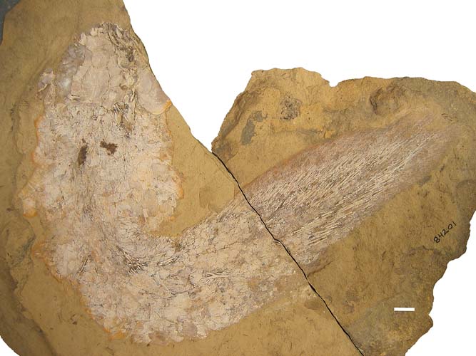
KUVP 84201. Sagenodus copeanus. Hamilton Quarry, Kansas. Virgilian (Gzhelian) Upper Pennsylvanian. Complete specimen in dorsal and lateral view.
Sagenodus quinquecostatus
NMS G 1886 82.11. Burghlee Ironstone, Loanhead district, Scotland, lower Carboniferous, Serpukhovian (Pendleian). Skull roof and partial mandible, including vomerine and other tooth plates.
NMS G 1993.56.118. Dora Bone Bed, Cowdenbeath, Fife, Scotland, lower Carboniferous, Serpukhovian (Pendleian). Right pterygoid tooth plate. Figured by Smithson et al. (Reference Smithson, Challands and Smithson2019).
NMS G 1993.56.115. Dora Bone Bed, Cowdenbeath, Fife, Scotland, lower Carboniferous, Serpukhovian (Pendleian). Right prearticular tooth plate.
NMS G 1993.56.117. Dora Bone Bed, Cowdenbeath, Fife, Scotland, lower Carboniferous, Serpukhovian (Pendleian). Left pterygoid tooth plate.
NHMUK (BMNH) P 11506. Burghlee Ironstone, Loanhead district, Scotland, lower Carboniferous, Serpukhovian (Pendleian). Left pterygoid with tooth plate.
2. Systematic palaeontology
Osteichthyes Huxley, Reference Huxley1880
Sarcopterygii Romer, Reference Romer1955
Dipnoi Müller, Reference Müller1845
Sagenodontidae Jaekel, Reference Jaekel1911
Genus Sagenodus Owen, Reference Owen1867
Type species Sagenodus inaequalis Owen, Reference Owen1867
Synonyms. Ctenodus applantus (partim) Romer, Reference Romer1945; Ceratodus barrandei Frič, Reference Frič1874; Ctenodus caudatus Barkas, Reference Barkas1877; Ctenodus complantus Fritsch, Reference Fritsch1888; Ctenodus elegans Atthey, Reference Atthey1868; Ctenodus ellipticus Atthey, Reference Atthey1868; Ctenodus imbricatus Atthey, Reference Atthey1868; Ctenodus monoceras Barkas, Reference Barkas1873a; Ctenodus obliquus Atthey, Reference Atthey1868; Ctenodus obtusus Barkas, Reference Barkas1870; Ctenodus quadratus (partim) Barkas, Reference Barkas1873b; Petalodopsis miribalis Barkas, Reference Barkas1874; Hemictenodus obliquus Jaekel, Reference Jaekel1890; Sagenodus barrandei Romer, Reference Romer1945.
Other species (after Schultze Reference Schultze1992). Sagenodus applantus Fritsch, Reference Fritsch1879*; Sagenodus carbonarius Romanowsky, 1864*; Sagenodus copeanus Williston, Reference Williston1889*; Sagenodus corrugatus Atthey, Reference Atthey1868; Sagenodus gaudryi Author unknown*; Sagenodus octodorsalis Barkas, Reference Barkas1869; Sagenodus ohioensis Cope, Reference Cope1874*; Sagenodus periprion Cope, Reference Cope1878*; Sagenodus serratus Newberry, Reference Newberry1875.
Sagenodus carbonarius and S. gaudryi are considered here to be nomina dubia due to poor initial descriptions and lack of type material. The asterisks show those species that are not present in the UK, and which will not be considered further.
Diagnosis. Emended diagnosis after Schultze & Chorn (Reference Schultze and Chorn1997). Tooth-plated lungfish attaining a length of approximately 1m. Apomorphic characters: massive B-bone pentagonal or septagonal, often with concave anterior margin, unpaired B-bone pentagonal with the following relations: bounded laterally by the paired I-, J-, and LM-bones, anteriorly by the C-bone. Pentagonal unpaired C-bone, bounded laterally by the LM-bones and anteriorly by the paired E-bones. Anterior to those, a single unpaired F-bone. J-bone does not contact C-bone. Tooth plates with a high length-to-width ratio relative to other genera, and between five and 13 ridges diverging lingually. Prearticular tooth plates narrower than pterygoid plates, with tooth ridge 1 angled dorsomedially, and widely separated from tooth ridge 2. Posteriorly placed ridge on cleithrum separating the branchial and postbranchial laminae.
Remarks. Schultze & Chorn (Reference Schultze and Chorn1997) gave a detailed diagnosis of Sagenodus, after Hussakof (Reference Hussakof1911). There seems to be little inter-specific variation in the structure of the skull roof, and any differences are very subtle. Sagenodus seems to have one of the most invariant skull roofs among Palaeozoic dipnoans. Specific differences are mainly to be found in their dentition. The lack of contact between the J- and C-bones is unusual among Palaeozoic lungfishes.
Type species. Sagenodus inaequalis Owen, Reference Owen1867.
Holotype. NHMUK (BMNH) P 6246. Thin horizontal section of a mandibular tooth plate regarded here as non-diagnostic. Case 3800 was submitted on 30 April 2019 to the ICZN to change the holotype to NMS G 1878.45.7.
Proposed neotype. NMS G 1878.45.7. Articulated skull roof of Sagenodus.
Proposed neotype locality and horizon. Low Main Seam, Newsham, Northumberland, Duckmantian (Westphalian B), late Carboniferous.
Stratigraphic range of genus. Viséan to lower Permian.
Remarks. Tooth plates attributed to Sagenodus by Carpenter et al. (Reference Carpenter, Falcon-Lang, Benton and Henderson2014) from the Tournaisian of the Isle of Bute may not belong to this genus (Smithson et al. Reference Smithson, Richards and Clack2015). The tooth plate from the Tournaisian of Coldstream, Berwickshire, Scotland, thought by Smithson (pers. comm. in Schultze & Chorn Reference Schultze and Chorn1997, p. 57) to be Sagenodus, has recently been named Ballagadus caustrimi (Smithson et al. Reference Smithson, Richards and Clack2015) and the two taxa are probably not closely related.
A number of specimens listed as ‘Sagenodus' were collected from Jarrow colliery in Ireland during the 19th Century, but they have never been examined or described. Specimens NHMUK PV OR41851 (a tooth plate) and NHMUK PV P41684 (B&C bones), and British Geological Survey GSL 1721 (rib and tooth plate) have been identified as Sagenodus, but their specific identity remains to be established.
Sagenodus inaequalis Owen, Reference Owen1867
Differential diagnosis. Differs from S. copeanus in B-bone less posteriorly constricted and J-bones of a more angular and variable shape. Tooth plates never more than eight ridges. Teeth are fused into shearing ridges, with little evidence of individual teeth except in the most labial regions, where there are typically one or two unfused teeth. Pterygoid tooth plates broadly ovate in shape, with a length-to-width ratio of between 1:7 and 2:8, tooth ridge angle between 50° and 70°. Tooth ridges of equal lateral extent. Prearticular tooth plates elongate, with length-to-width ratio of between 2:9 and 3:8, tooth ridge angle c.90°. Ridge 1 is longer than the other ridges. Typically fewer tooth ridges than American species and length-to-width ratio of tooth plates is also less.
Stratigraphic range of species. ‘Middle Coal Measures', upper Carboniferous, Langsettian–Bolsovian (Westphalian A–C) of England and Scotland.
UK localities. Low Main Seam, Newsham, Northumberland (type locality); Newarthill, Lanarkshire; Fenton and Longton, Staffordshire (Figure 1b; see also Panchen Reference Panchen and Kuhn1970 for a map of UK early Late Carboniferous localities).
Remarks. Schultze & Chorn (Reference Schultze and Chorn1997) described the American species of Sagenodus as having tooth plates with the following approximate proportions:
Sagenodus copeanus: upper tooth plates 8–12 ridges, length-to-width ratio 3:8; lower tooth plates 8–10 ridges, length-to-width ratio 4:6.
Sagenodus serratus: upper tooth plates 6–9 ridges, length-to-width ratio 2:6; lower tooth plates 6–9 ridges, length-to-width ratio 3:9.
Sagenodus periprion: upper tooth plates 10–12 ridges, length-to-width ratio 3:7; lower tooth plates 11–13 ridges, length-to-width ratio 4:1.
Sagenodus quinquecostatus Traquair, Reference Traquair1883
Synonyms. Ctenodus obliquus var. quinquecostatus Traquair, Reference Traquair1883; Hemictenodus quinquecostatus Traquair, Reference Traquair1890.
Differential diagnosis. Differs from S. inaequalis in smaller estimated body length up to 0.4m. Pterygoid tooth plates fan-shaped, tooth ridge angle up to c.160°. Length-to-width ratio 3:3. Typically five tooth ridges present, separated by deep grooves; some specimens have an accessory sixth ridge immediately behind the fifth with no groove between. Unworn teeth are small, laterally compressed, and recurved. They are often fused and worn to narrow ridges on the lingual part of the tooth plate. Limited increase in tooth size along the tooth ridges. Prearticular tooth plates long and narrow, tooth ridge angle c.90°. Length-to-width ratio 3:8. Typically four tooth ridges. Tooth ridge 1 long, remaining tooth ridges short, separated by shallow grooves, with little divergence between them and set almost at right angles to tooth ridge 1. Teeth on tooth ridge 1 fused to a worn, sharp blade lacking enamel, bearing two or three teeth at the labial end. Remaining tooth ridges bear up to four small, laterally compressed, recurved teeth showing little wear.
Lectotype specimen. NMS G 1886.32.11 (Figs 2, 3). Skull preserved in dorsal view, showing full complement of skull roofing bones, right operculum, mandibles, and upper and lower tooth plates. Vomerine tooth plate present with three teeth, the central of which is larger.

Figure 2 Sagenodus quinquecostatus. Skull of lectotype NMS G 1886 82.11: (A) photograph of dorsal surface showing skull roof and tooth plates; (B) image from micro-CT scan showing parasphenoid, left, internal (visceral) view, right, external (buccal) view; (C) drawing of holotype skull; (D) close-up of holotype skull showing tooth plates. Abbreviations: L=left; Prart t.p=prearticular tooth plate; Pte t.p=pterygoid tooth plate; R=right; t.p=tooth plate; Vom=vomer. Scale bars=10mm.

Figure 3 Sagenodus quinquecostatus. Slices from micro-CT scans from ImageJ of lectotype NMS G 1886 82.11: (A) slice 42 showing parasphenoid stalk, tooth plates, and unidentified bones; (B, C) slices 54 and 59 showing ribs and unidentified bones; (D) slice 89 showing ribs and fin rays; (E, F) slices 137 and 148 showing fin rays. Slices go from dorsal to ventral horizontally through the specimen. Abbreviations: Psph=parasphenoid; Prart t.p=prearticular tooth plate; Pte t.p=pterygoid tooth plate; R=right; L=left. Scale bars=10 mm.
Type locality and horizon. Burghlee Ironstone (also known as Blackband Ironstone of Borough Lee, Rumbles Ironstone, or Loanhead Ironstone Number 3 (see Smithson Reference Smithson1985; Andrews & Carroll Reference Andrews and Carroll1991; Smithson et al. Reference Smithson, Challands and Smithson2019)), Loanhead (Burghlee Colliery), Midlothian, Scotland. Pendleian, early Serpukhovian, early Carboniferous.
Stratigraphic range of species. Brigantian (Viséan) to Pendleian (Serpukhovian), early Carboniferous.
Other localities. Relatively common lungfish found at four localities in eastern Scottish Midland Valley–Gilmerton, Loanhead, Dora, and Niddrie (Figure 1b; see also Smithson et al. Reference Smithson, Challands and Smithson2019 for a map of late Early Carboniferous localities).
Remarks. From the collections of specimens nominated by Traquair (Reference Traquair1883, Reference Traquair1890) as S. quinquecostatus, we have chosen an almost complete skull with its four tooth plates as a lectotype. The additional specimens become paratypes. We are satisfied that the material represents the genus Sagenodus, but the associated tooth plates differ sufficiently from those of S. inaequalis to be recognised as a separate species. We retain the name given to this species by Traquair (Reference Traquair1883). Sagenodus quinquecostatus occurs in the early Carboniferous, whereas S. inaequalis occurs in the late Carboniferous.
Sagenodus quinquecostatus is the earliest known occurrence of the genus. It displays the diagnostic Sagenodus pattern of skull roofing bones, and is similar to S. inaequalis in parasphenoid shape, lower jaw morphology, and the narrow prearticular tooth plates with angled tooth ridge 1. Only on this ridge are the teeth fused into a sharp blade; the other ridges bear laterally compressed teeth showing little wear. The orientation of tooth ridge 1 suggests that during food processing each mandible could rotate either medially along its long axis (clockwise on the left, anticlockwise on the right), to bring the ridge 1 into occlusion with the ridges 1 and 2 on the pterygoid plate, or the reverse, to bring the more posterior ridges into occlusion with those on the pterygoid. This is a form of kinesis not previously described in lungfish, and is discussed in more detail below.
Sagenodus quinquecostatus is one of six lungfish taxa found in the Burghlee Ironstone (Smithson et al. Reference Smithson, Challands and Smithson2019) and, judging by the number of specimens in the Natural History Museum, London, and National Museums Scotland, was probably the second most common after Ctenodus interruptus. At Dora it is found with Uronemus splendens (Smith et al. Reference Smith, Smithson and Campbell1987) and at Gilmerton and Niddrie it is found with C. interruptus (Henrichsen Reference Henrichsen1972).
As well as an almost complete skull roof with four tooth plates, the specimen also preserves a parasphenoid, an operculum, and some postcranial material revealed by micro-CT scan (Figs 2, 3). The latter consists of a series of ribs at a mid-level in the specimen, and, more externally, an array of fin rays. We find it hard to envisage how this distribution could come about, unless the specimen represented a compacted skeleton that was perhaps enclosed in a burrow at the time of death. We discuss this further below.
3. Description
The skeletal elements described below pertain to both UK species of Sagenodus unless otherwise stated. The tooth plates are described separately, and differ between S. inaequalis and S. quinquecostatus.
3.1. Dermal skull roof
The skull roof of Sagenodus is somewhat square and the bones form a slightly convex surface. The bones of Sagenodus are much less ornamented than those of Ctenodus (Sharp & Clack Reference Sharp and Clack2013), and in most specimens the union between the cranial bones is less secure, although the posterior series are often found in articulation. In many articulated skulls, the ventral lappets that underlie the dermal skull roof are rarely seen. As in many dipnoans, there is a degree of intraspecific variation in the configuration of the skull roof, although Sagenodus seems to be the most consistent.
The most prominent element of the skull roof is the B-bone, the midline element, which lies at the posterior margin of the skull roof and bears the occipital sensory canal. There is no A-bone. Anterior to the B-bone lies the single C-bone. This five-sided element is highly characteristic of the genus, as is the shape of the contact it makes with its surrounding bones. In S. quinquecostatus, the C-bone is relatively wider and the B-bone relatively shorter than in S. inaequalis (Fig. 2). In other respects, the bone relationships are similar as far as can be judged. In front of the C-bone are the paired E-bones, and anterior to this lies the single unpaired F-bone. Lateral to this midline series are the paired I-, J-, and LM-bones. These are similarly large elements and, together, these bones form the majority of the skull roof area. Lateral to the E-bones lie the small N- and P-bones, which carry the supraorbital canal forwards onto the rostral mosaic. Lateral to the I-bone lies the Z-, Y-, and KX-bones. These bones form the lateral margins of the posterior part of the skull roof. The KX-bone bears a prominent notch laterally, continuous with the margin of the Y-bone, where it bears the operculum. A similar feature is seen in Limanichthys, where it occurs on the separate X-bone (Challands et al. Reference Challands, Smithson, Clack, Bennett, Marshall, Wallace-Johnson and ill2019). Anterior to the E-, N-, and P-bones is a series of small elongate bones, which do not conform to a conserved pattern. In those specimens that preserve them, they vary in shape, number, and arrangement; they are sometimes found united together, independent of the named skull roof bones, and are, therefore, termed the ‘prenasal bones' or rostral mosaic.
The circumorbital bones are poorly known from British deposits, although they have been described for the American species S. copeanus, and Schultze & Chorn (Reference Schultze and Chorn1997) claimed the genus had a smaller orbit than reconstructed by Watson & Gill (Reference Watson and Gill1923). The following description of the skull roof of Sagenodus is based on data mainly from NMS G 1878.45.7 (Fig. 4a, b) and a number of others such as NMS G 1897.112.5 (Fig. 4c, d). Because Sagenodus is based only upon the cross section of a tooth plate of uncertain generic identity, we have applied to the International Commission on Zoological Nomenclature to designate NMS G 1878.45.7 as the lectotype of the genus Sagenodus. This constitutes Case 3800 submitted to the Commission.

Figure 4 Sagenodus inaequalis skull specimens. (A, B) NMS G 1878.45.7 proposed neotype: (A) photograph of specimen in dorsal view; (B) interpretive drawing. (C, D) NMS G 1897.112.5: (C) photograph of specimen in dorsal view; (D) interpretive drawing. Scale bar=10mm.
The B-bone is the largest bone of the skull roof and its relative dominance makes it a unique characteristic of this genus. It is roughly rectangular in shape and the bone is constricted in three places: at its posterior point, around its mid-length, and anteriorly. This posterior constriction is less prominent in specimens of S. inaequalis than in S. copeanus. The anterior margin of the B-bone tapers into two curved prongs, and the concave anterior margin so formed could be a derived feature in dipnoans relating to the acquisition of a single C-bone. This is more obvious in NMS G 1878.45.7 (Fig. 4a, b) than in some other specimens (e.g., GNMHM G 60.99, Fig. 5a–d). The contact between the B-bone and C-bone is one of the more variable features within Sagenodus (e.g., Fig. 5e, f). The relative extent of the anterior part of the B-bone compared with the total length of the bone is also variable.

Figure 5 Sagenodus inaequalis skull specimens. (A–D) GNMHM 60.99 B-bone: (A) dorsal view; (B) interpretive drawing; (C) ventral view; (D) interpretive drawing. (E, F) GNMHM 60.91 skull roof: (E) dorsal view; (F) interpretive drawing. Abbreviations: B.vl=ventral lappet of B-bone; l.oa=I-bone overlap; J.oa=J-bone overlap; LM.oa=LM bone overlap. Scale bars=10mm.
The posterior margin of the B-bone is gently convex, curves ventrally, and, in some specimens, there is evidence of an occipital flange projecting posteriorly from its ventral margin, although in NMS G 1878.45.7 (Fig. 4) it is somewhat damaged. The posterior region of the B-bone carries the occipital sensory line canal (occipital commissure of Schultze & Chorn Reference Schultze and Chorn1997). This is typically seen opening as pores on the posterior third of the surface of the B-bone. This is illustrated in Figure 5e, f, although the B-bone is often broken along the path of this canal, which, in some cases, is clearly visible. Pores or pits are often found at the centres of radiation of many bones, and some are probably related to lateral lines passing beneath. Parallel and sub-parallel grooves and ridges radiate from this position to the J- and I-bones, indicating direction of growth. There is considerable variation in the extent to which the path of the lateral line canals can be traced. As in all Palaeozoic dipnoans (Miles Reference Miles1977), the lateral line canals pass through the centre of ossification of the bone and then give rise to a network of tubules, which disseminate throughout the bone.
The B-bone is almost unique for bones of the skull roof in that it has been found isolated, allowing the visceral surface to be seen in relief. Figure 5a–d illustrates GNMHM G 60.99, which shows the visceral surface of the B-bone as significantly different in both shape and anterior extent to that seen from the dorsal surface of the bone. The anterior expansion of the visceral surface exceeds that of the dorsal surface and, laterally, the bone also occupies a slightly greater area. In addition, there are overlap areas at the antero- and posterolateral corners of the bone where the lappets of the adjacent bones such as the I-bone and LM-bone overlap with the B-bone.
It is in the anterior portion of the bone that the discrepancy in extent between dorsal and ventral surfaces is most obvious. The anterior third of the ventral surface of the bone is highly ridged and grooved (NMS G 1898.17.36, Fig. 6a). In those specimens that show an impression of the ventral surface of the skull roof, the suture between the B- and C-bones is very difficult to see clearly (Fig. 6b, c) because the degree of interdigitation is so tight.

Figure 6 Sagenodus inaequalis skull specimen. (A) NMS G. 1898.17.36, inner surface of skull roof. (B, C) NMS 1898.162.2.1: (B) impression of surface; (C) interpretive drawing of same. Scale bars=10mm.
Schultze & Chorn (Reference Schultze and Chorn1997) noted a trough in the mid-sagittal plane of the ventral surface of the B-bone of S. copeanus, which was bordered, to a greater or lesser extent, by two radiating ridges. This depression is also found in specimens of S. inaequalis and, as reported for the North American genus, its bounding ridges are of variable prominence and extent. GNMHM G 60.99 (Fig. 5a–d) has a subtle trough but no evidence of parasagittal ridges, whereas the impression of the skull roof seen in NMS G 1897.110.34 (not figured, ELS, pers. obs. 2006) suggests that the trough was more pronounced. Only in NMS G 1898.162.2.1 (Fig. 6b, c) are the sagittal ridges well developed. It seems likely that these troughs and ridges in life would have contacted the median cristae, by which the braincase would have been suspended from the ventral surface of the skull roof. This cannot be confirmed as the form of the braincase in most post-Devonian dipnoans is not known.
Anterior to the B-bone is the single median C-bone. In contrast with Ctenodus, this bone is unpaired in Sagenodus, a derived feature within the lungfishes. It is slightly larger than half the size of the B-bone and, where the E-bones are preserved, the C-bone is slightly longer. While the junction between the posterior and lateral margins is fairly smooth, that between the anterior and lateral margins is at a distinct angle. The lateral margins taper slightly posteriorly, meaning that the bone is broader anteriorly. Again, the overall shape of the bone in S. inaequalis does seem much more variable than that described for S. copeanus, some examples being more rounded in their proportions (Fig. 4c, d) while others are longer than they are broad (e.g., Fig. 4a, b). In keeping with the B-bone, the posterior margin of the C-bone is convex, although the degree of curvature varies. The anterolateral margin forms an obtuse angle and the anterior margin forms a similar angle, which nestles between the posteromedial margins of the paired E-bones.
There are pores visible at the centre of the C-bone in some specimens, as well as lines from the centre of radiation, which straddle the boundary between the B- and C-bones. The visceral surface of the bone is seen in NMS G. 1898.17.36 and NMS 1898.162.2.1 (Fig. 6). Although there is a tightly knit association between the B- and C-bone, the contact between the two bones is difficult to define in that view (Fig. 6). There is no sufficient evidence of the pit for the pineal organ described by Schultze & Chorn (Reference Schultze and Chorn1997).
The E-bones lie anterior and slightly lateral to the C-bone. Together, these paired elements occupy the same width of the skull roof as the single B-bone and are approximately five-sided, although the shape of the anterior margin is extremely variable. It is not common for the E-bones to be preserved. Only two specimens (Figs 6, 7) show them in articulation with the rest of the skull roof, although they are quite fragmentary. These paired bones meet in a straight line medially and then diverge posteriorly, as they are separated by the anterior portion of the C-bone. Both the posteromedial and posterolateral margins are fairly straight in dorsal view. The lateral margin has two embayments to receive, posteriorly, the N-bone and, anteriorly, the P-bone. The anterior margin also receives the ‘prenasal bones' of Watson & Gill (Reference Watson and Gill1923, fig. 1, p. 164) and, centrally, the single F-bone. Watson & Gill (Reference Watson and Gill1923, p. 167, fig. 3) illustrated an isolated bone that they considered as an E-bone (their ‘nasal'). The E-bones carry branches of accessory canals from the LM-bones; accessory branches also pass from the E-bone into the anterior prenasal bones (Fig. 4).
Anterior to the E-bones are the prenasal bones. Only one specimen has these bones in articulation with a partially complete skull roof (NMS 1878.45.7, Fig. 4a, b). Watson & Gill (Reference Watson and Gill1923, p. 167, fig. 3) also illustrated what they considered to be isolated or partially isolated prenasal bones of Sagenodus from Newsham, although there is no way to associate these bones unequivocally with Sagenodus, because Ctenodus also occurs at this locality. The prenasal bones and the F-bone carry the supraorbital canal, evidenced by the presence of a series of sensory pores visible on the dorsal surface. They have been reported as being fused in some specimens (Watson & Gill Reference Watson and Gill1923) or replaced by a single bone in one specimen of S. copeanus (Schultze & Chorn Reference Schultze and Chorn1997).
The extent of these rostral bones can be quite large, as shown by a specimen of S. copeanus from the upper Pennsylvanian Hamilton Quarry in Kansas (Fig. 7). This specimen lacks the lateral bones of the skull roof, but it is clear that the skull has a considerable anterior extent, making it appear longer than it is wide, in contrast to square skull roof typically described for Sagenodus (Schultze & Chorn Reference Schultze and Chorn1997). Not only does the specimen illustrate the marginal N- and P-bones, but it also illustrates the crescentic O-bone, not known in S. inaequalis. In addition, it is clear that there are multiple rows of small elements present anterior to the E-bones, and that more than one row of these bones carries the sensory pores of the supraorbital canal.

Figure 7 Sagenodus copeanus skull roof. KUVP 103259 with bone lettering and numbering applied. Scale bar=10mm.
Lateral to the B-bones lie the paired I-bones (‘tabulars' of Watson & Gill Reference Watson and Gill1923). These are relatively large bones, smaller only than the B- and LM-bones (although in S. copeanus, the I-bones are consistently larger than the LM-bones). These elements form most of the posterior width of the skull roof and only the lateral corner of the posterior margin is occupied by bone Z. As with the B-bone, the I-bones also have a ventral occipital flange on the posterior margin of the skull, which articulates with the anocleithrum (‘tabular horns' of Watson & Gill Reference Watson and Gill1923). While in NMS G 1878.45.7 (Fig. 4) this flange is equal in posterior extent to that of the B-bone, in most specimens it is usually large and extends further in a posterior direction (Fig. 5e, f), though they are never as extensive or of the same size as those found on the I-bones of Ctenodus.
Each I-bone is approximately equal in width to the B-bone, and at maximum length extend forward for more than half the length of this median element. The anterior margin of the I-bone begins at the most lateral extent of the B-bone and turns laterally at about 90°. It continues for about half the width of the bone and then turns forward at an oblique angle. The most anterior point of the I-bone is where it meets the posteromedial corner of the KX-bone and the posterolateral corner of the J-bone. From this point the margin follows that of the KX-bone until it meets the Y-bone, where it turns posteriorly (Fig. 4). The lateral margin of the I-bone is undulating, with two shallow embayments to receive the Y- and Z-bones. Although the specimens of I-bones are broadly similar, the shape of the anterior margin varies between specimens and within a single specimen, from almost straight to undulating.
Continuing from B-bone, the I-bones carry the occipital commissure, which traverses the posterior third of the bone into the Z-bone (Fig. 8). There are numerous pores on the surface of the posterior half of the bone (Fig. 4). The bone also has branches of the occipital lateral line shown in Figure 8e, a composite reconstruction based on the part and counterpart specimens NMS 1898.154.23 and 24. Overall, the I-bone of Sagenodus is much larger and more prominent a member of the skull roof than the equivalent bone in Ctenodus.

Figure 8 Sagenodus inaequalis skull specimens. (A, B) NMS G 1897.110.30: (A) photograph of skull roof; (B) interpretive drawing. (C–E) NMS G 1898.154.23/24 part and counterpart: (C, D) visceral skull roof in part and counterpart; (E) interpretive drawing. Abbreviations: c.occ=occipital canal; pl.p=posterior pit line; Rmd=right mandible; Lmd=left mandible; RPte=right pterygoid; LPte=left pterygoid; pn=prenasal bones; Rt.p=right prearticular plate; Lt.p=left prearticular plate; c.so=supraorbital canal. Scale bars=10mm.
Anterior to the I-bones are the paired J-bones (‘intertemporals' of Watson & Gill Reference Watson and Gill1923). Of the major bones of the skull roof, these seem to be the most variable in their shape and relationship with the surrounding bones. Those in S. copeanus are described as being ‘shield shaped' (Schultze & Chorn 55, p. 22) and resemble a lozenge, but those of S. inaequalis are more rounded and vary considerably in the extent to which they penetrate the KX- and LM-bones (Figs 4, 8). Medially, they occupy the anterior half of the lateral margin of the B-bone, which they contact via a curved margin. In anterior extent the J-bones can be slightly shorter, equal in length, and, in some cases, extend further forward than the B-bone (Fig. 4).
Posteriorly, the contact with the I-bones can be tightly interdigitated or a simple contact. Laterally, the margin of the bone bows outwards to form an angular convex margin with the medial margin of the KX-bone. Similarly, the anterior margin forms an obtuse angle with the posterior margin of the LM-bone and it is these lateral and anterior margins that are the most variable. For example, in NMS G 1878.45.7 (Fig. 4) the lateral margin forms a gentle curve with the KX-bone and a sharp prong penetrates the LM-bone, whereas GNMHM G 60.91 (Fig. 5e, f) demonstrates a lateral margin which invades the KX-bone to a much greater extent. The J-bones of NMS G 1897.112.5 (Fig. 4) are sub-circular and penetrate very little into the LM-bones.
Although there are no isolated examples of J-bones, evidence from impressions of the visceral surface of the skull roof suggests that the ventral anterior and lateral extent of the bones is somewhat greater than that seen in dorsal view. In some specimens the J-bone carries an extension of the supraorbital canal from the KX-bone.
The J-bone represents a major difference between the morphology of Sagenodus and Ctenodus. As noted, Sagenodus has a single median C-bone, in contrast with the paired C-bones of Ctenodus. In Ctenodus, the J-bones contact the C-bone over a large area. This is not the case in Sagenodus: here the J-bones and C-bone are prevented from contact by the large anterior extent of the B-bone, which is in contact with the LM-bone. Whether this is a result of the increased size of the B-bone, or a result of the loss of paired C-bones, is impossible to know.
The LM-bones are the second largest elements in the skull roof of S. inaequalis. They are three-lobed and bounded by the B-, C-, E-, J-, KX-, N-, 3a-, and 3b-bones. The largest of the three lobes points posteromedially and is bounded by the J- and C-bones, forming a junction with the B-bone. The anterior lobe meets the E-bone and the posterolateral lobe meets the KX-bone. Posteriorly, the bone is embayed where it receives the J-bone (Fig. 4). Laterally and anteriorly the bone is bordered by the N-bone and two bones of the orbital series, 3a and 3b; these bones are contacted via three shallow embayments, which give this margin a variable and zig-zig appearance. The only specimen that illustrates these contacts in full is NMS G 1897.112.5 (Fig. 4c, d).
The supraorbital canal is prominent in the LM-bone; large pores offset from centre towards the KX-lobe are visible. In larger specimens these pores are raised relative to the rest of the bone (Fig. 4c, d). Specimens NMS G 1897.110.34 and NMS G 1898.154.23/24 (Fig. 8) show that the supraorbital line traverses the LM-bone and that from it the lateral line passes into the N-, KX-, J-, and B-bones, where it splits into the postorbital and infraorbital canals.
Westoll (Reference Westoll, Jepsen, Mayr and Simpson1949) described a specimen lacking the L-portion of the LM-bone, probably NHMUK (BMNH) P 7719, which has an extra bone between the Y- and LM-bones. There are some lateral line pores on this accessory bone, suggesting that it could indeed be a separate L-bone. It is not unusual for the skull roofs of dipnoans to be polymorphic, though it is less common in the post-Devonian dipnoans.
The N-bone is the most anterior bone in the lateral series that can be readily characterised, although it is only known from a minority of specimens of S. inaequalis, being more commonly preserved in the American S. copeanus. Only two specimens with the N-bone in articulation with other skull roof bones have been examined in the course of this study (Figs 4c, d, 8a, b). The posterior margin of the N-bone forms a V between the 3a- and LM-bones, the margins with both being gently undulating and convex with respect to the N-bone. Medially, it is bordered by the E-bone and fits into the posterior of two notches on the lateral margin. The anterior margin of the N-bone is straight, as far as it is possible to tell, at its junction with the P-bone.
The N-bone carries the supraorbital canal forwards onto the P- and rostral bones, confirmed by the presence of sensory pores at the centre of the bone. Both Westoll (Reference Westoll, Jepsen, Mayr and Simpson1949, p. 151, fig. 8A) and Schultze & Chorn (Reference Schultze and Chorn1997, p. 18, fig. 3) illustrated the N-bone as being entirely separated from the orbital margin by the 3a-bone. This does not appear to be the case in NMS G 1897.112.5 (Fig. 4c, d), in which the 3a- and N-bones are of roughly similar size and shape. Because the areas lateral to these bones are not present on either specimen, it is not possible to comment on the status of the anterior orbital bones.
The P-bones lie anterior to the N-bones. The presence of these bones is suggested by two distinct notches in the lateral margin of the E-bones, the posterior of which accommodates the N-bone and the anterior P-bone. Unfortunately, there is no fully articulated evidence of the P-bone in any specimens of S. inaequalis. NMS G 1897.110.30 (Fig. 8) has a bone preserved anterior to the N-bone, but it is slightly disarticulated. The bone is slender, with a length 1.5 times that of its width. It is a little constricted about its centre, but has a posterior margin which fits roughly with that of the N-bone behind it. The medial margin seems straighter than might be supposed by examining the E-bone.
Lateral to this bone is a slightly shorter and stouter element of unknown identity. It is probable that it is the O-bone but, as this bone is unknown in all other specimens of S. inaequalis, it cannot be confirmed. This identification is supported by its position anterior to the N-bone and in close association with the P-bone. The alternative bone, which might be seen to lie lateral to the P-bone, is the 3a-bone, although it would be expected for the bone to be placed equidistantly between the N- and P-bones. The lack of information regarding these anterolateral skull roof bones means that it is useful to examine the conditions found in specimens of other species of Sagenodus, in particular that of the better known S. copeanus.
Observation of S. copeanus (KUVP 103259, Fig. 7) illustrates a distinct individual bone occupying the anterior half of the E-bones and lying lateral to them, probably a P-bone. This bone is twice the length of the N-bone and slightly wider than its neighbour. Anteriorly, it is associated with two ‘prenasal bones', and, laterally, it contacts the O-bone. Posteriorly, it has a small area of contact with orbital bone 3a, comprising half of the anterolateral and lateral margin of this bone, together with the N-bone. The P-bone carries the supraorbital canal forwards into the ‘prenasal' bone, and this is supported by the presence of sensory pores on its surface in this specimen.
The Z-bones are the most posterolateral bones in the dermal skull roof of Sagenodus, forming the back corners of the skull roof of this genus (Fig. 4a, b). The Z-bones are small but thick elements of a generally rounded shape, having a convex, crenelated posterior margin and a pitted dorsal surface. The Z-bones nestle between I- and Y-bones. It is through the Z-bones, on their ventral surface, that the main lateral line canal enters the skull and bifurcates into two branches. That passing medially, and parallel with the posterior margin of the skull roof, is the occipital commissure; that passing anteriorly through the marginal bones of the skull roof is the cephalic division of the main lateral line.
Because of the bulk of the lateral line canal, the dorsal surface of the Z-bones are pocked with numerous sensory pores. There are no known examples of the ventral surface of a Z-bone of S. inaequalis so it is not possible to comment on its morphology. Personal observation (ELS, 2006) of specimens of S. copeanus confirms the description given by Schultze & Chorn (Reference Schultze and Chorn1997), which stated that the sensory canals were carried on the ventral surface enclosed within bone. This gives the ventral surface of the bone the appearance of a flat surface with a Y-shaped cylinder on it.
The Y-bone lies anterior and slightly lateral to the Z-bone, forming part of the lateral margin of the skull roof. It is generally trapezoid in shape, its lateral margin being the longest (Fig. 4). As described, it articulates posteriorly with a Z-bone and this margin is concave with respect to the Y-bone. Medially, the border with the I-bone is straight and the union between the two takes up the anterior half of the I-bone. The anterior margin of the Y-bone is formed by the junction with the KX-bone, and this is a straight but oblique border, moving anterolaterally. Thus, the most anterior extent of the Y-bone is pointed, but also somewhat laterally placed. The lateral margin, crenelated as in the B-bone, is gently convex. It passes posterolaterally, and then posteriorly in a curve, which is confluent with the lateral margin of the X-bones.
The anterior portion of the lateral margin is confluent with the notch in the KX-bones, which receives the tabulate process of the operculum. On the dorsal surface the bone is highly pitted and dimpled (Fig. 4), evidence of the high density of sensory canal elements present in this bone. The ventral surfaces of the Y-bones are poorly known for S. inaequalis, but the lateral extent seems to be greater in ventral view, presumably forming a region of overlap for the operculum. Observation of the visceral surface of the Y-bone of S. copeanus shows a distinct bony structure running anteroposteriorly down the mid-point of the bone, which exceeds both the anterior and ventral extent of the dorsal surface: it would have contained the main sensory line.
The KX-bones (‘squamosal', Watson & Gill Reference Watson and Gill1923; ‘X-intertemporal', Ahlberg Reference Ahlberg1991; ‘X+Y2', Westoll Reference Westoll, Jepsen, Mayr and Simpson1949) are three-lobed and similar in shape and orientation to the LM-bones, although they are smaller (Fig. 4). They form the most lateral extent of the skull table, articulating laterally with the tabulate process of the operculum. The three lobes have straight, truncated distal margins, and the posterior lobe fits between the J- and Y-bones. This lobe has a straight margin with the I-bone (Fig. 4). All margins are smooth with little interdigitation. Posterolaterally, there is a distinct notch in which the operculum is housed, and the lateral border also abuts this bone with a concave boundary. The lateral margin of KX- is posteriorly excluded from the lateral wall of the skull by the anterior projection of Y-, although these bones form a smooth notch in the skull wall.
Anterolaterally, the KX-bone meets the orbital bone 4 in a straight margin, although this bone is not known in articulation for S. inaequalis. Anteriorly, there is a concave margin into which fits the 3b-bone, and, anteromedially, the bone contacts the LM-bone. The medial contact with the J-bone occupies the entire lateral extent of J- and this contact is angular with the margin of the KX-bone, forming a concave surface of more than 90°.
The KX-bone is the point of bifurcation of the lateral line ventrally into the infraorbital lateral line canal, and continuing anteriorly into the LM-bone as the supraorbital lateral line canal. Accessory canals also penetrate into the J-bones (Fig. 5), something which is characteristic of the K-bone in Dipterus (Forster-Cooper Reference Forster-Cooper1937; Westoll Reference Westoll, Jepsen, Mayr and Simpson1949), hence its identification with the X-bone here. NMS 1898.17.36 (Fig. 6) shows a ventral thickening, which suggests that the lateral line was buried deep within the KX-bone. There are numerous pores on the dorsal surface, which are set slightly off-centre towards the opercular notch (Fig. 4).
3.2. Orbital region
Until the description of S. copeanus, the orbital region of Sagenodus was very poorly known. This is still the case for S. inaequalis, with few circumorbital bones known in detail and only two known in articulation with the skull roof – i.e., the dorsal elements 3a and 3b. The specimen that preserves these elements best is NMS G 1897.112.5 (Fig. 4c, d), although 3a is seen in NMS G 1897.110.30 (Fig. 8a, b). Even in the well-known North American species the reconstruction of the orbit from Schultze & Chorn (Reference Schultze and Chorn1997) was based on isolated elements, making any reconstruction tentative. There is no evidence of cheek bones in any specimens known for this genus and they are, therefore, assumed not to have been preserved.
Of the two orbital bones known with certainty for S. inaequalis, 3a and 3b are both in articulation with the LM-bone (Fig. 4c, d). The posterior element, the 3b-bone, is associated along its posterior margin with the KX-bone and is concave here. It seems that the union between these bones was not strong and they are usually disarticulated. The sutures seem to lack interdigitation and are found with some matrix between the bones. The medial margin meets the LM-bone and is broadly straight and anteromedially directed. The area around this junction is elevated relative to the more proximal areas of the bones, and this is seen, to a smaller degree, on the sutures between the other orbital elements and their neighbours. Although the dorsal surface of the bone is obscured by matrix in NMS G 1897.112.5 (Fig. 4a, b), there is a pitted and rugose surface, which becomes depressed laterally, and probably represents overlap areas for the adjacent 3-bones.
The anteromedial margin of the 3b-bone meets the 3a-bone in an oblique, straight junction. It is the anterodorsal element of the skull roof and was described by Westoll (Reference Westoll, Jepsen, Mayr and Simpson1949, p. 151, fig. 8) as bone 2. Its medial margin fits within a concavity in the LM-bone and is consequently angularly convex. The anteromedial margin meets the N-bone and this border is undulating. The anterolateral margin, presumably with the 2-bone, is oblique and seems straight in its most anterior portions, turning posteriorly and becoming confluent with the lateral margin of the 3b-bone. The 3a-bone is also pitted and grooved in the visible areas. Both the 3a- and 3b-bones carry pores of the sensory canal, the 3b-bone carrying a short dorsal branch of the infraorbital sensory canal. GNMHM G 60.91 (Fig. 5e, f) suggests that the 3a-bone carries a small accessory branch of the supraoccipital canal, which enters it from the LM-bone.
The size and position of the left 3a-bone in NMS G 1897.112.5 is somewhat unusual. In other reconstructions of the species, as well as in examined specimens of S. copeanus, S. serratus, and S. ohioensis, the anterior extent of the 3a-bone causes it to sit lateral to the N- and P-bones, thus excluding them from the orbital margin (Fig. 4c, d). In NMS G 1897.112.5, however, the N-bone is larger than reconstructed elsewhere, and the 3a-bone is offset posterolaterally, with its anterior point reaching only to the midpoint of the N-bone. Although the lateral margin of the 3a-bone is broken, there is no reason to suppose that much bone is missing. It seems that Sagenodus is the only known dipnoan in which the dorsal margin of the orbit is composed of two bones, both distinct from those at the anterior and posterior orbital margins.
The other bones of the orbit are unknown in articulation with skull roof material of S. inaequalis; however, there is little reason to suppose that the orbital region of S. inaequalis is significantly different from that of S. copeanus or S. serratus (Schultze & Chorn Reference Schultze and Chorn1997).
3.3. Palate
The palate of Sagenodus, as in Ctenodus (Sharp & Clack Reference Sharp and Clack2013), is composed of paired pterygoids, the upper tooth plates, and the median parasphenoid. Quadrates are not preserved. In addition, there is evidence of the presence of vomerine tooth plates in S. quinquecostatus (see below). The tooth plates will be described separately below.
The pterygoids of Sagenodus conform closely to the form of those seen in Ctenodus, and good examples of pterygoids of Sagenodus and its associated tooth plate are shown in Figure 9. Anteriorly, the medial margin is sub-parallel with that of the tooth plate, where the two pterygoids articulate in the midline of the palate. Posterior to this, the medial margin turns laterally at an angle of approximately 140°, and from this point the two pterygoids are separated by the parasphenoid.

Figure 9 Sagenodus inaequalis pterygoids and associated tooth plates. (A, B) GNMHM G 61.34 left pterygoid: (A) photograph; (B) interpretive drawing. (C) Drawing of tooth plate showing wear pattern. (D, E) GNMHM G 172.32: (D) photograph; (E) interpretive drawing. Abbreviations: Pte.t.p=pterygoid tooth plate; r.i.acc = accessory ridge; t.r.1=tooth ridge 1; t.r.7=tooth ridge 7. Scale bars=10mm.
The posterior portion of the pterygoid is offset laterally with respect to the tooth plate and is a characteristic ‘delta-shape'. The medial region here has an ‘unfinished' surface, and at this point the pterygoid would have been underlain by the parasphenoid. The pterygoids of Sagenodus have a less curved medial margin than those of Ctenodus (Sharp & Clack Reference Sharp and Clack2013). Anterior to this, however, the body of the parasphenoid would overlie the paired pterygoids to a degree. Lateral to this region is the quadrate ramus, and although it is always preserved in a single plane, in life this lateral corner of the pterygoid would be angled ventrally and abut the quadrate. The anterior portion of the pterygoid near the midline of the palate seems quite thin (although the bones are not known in isolation for the British genera), but posteriorly the quadrate ramus is very robust.
The parasphenoid of Sagenodus is relatively smaller and less extensive than that of Ctenodus. Again, it is divided into the distinct corpus and stem, and in Sagenodus the stem is narrower than that of Ctenodus (Sharp & Clack Reference Sharp and Clack2013), and the lateral margins are scarcely flared. At their point of contact there is a slight constriction of the stem. The parasphenoid of Sagenodus is illustrated in buccal view in Figure 10a, b. There were no available specimens that preserve the visceral surface of the parasphenoid for S. inaequalis, and that in Figure 10c is of S. copeanus. The parasphenoid of S. quiquecostatus is similar as far as can be seen (Fig. 2b) from micro-CT scan reconstructions, except that the stem has much more extensive lateral flare on the margins and a much more strongly tapering component on the visceral surface of the corpus.

Figure 10 Sagenodus spp. parasphenoids. (A, B) Sagenodus inaequalis NMS G 1878.45.13 parasphenoid in buccal view: (A) photograph; (B) interpretive drawing. (C) Sagenodus copeanus KUVP 70738 parasphenoid in visceral view. Abbreviations: Psph=parasphenoid; c.Psph=corpus of the parasphenoid; m.d.Psph=median depression of the parasphenoid; m.r.Psph=median ridge of the parasphenoid on the buccal surface; Pte.oa = pterygoid overlap area; s.Psph=stalk of the parasphenoid; m. ri=median ridge on the visceral surface. Scale bars=10mm.
The corpus is roughly diamond shaped, with somewhat concave lateral margins. The anterior margin of the corpus makes an angle of between 85° and 90°, and the anterior tabulate process has a gently curved anterior margin. Similarly, the lateral angles of the parasphenoid corpus are at approximately 90°, making the body more square than described for S. copeanus (Schultze & Chorn Reference Schultze and Chorn1997). The margins of the corpus are irregular and ‘unfinished' at the area of overlap with the pterygoids; the surface of the tabulate process bears anteroposteriorly oriented ridges.
The buccal surface of the parasphenoid varies in its texture: some specimens show a smooth surface, others are broadly smooth but ornamented with pits and grooves, something that is prominent in GNMHM G 61.44, which has an area of dense pustulate ornament on the posterior half of the corpus. The lateral ‘wings' of the corpus are less ornamented, where the pterygoids would overlap (Fig. 10). The overlapping pterygoids would arch ventrally, allowing the quadrate ramus to contact the quadrate. Similarly, the lateral angles of the parasphenoid arch downward, although the preservation does not permit this to be seen. Instead, many specimens of parasphenoid are broken in the lateral regions.
Where the stem and the corpus meet there is a ridge, as the posterior corner of the corpus is elevated relative to the rest of the bone. This ridge thins anteriorly and continues forward onto the corpus for a third of its length, and is bordered laterally by two depressions. This is in contrast to the prominent ridge of the corpus seen in Ctenodus (Sharp & Clack Reference Sharp and Clack2013). The median ridge in S. copeanus forms a smooth transition with the parasphenoid stem.
The stem of the parasphenoid makes up almost half of the total length of the bone, and is ornamented with small pits and grooves parallel to the orientation of the stem. Its margins flare laterally for part of its length, after which they begin to converge again, meeting in a rounded posterior termination. The most conspicuous feature of the parasphenoid is the median depression, bordered by parallel ridges (Fig. 10). This depression is not seen to an equal extent in all specimens but is present, to some degree, in all of those studied.
The visceral surface of the parasphenoid is not known for S. inaequalis but is known for other species. A poorly preserved example is seen in S. quinquecostatus (Figs 2, 3). The anterior portion of the stem of S. copeanus (ELS, pers. obs. 2006) bears very prominent ridges on the lateral margins, which continue onto the corpus and converge near the centre. The stem is characterised by a number of somewhat radiating ridges, running anteroposteriorly, for most of its length (Fig. 10). It is interesting to note that the parasphenoid stem of S. copeanus is longer in relation to the body than seen in S. inaequalis. Similarly, the anterior stem is more flared than found in S. inaequalis but similar to those seen in Ctenodus (Sharp & Clack Reference Sharp and Clack2013) and S. quinquecostatus.
3.4. Lower jaw
The lower jaw of Sagenodus comprises three paired bones, angular, splenial, and prearticular, and the paired tooth plates. Laterally, the angular is a curved bone that tapers anteriorly and articulates with the splenial. Medially, the tooth plate is borne on the prearticular. Both the mandibular and oral lateral lines are carried on the lower jaw. It is very uncommon for these three elements of the lower jaw to be found in articulation and, for S. inaequalis, such preservation has not been observed.
The angular is the largest element of the lower jaw and is situated laterally in the mouth, forming most of the external area (Figs 11, 12). It is a curved triangle in shape, being deepest posteriorly and tapering to a truncated point anteriorly. Its posterodorsal margin is divided into two areas by a subtle angle on the margin. The most posterior region of this margin forms a gentle and shallow notch into which the unossified articular would have fitted. Moving dorsally from here is a second region, found as a notch in some specimens or as a plain surface in others. This margin leads to the highest point of the angular, the ascending process situated at approximately one quarter of the length of the bone. From here, the dorsal margin of the bone descends, steeply at first, and then in a more shallow concave arc, tapering to its most anterior point. This margin is notably more curved than the dorsal margin of the angular of Ctenodus (Sharp & Clack Reference Sharp and Clack2013).

Figure 11 Sagenodus inaequalis lower jaw elements. (A, B) NMS G 1888.33.19 right angular in lateral view: (A) photograph; (B) interpretive drawing. (C, D) GNMHM G 61.36 right angular and splenial in lateral view: (C) photograph; (D) interpretive drawing. Abbreviations: Ang=angular; add.m.ar=area of attachment for the adductor mandibulae; pr.asc.Ang=ascending process of the angular; c.o=oral sensory canal; f=foramen; r.Ang=ridge of angular; Spl=splenial. Scale bars=10mm.

Figure 12 Sagenodus inaequalis lower jaw elements. (A–D) GNMHM G 61.35 right angular, prearticular, and prearticular tooth plate in lateral view: (A) photograph; (B) interpretive drawing; (C, D) specimen in lingual view: (C) photograph; (D) interpretive drawing. Abbreviations: Ang=angular; glen.a=area of glenoid; fu.Ang=furrow of the angular; md.av.m=path of the mandibular ramus of the anteroventral lateral line nerve; mk.c.r=area that receives the Meckelian cartilage; Prart=prearticular; Prart t.p=prearticular tooth plate; sym=symphysial region of mandible; v.r.Ang=ventral ridge of the angular. Scale bar=10mm.
The dorsal margin of the bone anterior to the ascending process is the region for the attachment of the adductor mandibulae. Schultze & Chorn (Reference Schultze and Chorn1997) described some specimens of S. copeanus with a deep pit in this region. GNMHM G 61.36 (Fig. 11) does have an expanded flattened region in front of the ascending process, which might be equivalent in function. The internal face of the angular is unknown in that specimen. Posterior to this region, and anterior to the ascending process of the angular, is a small pit, seen only in GNMHM G 61.36 (Fig. 11). While it is difficult to interpret, its position corresponds to that described by Jarvik (Reference Jarvik1980) as the foramen for the ramus intermandibularis and mandibularis trigemini.
The ventral margin of the angular forms either a shallow curve or is sub-horizontal. The most posterior point of the bone is a rounded prong, confluent dorsally with the articular facet. The curved ventral surface turns dorsally upon the point of contact with the splenial. If the angular is isolated this region can be easily identified by this change of direction of the margin of the bone, as well as the somewhat ‘unfinished' surface on the anteroventral regions of the bone.
The angular possesses two branches of the lateral line: the oral canal, which passes dorsally through the middle of the bone onto the splenial, and the mandibular canal, which traverses the ventral portion of the angular onto the splenial. These lateral line canals open to the surface via pores which are seen to a lesser or greater extent on different specimens. The oral canal is the most prominent, and is usually represented by six or seven pores on the angular, whereas the mandibular canal is more elusive on the angular, only represented by a few thin and elongate pores on the ventral margin of GNMHM G 61.35 (Fig. 12). Schultze & Chorn (Reference Schultze and Chorn1997) refer to the mandibular canal as being found in a gutter, but there is no evidence for that here.
The lateral face of the angular is, broadly speaking, convex, but there is a notable thickening ventrally in the region of the lateral line canals. This is bordered by a distinct dorsal undulating margin, above which the bone becomes distinctly less thick. It is presumed that this thickening contained the lateral line canal. This ridge is much more developed in the two larger specimens illustrated here. The smaller example of an angular (Fig. 11) from a presumed sub-juvenile shows some thickening, but to a lesser extent. This thickened ridge was not described for S. copeanus, and was not noted upon personal investigation (ELS 2006) to be as well developed as that in S. inaequalis.
The medial face of the angular is concave (Fig. 12). There is a distinct medial furrow, bordered ventrally by a large and well-defined ridge, which is thickest just ventral to the furrow, thinning into another smaller furrow below that. Schultze & Chorn (Reference Schultze and Chorn1997) described this as carrying the mandibular canal. As the ridge extends posteriorly it becomes confluent with the ventral margin of the angular, eventually forming the posterior prong of the angular.
Schultze & Chorn (Reference Schultze and Chorn1997) also described the path of the mandibular ramus of the anteroventral lateral line nerve. It is not possible to confirm the presence of that path on GNMHM G 61.35, although there is some evidence of a canal (subsequently filled with matrix), which may represent the path of this nerve in S. inaequalis (Fig. 12).
The junction of the angular with the splenial is illustrated in a single specimen, GNMHM G 61.36 (Fig. 11), which was figured by Watson & Gill (Reference Watson and Gill1923, fig. 14, p. 180). Posteriorly, the junction is straight, becoming more intimate as the margin rises anterodorsally. Halfway along this junction it becomes difficult to see the detail of the relationship. Schultze & Chorn (Reference Schultze and Chorn1997) described the angular as overlying the splenial, but that does not seem to be certain in S. inaequalis.
The splenial is situated anterior to the angular. It is a triangular bone that tapers posteriorly, articulating with the anterior taper of the angular. Its anterior margin is roughly straight, with an ‘unfinished' margin that articulates with its partner ventrally at the symphysis of the lower jaw (Fig. 12), indicating that the symphysis was not firmly sutured. The posterior point of the splenial is truncated and articulates incompletely with the most anteroventral point of the angular. From there, its dorsal and ventral margins rise dorsally in parallel, although the splenial remains ventral to the articular throughout its length. The ventral border of the splenial is asymmetrically convex, the longer ascending portion of the arc being posterior in position and the shorter descending portion being anterior.
The anterior margin of the bone is anteriorly inclined. This angle varies between specimens, being between 110° and 120° from the ventral margin. While it appears from the specimens that both the angular and splenial are on the same plane, this is not the case. The splenial is oriented at an oblique angle to the angular so as to meet its antimere at the anteroventral midline of the jaws such that the splenial is positioned anteriorly, ventrally, and medially to the angular.
As with the angular, the splenial carries the mandibular and oral branches of the lateral line (Fig. 11). The mandibular canal continues along the ventral margin of the bone, received from the angular at its most posterior point, and extends forward until it meets the canal of its antimere at the symphysis. The oral canal enters the splenial approximately halfway along its union with the articular, and the splenial is also thickened in the region of this canal. Both these bones have incomplete pores, which unite at the suture. The oral canal passes into the splenial and then extends dorsally, subparallel to the junction of the two and opening through a pore in the dorsal corner of the splenial. There is an accessory branch connecting the mandibular and oral canals together, shown by the presence of one large sensory pore between the two. The inner face of the splenial is unknown in this species, though in S. copeanus it is concave. Only one splenial of S. inaequalis has been found in articulation with any other bone of the lower jaw (Fig. 11).
The prearticular is a long narrow, almost parallel-sided bone, which occupies the whole of the inner surface of the lower jaw. It lies lingually to the angular and splenial and slightly dorsal to both, forming the pocket in which the Meckel's cartilage would have been situated. GNMHM G 61.35 (Fig. 12) shows the angular and the prearticular, although the latter bone is dorsally and anteriorly offset to its orientation in life.
The prearticular has a tight union with its tooth plate and in most cases it is not possible to see any suture between them due to fusion during development. It is less common to see these tooth plates disarticulated than it is to find disarticulated pterygoid tooth plates. The prearticular has a dorsal shelf onto which the tooth plate sits, although the tooth plate extends considerably further labially than the bone itself, forming a dorsal overhang between the ventral surface of the tooth plate and the lateral wall of the prearticular. Internally, the extent of the tooth plate is not so marked, and the lingual margin of the tooth plate is confluent with that of the prearticular, forming a smooth curve between the two elements.
Posterior to the tooth plate, the prearticular is extremely thin, flaring a little at its most posterior point. The dorsal margin has one emargination directly posterior to the tooth plate, which ends posteriorly in a pointed dorsal spur. Behind this, the dorsal margin has another, much more distinct, emargination in which the dorsal margin drops to below the level of the tooth plate (Fig. 12). This deep notch is associated with a similar one at the posterior extent of the angular and, together, they receive the unossified articular (Schultze & Chorn Reference Schultze and Chorn1997), forming the articular surface of the lower jaw. Posterior to this, there is another small dorsal spur, and the margin then proceeds posteriorly until forming the tapered posterior margin of the prearticular.
The ventral margin of the bone is gently undulating and broadly concave until meeting its anterior margin. When viewed laterally, the margin of the bone turns anterodorsally at an oblique angle, eventually meeting the dorsal margin of the bone. If viewed medially, the ventral margin meets the symphysial surface. The symphysial surface is an elongated oval in shape and, like that of the splenial, is ‘unfinished'. In the region of the symphysis, the whole of the prearticular turns inwards and, to a small extent, ventrally. The result of this is that, if viewed from the front, the symphysial margin (Fig. 12) is oriented so that the most anterior portion is dorsally and laterally situated, whereas the posterior end is ventrally and medially located.
The prearticular tooth plate is approximately two thirds the length of the prearticular. It does not sit symmetrically along the length of the prearticular, but instead occupies an anterior position, much as the pterygoid tooth plate does on the pterygoid.
3.5. The branchial region
The branchial region of Sagenodus is poorly known and the only elements described and identified with certainty here are the operculum and ceratohyal. Schultze & Chorn (Reference Schultze and Chorn1997) state that these are the only elements of the branchial skeleton to ossify in Sagenodus. As with Ctenodus, it is assumed that the branchial region of Sagenodus conforms in its nature closely to the configuration seen in Neoceratodus (Rosen et al. Reference Rosen, Forey, Gardiner and Paterson1981).
There is little to distinguish the operculum of Sagenodus from that of Ctenodus (Sharp & Clack Reference Sharp and Clack2013) and the basal Carboniferous genus Limanichthys (Challands et al. Reference Challands, Smithson, Clack, Bennett, Marshall, Wallace-Johnson and ill2019). The bone is large and robust; it is outwardly convex and its posterior margin forms a regular curve. For this genus the only specimen in which the operculum has been found in natural articulation with the KX-bone is NMS G 1886.82.11, the lectotype specimen of S. quinquecostatus (Fig. 2). Although this operculum is not entirely complete, it is more regularly rounded than those of S. inaequalis, which typically have a slightly greater dorsoventral diameter than anteroposterior diameter. The outer surface is pitted and there are striations radiating outwards from the tabulate process. The posterior portion also exhibits a triangular segment of finer ornament more similar to that found on the operculum of S. inaequalis.
Figure 13 illustrates adult opercula of Sagenodus, identified in association with other elements of Sagenodus. Similar to Ctenodus, the operculum articulates anteriorly with the KX- and Y-bones via the tabulate process, a rounded protuberance that sits in the notch between the former bones of the skull roof. The tabulate process is positioned in a slightly dorsal position on the anterior margin of the operculum (Fig. 13). Dorsal to it, the margin is straight, and above this the margin is inflected again before the major curve of the operculum begins. It is in this region, dorsal to the tabulate process, that the KX- and Y-bones overlie the operculum.

Figure 13 Sagenodus inaequalis opercular bones. (A) MM L10453 left operculum in external view; (B) GNMHM G 61.34 right operculum in internal view. Abbreviations: art. Subop=articulation for subopercula; Op=opercular bone; t.p.Op=tabulate process of the operculum. Scale bars=10mm.
Ventral to the tabulate process the margin becomes straight and turns posteroventrally at a shallow angle, and then posteriorly, forming an angle of approximately 120°. On the internal face of the operculum these margins border a depressed triangular region, which is the region of articulation between the operculum and the suboperculum.
The ceratohyal is the only known element of the hyoid arch of Sagenodus because, as with Neoceratodus, it is the only element to ossify. It was well described for S. copeanus (Schultze & Chorn Reference Schultze and Chorn1997), and here is identified and described for S. inaequalis for the first time. The identity of the ceratohyal in Sagenodus spp. has been debated. Examples have been described by, among others, Watson & Gill (Reference Watson and Gill1923) as a quadrate (Fig. 14a) and, according to them, by Fritsch (Reference Fritsch1885–1889) as both a scapula and a femur. Williston (Reference Williston1899) gave it its correct identification. As a member of the hyoid arch, the ceratohyal is principally an endoskeletal ossification. Its preservation is based upon the fact that it is surrounded by a perichondral ossification. For S. copeanus, for which there is 3D acid-etched preparation, this can be observed in great detail; the anterior and posterior ends of the bone are hollow (ELS, pers. obs. 2006). This structure can be inferred, rather than observed directly, in British specimens, as illustrated in Figure 14.

Figure 14 Sagenodus inaequalis ceratohyals. (A, B) NMS G 1888.33.4 right ceratohyal in oblique view: (A) photograph; (B) interpretive drawing. (C, D) GNMHM G 61.46 left ceratohyal in lateral view: (C) photograph; (D) interpretive drawing. Abbreviations: Chy=ceratohyal; l.r.Chy=ridge on left ceratohyal; unf=unfinished region. Scale bars=10mm.
Figure 14 illustrates the external/lateral face of the ceratohyal. That illustrated in Figure 14a, b (NMS G. 1888.33.4) is the only specimen associated with other good Sagenodus material but is less well preserved, missing both the anterior and posteriormost portions. It is also seen in an oblique view, broadly dorsomedially, and tentatively interpreted as a right ceratohyal. The other specimen, GNMHM G 61.46 (Fig. 14c, d) was illustrated by Watson & Gill (Reference Watson and Gill1923, p. 177) and is cautiously identified here as a left ceratohyal in lateral view.
Although it is common for such material to be compressed and distorted, these specimens are both crushed and collapsed, evidence of the hollow nature of the bone without its cartilaginous interior. The only region of the bone that is relatively unaffected is the middle portion and, in particular, the lateral ridge that runs along the constricted region between the two expanded ends. That the bones are somewhat crushed shows how thin the perichondral layer of bone was. The form of the bone is that of an hourglass – both the anterior and posterior ends are flared and the middle constricted. The posterior end is considerably more flared, particularly in what is believed to be the dorsal direction.
The lateral face of the ceratohyal is characterised by the presence of a prominent ridge (Fig. 14d). Although this ridge is incomplete and damaged, it appears that its posterior extent is true. How far this ridge proceeded anteriorly is difficult to interpret, but it may have reached halfway onto the anterior flare and was probably approximately half the total length of the bone. Miles (Reference Miles1977) described a distinct ‘crest' on the lateral face of the ceratohyal of Chirodipterus australis, which he interpreted as separating the insertion areas for the levator and interhyoideus muscles. Despite this similarity there is no clear region of insertion of muscles either above or below this ridge and, as a consequence, an inference of muscle insertions must be regarded with some caution.
The posterior region of the ceratohyal is the most compressed region of the bone, although it is not clear to what extent this region would have been flattened in life. The expanded posterodorsal process appears to have a depressed and ‘unfinished' region indicating continuation in cartilage or other tissue; Miles (Reference Miles1977) suggested that the hyosuspensory ligament would have inserted here, a possible interpretation of this feature. The ceratohyal was probably suspended by such a structure from the braincase, and the interpretation of this region for attachment does not seem unreasonable. It is, however, impossible to infer to what posterior extent the ceratohyal was composed of cartilage. Similarly, the anterior region of the ceratohyal would have articulated with the endochondral hypohyal, and would itself be partly endochondral and lacking in perichondral ossification. This area of bone is also partly crushed in the specimens. The dorsal surface of the ceratohyal is smooth, and is ornamented with fine pits and small grooves.
3.6. Postcranium: appendicular skeleton
The first described element of pectoral girdle of Sagenodus inaequalis was the clavicle, by Hancock & Atthey (1872). After this, descriptions of the pectoral girdle were by Miall (Reference Miall1880), Fritsch (Reference Fritsch1885–1889), and by Watson & Gill (Reference Watson and Gill1923) somewhat schematically. Schultze & Chorn (Reference Schultze and Chorn1997) described the pectoral girdle of S. copeanus in some detail on the basis of three-dimensionally preserved material. There are no such well-preserved specimens of the pectoral girdle of S. inaequalis, although, generally, the elements agree with those described in the latter. The biggest problem of identifying elements is that of association, especially in cases where more than one genus is known from a locality. In the case of Sagenodus, it is possible to compare material that is in doubt with better preserved material from other localities.
There are no complete specimens of anocleithra (‘post-temporal' of Watson & Gill Reference Watson and Gill1923) known for Sagenodus, and incomplete ones are rare. The anocleithrum is the only element of the pectoral girdle that has not been found in articulation with other undoubted elements of Sagenodus and, therefore, the identification of the following element (Fig. 15a, b) GNMHM G 61.64 is made by comparison with the anocleithrum of S. copeanus (Schultze & Chorn Reference Schultze and Chorn1997). A similarly shaped lungfish anocleithrum from the Tournaisian of Scotland is figured in Clack et al. (Reference Clack, Bennett, Davies, Scott, Sherwin and Smithson2019, fig. 8D).

Figure 15 Sagenodus inaequalis shoulder girdle elements. (A, B) GNMHM G 61.64 left anocleithrum in external view: (A) photograph; (B) interpretive drawing. (C, D) NMS G 1898.174.16 left cleithrum in internal view: (C) photograph; (D) interpretive drawing. (E, F) NMS G 1878.45.21 left cleithrum in external view: (E) photograph; (F) interpretive drawing. Abbreviations: ad.r=dorsal ramus of the anocleithrum; Ano = anocleithrum; art.f.Cla=articular facet for clavicle; b.l.Cle=branchial lamina of the cleithrum; Cle = cleithrum; l.l.Cle=lateral lamina of the cleithrum; o.a.Cle=overlap area for the cleithrum; ri=ridge; sp=spur of Schultze & Chorn (Reference Schultze and Chorn1997); r=rib. Scale bars=10mm.
The anocleithrum is a large, flat bone consisting anteriorly of a single projecting ‘ramus', which articulates with the I-bone and, posteriorly, of a broad, ventrally projecting ‘corpus' (Fig. 15a, b). The anterior part of the ramus is missing and, consequently, the attachment area for the ligament is not found. Unlike that illustrated by Schultze & Chorn (Reference Schultze and Chorn1997), there is no marked difference in the angle between the dorsal margins of the anterior ramus and the corpus of the anocleithrum. This margin approximates to horizontal for two thirds of its length, after which it turns dorsally briefly and then makes a rounded posterior margin. There is also no evidence of the posterior notch described by Schultze & Chorn (Reference Schultze and Chorn1997).
The anterior ramus bears a ridge, broken in places, which extends posteriorly, becoming less prominent and more rounded towards the posterior region of the corpus in external view. The whole dorsal region of the anocleithrum is raised relative to the ventral region, on which the cleithrum overlapped. Ventral to the anterior ramus is a large notch, below which the margin of the bone turns ventrally, and then posteriorly, to meet the posterior margin of the corpus in what is reconstructed as a smoothly rounded apex.
The lateral (external) face of the corpus preserves a number of ridges of varying prominence. The most pronounced of these is that which lies parallel with the dorsal ridge, and between the two is a striated trough, which may have borne some form of blood vessel. It is possible that this ridge marks the most dorsal extent of the overlap of the cleithrum, which has been identified in some Devonian lungfishes (ridge 1 of Jarvik Reference Jarvik1980). Another ridge is seen on the posteroventral margin of the corpus. Two smaller, parallel ridges at the centre of the corpus make an anteroventral-posterodorsal angle. The anocleithrum has a smooth surface, which indicates (Jarvik Reference Jarvik1980) deep burial of the bone within the skin.
A more commonly found element is the cleithrum – a large, triangular bone, whose large blade is orientated laterally and lies against the back wall of the gill chamber. As with the anocleithrum, it is sometimes difficult to assign specimens to a genus – for example, in material from Newsham, which preserves both Sagenodus and Ctenodus (Sharp & Clack Reference Sharp and Clack2013). There are a few examples of cleithra known in association with identifiable specimens of S. inaequalis. Specimen GNMHM G 174.78 (not figured) includes a poorly preserved cleithrum associated with well-defined skull roof material from Newsham. It provides the general shape of the cleithrum and, although showing no further detail, allows identification of other cleithra from this species. On one face of the block is a prearticular tooth plate that is readily attributable to Sagenodus. NMS G 1898.174.16 (Fig. 15c, d) is a better-preserved left cleithrum in posterior/internal view, from the Virtuewell Coal Shale from Newarthill in Lanarkshire from which there is no record of Ctenodus. The bone is broadly triangular in shape, the most conspicuous feature being the broad branchial lamina (Fig. 15d). This thin blade broadens laterally into a thickened ridge, which in life would be confluent with a similar ridge laterally placed on the external portion of the clavicle. Lateral to this is a smaller partially exposed ‘lateral' lamina (Fig. 15d). The dorsal margin of the bone is rounded, and for a quarter of the height of the branchial lamina the lateral and medial margins are parallel. The medial margin then broadens in two steps, until it turns laterally at approximately 45°, meeting the articular process of the cleithrum. The branchial lamina is broadly concave in posterior view with the surface gently undulating; it has small, radiating ridges on the surface of the bone, which become more ornamented ventrally. This ornament continues onto the articular process of the cleithrum, suggesting that the bone was not deeply embedded but close to the skin.
In S. copeanus, the articular process of the cleithrum turns slightly medially, and Schultze & Chorn (Reference Schultze and Chorn1997, p. 39) described it as being ‘inflected anteriorly', but that feature is not seen in our specimens. The ventral part of the articular process is incomplete in NMS G 1898.174.16 (Fig. 15c, d), although in life this would articulate with the clavicle. As illustrated by Schultze & Chorn (Reference Schultze and Chorn1997, fig. 34, p. 40), there is what appears to be a ‘small spur' (Fig. 15d) on the lateral ridge of the cleithrum, which the authors claim is the most dorsal extent of the cartilaginous scapulocoracoid.
The external aspect of the cleithrum is shown in GNMHM G 61.64 (Fig. 15a, b) and the internal in NMS G 1878.45.21 (Fig. 15e, f). The dorsal part of the bone is rounded and the shape of the branchial lamina conforms broadly to that of a scalene triangle. From this view the ventral margin of the branchial lamina is thickened, and in the area where the blade meets the lateral ridge there are two distinct hollows, presumably for the insertion of a ligamentous connection to the hyoid apparatus and cranial ribs. The anterior surface of the branchial lamina is unornamented, and thickens laterally to form a second lateral ridge, in S. copeanus, which Schultze & Chorn (Reference Schultze and Chorn1997) claimed was sub-parallel to the ridge visible from posterior view. It has not been possible to verify this in our material.
The ventral ridge of the branchial lamina becomes confluent with that of the lateral ridge, and extends laterally onto the articular process. Medially, this thickened ridge ceases abruptly and the rest of the bone is thin. The concavity made at this junction is the articular facet, and it is here that the cleithrum receives the clavicle.
The clavicle is also poorly known from articulated specimens of Sagenodus. There are two poorly preserved clavicles present on the composite specimen GNMHM G 174.78, giving the general bone form and illustrating that the clavicle is of roughly the same length as the cleithrum. There are also poorly preserved specimens in disarticulation with material attributable to Sagenodus from a number of other specimens, but those best preserved are found in isolation. The clavicle is the pectoral girdle element, which is typically most deformed by preservation. The 3D nature of the bone is missing in most specimens, and the blade appears flat rather than curved. The clavicle of Sagenodus is illustrated in Figure 16 (right internal view).

Figure 16 Sagenodus inaequalis right clavicle. UMZC GN 159: (A) photograph; (B) interpretive drawing. Abbreviations: art.f.Cle = articular facet for clavicle; pb.l.Cla=postbranchial lamina of the clavicle; v.bl.Cla=ventral blade of the clavicle. Scale bar=10mm.
The most prominent feature of this bone is the large anterior and ventrally directed blade. It is broad and diverges along its length, terminating in an abrupt and straight margin, in contrast to Ctenodus whose blade has roughly parallel margins. The internal surface of this distal margin is gently scalloped and the surface of the bone lightly ornamented with striations. In life the blade would be curved so that the distal margin is rotated by approximately 150° relative to the articular surface. Dorsoposteriorly, the clavicle thickens and the margin of the blade is raised above the small, medially oriented branchial lamina. The articular facet, which contacts the cleithrum, is posteriorly placed, and the bone is ornamented here. Two ridges on the medial edge of the clavicle would be confluent with those found on the cleithrum.
3.7. Postcranium: axial skeleton
The axial skeleton of Sagenodus is poorly known. There are few known ‘whole-body' specimens, none of which are of the British species, although the lectotype of S. quinquecostatus retains an array of associated ribs, revealed by micro-CT scans (Fig. 3). The best of the articulated specimens is a small individual from the Hamilton quarry in Kansas: KUVP 84201 (Fig. 17). It was described by Chorn & Schultze (Reference Chorn, Schultze, Mapes and Mapes1989), reconstructed by Schultze & Chorn (Reference Schultze and Chorn1997, p. 44, fig. 39). There is little reason to suppose that the postcranial skeleton of British Sagenodus species are different in any important respect from that specimen, and cannot be described further.

Figure 17 Sagenodus copeanus KUVP 84201, entire fish. Scale bar = 10mm.
3.8. The tooth plates
The tooth plates of Sagenodus are probably the most widely known of any genus in the Palaeozoic in terms of numbers. This is evidence not only of the geographically wide distribution of this genus but also of the highly hard-wearing and durable nature of these plates, much more so than genera with tooth plates composed of denticle fields or less fully consolidated tooth rows. Although Sagenodus was defined on the basis of tooth plate histology, it is relatively uncommon for a tooth plate to be found in articulation with skull roof material: rather, these elements are often found disarticulated and scattered on blocks with other skull roof elements, and it is through such associations that the tooth plates of Sagenodus are identified.
We present descriptions of the tooth plates of S. inaequalis and S. quinquecostatus as examined in this study. The tooth plates of S. quinquecostatus differ significantly from those of S. inaequalis.
3.8.1. Tooth plates of S. inaequalis
The pterygoid tooth plates of S. inaequalis are the most widely known, although prearticular tooth plates have occasionally been mistaken for them. Examples of the pterygoid tooth plates are illustrated in Figure 9. They are ovate, with a length-to-width ratio of between 1:7 and 2:8. This is lower than the prearticular tooth plates, which are typically narrower than their opposing pair. The pterygoid tooth plates of S. inaequalis examined in this study have, on average, a lower length-to-width ratio than those Sagenodus species from the US.
The lingual margin forms a smooth curve, which extends from the tip of ridge 1 to that of the most posterior or accessory ridge. The anterior third of the lingual margin is almost straight and is parallel with the lingual margin of the pterygoid. The maximum medial extent of the lingual margin of the tooth plate is just posterior to half of the maximum length of the tooth plate, whereas the maximum lateral extent of the lingual margin is slightly posterior to this at approximately two thirds of the way back. The labial margin, while undulating by virtue of the tooth ridges, is not symmetrical with the lingual one as it forms a much less laterally extensive arc. The tooth ridges radiate from a point approximately two thirds along the length of the lingual edge, and their distal ends form a smooth convex curve from which no tooth ridge projects much further than any other. The tooth ridge angle varies between 50° and 70°, depending on the number of tooth ridges. The tooth ridge angle increases with increasing tooth ridge number.
The number of tooth ridges varies within the genus (Schultze & Chorn Reference Schultze and Chorn1997). In S. inaequalis, the ridge numbers vary from 6 to 8. The morphology of the ridges themselves also varies. In all examined specimens there are teeth remaining on the labial margin of the tooth plate (Fig. 9). Some specimens show almost complete tooth fusion into solid ridges (Fig. 9c, d), whereas other specimens retain a greater number of distinct teeth on the ridges (Fig. 9a, b). It is very unusual for teeth to remain on the most lingual areas of the tooth plate; this region of the occlusal surface is often entirely worn away, with little ridge detail remaining.
The occlusal surface is gently concave, although this is variable amongst specimens. Similarly concave pterygoid tooth plates are present in Ctenodus species, and relate to occlusal relations with convex prearticular plates (Smithson et al. Reference Smithson, Richards and Clack2015; Clack et al. Reference Clack, Challands, Smithson and Smithson2018). The most lingual areas are usually significantly more concave than the outer regions, and in some specimens the labial portion of the tooth plate is somewhat convex. Similarly variable is the level of tooth wear seen on the occlusal surface, and this does not seem to be correlated with either size or collection locality.
Prearticular tooth plates (Figs 12, 18) are often mistaken for pterygoid tooth plates when not associated with the prearticular bone itself. Their general shape is more variable than that of the pterygoid tooth plates, although they consistently have a higher length-to-width ratio of between 2:9 and 3:8. The tooth ridge angle is c.90°, slightly greater than that of the pterygoid tooth plates.
One characteristic feature of the prearticular tooth plates of Sagenodus is that tooth ridge 1 is longer than the other ridges and is often inclined anteriorly, so that the most anterior furrow is asymmetrical to a large degree (Fig. 18c–f). In S. inaequalis, tooth ridge 1 also tends to preserve the teeth along more of its length than the other ridges and is commonly broken with its anterior portion missing. A similar disparity in the size of ridge in some Late Devonian lungfishes was noted by Clack et al. (Reference Clack, Challands, Smithson and Smithson2018). However, the individual teeth remain preserved on all the prearticular tooth ridges more commonly than on the pterygoid tooth plates, although they are often worn. The prearticular tooth plates are more often found incomplete and seem to break or damage more readily than the pterygoid tooth plates.

Figure 18 Sagenodus inaequalis prearticular tooth plates. (A, B) GNMHM G 61.34 left prearticular tooth plate in buccal view: (A) photograph; (B) interpretive drawing. (C, D) UMZC GN 168 left prearticular tooth plate in buccal view: (C) photograph; (D) interpretive drawing. (E, F) GNMHM G 61.35 right angular, prearticular, and prearticular tooth plate in buccal view: (E) photograph; (F) interpretive drawing. Pale shading represents prearticular; mechanical stippling, angular; hatching, broken bone. Abbreviations: Ang=angular; Prart = prearticular; Prart.t.p=prearticular tooth plate; t.r.1=tooth ridge 1. Scale bars=10mm.
Although the prearticular tooth plates are also oval in outline, they tend to be a more elongate oval. The lingual margin forms a shallow curve, the middle section of which is almost straight in a number of specimens. This curve becomes more distinct at the lingual margin of tooth ridge 1. In contrast, with the exception of tooth ridge 1, the labial margin is straighter than that of the pterygoid tooth plate, making the maximum width of the tooth plate fairly constant throughout its length. While the ridges do radiate, this is less distinct in the more posterior ridges, and the angle between the anterior ridges is greater. It is only lingual to the more posterior ridges that there is any significant wear on the occlusal surface.
The number of ridges on the prearticular tooth plate is variable similarly to those on the pterygoid tooth plate. Again, the ridge number varies between 5 and 8, and there seems to be a fairly close correspondence between the number of ridges on both the upper and lower plates. Although this seems intuitive since there would have been a close occlusal relationship between the upper and lower tooth plates, it is not the case in some Devonian forms (Clack et al. Reference Clack, Challands, Smithson and Smithson2018). Both the depth, width, and angle of the furrows between ridges is greater for the more anterior ridges, and the furrows are also more symmetrical in the posterior regions of the tooth. The furrows often contain a row of isolated depressions, suggestive of occlusion with the pterygoid tooth plates, although no specimens have been studied for this genus where occluding tooth plates can be identified with certainty. The tooth plate is convex to occlude with the concave pterygoid plate.
3.8.2. Tooth plates of Sagenodus quinquecostatus
Individual pterygoid tooth plates of S. quinquecostatus are relatively common in the late Early Carboniferous of Scotland (Fig. 19a), and incomplete specimens are preserved on the lectotype NMS G 1886.82.11 (Fig. 2). They differ from those of S. inaequalis in never having more than six tooth ridges and typically having five. They are distinctly fan-shaped, with a greater length-to-width ratio of 3:3.

Figure 19 Sagenodus quinquecostatus tooth plates. (A) NHMUK (BMNH) P 11506 left pterygoid tooth plate. (B) NMS G 1993.56.118 right pterygoid tooth plate. (C, D) NMS G 1993.56.115 right prearticular tooth plate: (C) view with ridge 1 held approximately horizontally and more posterior ridges approximately vertically; (D) view with ridge 1 approximately vertical. (E, F) Diagrams of the two positions of the tooth plates in S. quinquecostatus based on the type specimen NMS G 1886.32.11 and the right prearticular of NMS G 1993.56.115: (E) jaws in position as in (C) for rear tooth plates to occlude (medial rotation); (F) jaws in position as in (D) for ridge 1 to occlude (lateral rotation). Tooth plates, dark grey; tooth cusps, black; prearticular, light grey; splenial, vertical hatch; angular, white. Scale bars=10mm (A–D); 5mm (E, F) (scale bars are placed above their respective images).
Two morphs of the pterygoid tooth plate are known. One morph has blunt cone-like teeth that are usually worn into blades, especially at the lingual end of the tooth ridge, and has a tooth ridge angle of c.90° (Fig. 19a). The other has more laterally compressed teeth showing less wear and with a much greater tooth ridge angle of up to c.160° (Fig. 19b). Both morphs have been found at Loanhead, the morph with blunt cone-like teeth has been found at Niddrie, and the morph with laterally compressed teeth has been found at Dora and Gilmerton. They may represent two different species but, in the absence of any additional evidence, we here treat all the material as belonging to S. quinquecostatus.
Prearticular tooth plates are very rare. They are preserved on the lectotype (Fig. 2) and a small individual tooth plate, NMS G 1993.56.115, was collected from the Dora Bone Bed, Cowdenbeath (Smithson Reference Smithson1985) (Fig. 19c, d). They differ from those of S. inaequalis in having no more than four tooth ridges. Tooth ridge 1 is long, orientated almost at right angles to the short posterior ridges. The teeth on tooth ridge 1 are fused into a sharp blade on the lectotype specimen, but some individual teeth are still present on the small Dora specimen. The posterior tooth ridges bear up to four unworn teeth. In the lectotype specimen, the mesial side of the dorsal surface of the tooth plate is covered with a layer of enamel. This extends from the lingual edge to cover approximately two thirds of the plate. The labial margin is very clear and forms a distinct boundary between the open texture of the bone on the lateral side of the tooth plate and the smooth enamel-covered mesial side. The enamel surrounds the teeth on the posterior tooth ridges but has been worn away along most of the length of tooth ridge 1 (Fig. 2). Specimen BGS GSM 4556, S. inaequalis, also shows distinctive enamel. Its length-to-width ratio of 3:8 is similar to that of some S. inaequalis specimens, and the tooth ridge angle of 90° is also similar.
Until now, the presence of vomerine teeth in Sagenodus has been assumed, as in Neoceratodus (Kemp Reference Kemp1977), and isolated vomerine tooth plates have been inferred to be those of Sagenodus (Watson & Gill Reference Watson and Gill1923; Atthey Reference Atthey1868). The only vomerine tooth plate associated with skull material is preserved on the lectotype specimen of S. quiquecostatus NMS G 1886.82.11 (Fig. 2). The specimen has been dorsoventrally compressed to such an extent that the prearticular tooth plates appear anterior to the dermal skull roof in semi-articulation with the pterygoid tooth plates. Anterior to the E-bones, and between the tooth plates, is a single element orientated similarly to the pterygoid tooth plates. This element has three distinct cusps in a row, the enamel clearly visible. A closer view is illustrated in Figure 2e.
Although the view of the bone is obscured, the vomerine tooth plate appears to differ from that described by Schultze & Chorn (Reference Schultze and Chorn1997) for S. copeanus, all teeth being in a single plane. It comprises a single row of three teeth and the associated bone. The central tooth is larger than those flanking it, although the tip is incomplete. The left lateral margin of the vomer is not visible and, therefore, it is not possible to say with certainty the total number of teeth that the vomer bears. Lund (Reference Lund1970) described the vomer of Sagenodus periprion as having five or more teeth (cusps).
4. Other taxa
The following British taxa are listed amongst Schultze's (Reference Schultze1992) currently valid species list. The first three were described as Ctenodus, but Woodward (Reference Woodward1891) synonymised them into Sagenodus. Most of these derive from the Low Main Seam, Newsham, Northumberland, and are late Carboniferous in age.
Sagenodus caudatus Barkas, Reference Barkas1877 (syn. Ctenodus caudatus)
This taxon was erected by Barkas (Reference Barkas1877) on the basis of a single tooth plate specimen from the Northumberland Coal Measures. He described it as resembling Ctenodus ellipticus (S. inaequalis), although smaller and with a long projection from one (the anterior) extremity. The plate had four ridges, the most anterior of which he described as being ‘broad and inclined forward, projecting beyond the outer margin'. Also noted is a ridge running from the back of the prolongation along the centre of the underside of the tooth plate.
Based on Barkas' (Reference Barkas1877) figure, as well as the presence of this horizontal ridge, it seems likely that the specimen is that of a lower tooth plate and that the ‘prolongation' is the ‘prearticular' bone. The small size of the specimen and the number of ridges is an indication that the specimen is that of a juvenile and almost certainly S. inaequalis and, therefore it is proposed that C. caudatus be synonymised with S. inaequalis. This is supported by the supposed resemblance to C. ellipticus, also a synonym of S. inaequalis.
Sagenodus corrugatus Atthey, Reference Atthey1868 (syn. Ctenodus corrugatus)
This species was also based upon a single tooth plate from the Collingwood Pit in Northumberland, and transferred to Sagenodus by Woodward (72). This specimen was described as large, slightly convex, with nine ‘somewhat irregular, rounded' ridges. The spaces between the ridges were noted as large, and Atthey commented on the fact that the ‘tubercles' along the external border were indistinctly and irregularly tuberculated. The specimen is held in the collections of the Great Northern Museum (Hancock Museum) and was examined in the course of this study. Its size, general shape, and number of ridges suggests that the specimen represents an example of Ctenodus – most likely Ctenodus cristatus, as it is from the Carboniferous of Northumberland – contrary to Woodward's (Reference Woodward1891) suggestion of Sagenodus.
Sagenodus octodorsalis Barkas, Reference Barkas1869 (syn. Ctenodus octodorsalis)
Sagenodus octodorsalis, previously C. octodorsalis, was described by Barkas (Reference Barkas1869) on the basis of a single tooth plate and was never illustrated. It is described as having eight ridges free from denticulations, with rudimentary tubercles at the extremities of ridges one to six. The grooves between the ridges are broad, and their bases are shallow and smoothly rounded. The presence of eight ridges suggests that it likely belongs to Sagenodus, and the fact that these ridges are without ‘tubercles' supports that. Sagenodus octodorsalis is, therefore, synonymised with S. inaequalis.
5. Discussion
5.1. Phylogenetic considerations
Sagenodus has been considered by some authors (e.g., Schultze & Chorn Reference Schultze and Chorn1997) to occupy an unusual position within the Dipnoi, being in many ways structurally intermediate between the Devonian and post-Palaeozoic lungfishes, and has been used as an outgroup for later taxa and for comparison on many occasions. However, several cladistic analyses of lungfishes have been prepared in recent years, in which the position of Sagenodus has been variable. Lloyd et al. (Reference Lloyd, Wang and Brusatte2011) were among the first to produce comprehensive parsimony analyses using various criteria to identify character changes throughout the group. In that study, only S. inaequalis was used, based on the 1923 work by Watson and Gill, rather than the more recent study of S. copeanus by Schultze & Chorn (Reference Schultze and Chorn1997). In addition, for Ctenodus, only Ctenodus romeri was included, which has subsequently been placed in a separate genus, Occludus (Smithson et al. Reference Smithson, Richards and Clack2015). This taxon, consisting only of isolated tooth plates, was used rather than Watson & Gill's (Reference Watson and Gill1923) descriptions of the more complete C. cristatus. In their analyses, Lloyd et al. (Reference Lloyd, Wang and Brusatte2011) found Sagenodus to fall below Ctenodus, the latter being clustered with Tranodis and Straitonia plus C. romeri.
Pardo et al. (Reference Pardo, Huttenlocker and Small2014), describing a new lower Permian genus, Persephonichthys from the USA, used a dataset consisting of this taxon with mainly Devonian and a few Carboniferous taxa. Whereas Persephonichthys clustered with Neoceratodus and Lepidosireniformes, S. copeanus clustered with a number of Devonian taxa such as Adelargo, Howidipterus, and Barwickia. Ctenodus was not included in their dataset.
Kemp et al. (Reference Kemp, Cavin and Guinot2017), using a different and less comprehensive dataset, found Sagenodus to fall one node above Ctenodus, both appearing basal to all their other included post-Devonian taxa.
In an analysis of most Devonian and a range of Carboniferous taxa, Clack et al. (Reference Clack, Challands, Smithson and Smithson2018) found Sagenodus to fall above a clade including Ctenodus (C. interruptus, C. allodens), Delatitia, and the newly described Devonian taxon Celsiodon. That study also included a newly discovered but then un-named taxon from the Tournaisian of Scotland. This taxon, named Limanichthys, was included in another analysis by Challands et al. (Reference Challands, Smithson, Clack, Bennett, Marshall, Wallace-Johnson and ill2019) in which Sagenodus was found to cluster most often with Carboniferous taxa, although in one of their analyses it occurred in a polytomy with many Devonian taxa and some Carboniferous ones. One conclusion of those two studies was that some Late Devonian taxa show characters more typical of Carboniferous ones. They also corroborate the impression that Sagenodus is more derived than Ctenodus in reducing the number of dermal skull bones, and in the reduction of individual cusps on the tooth ridges, in which it more closely resembles Neoceratodus. Nonetheless, its position among other taxa remains equivocal.
We hope that the new data provided by this study will assist further phylogenetic analyses of lungfishes in the future.
5.2. Comparisons with other Carboniferous taxa
In Britain in the Mississippian, Sagenodus is one of a number of lungfish taxa of varying sizes and dentitions. These range from those with strongly diverging tooth ridges, those with almost parallel tooth ridges, to those with a single main tooth ridge (Smithson et al. Reference Smithson, Richards and Clack2015). They seem to have been adapting to changing conditions following the end-Devonian mass extinction, and were beginning to exploit different food types and habitats. In the later Mississippian, at the locality known as Loanhead, at least six different taxa were present (Smithson et al. Reference Smithson, Challands and Smithson2019), again showing a variety of feeding mechanisms, of which S. quinquecostatus was one. By the Pennsylvanian, in the coal swamps of the UK, Sagenodus is one of just two lungfish taxa, the other being Ctenodus, which has a very different pattern of skull roofing bones and tooth plates from Sagenodus. Elsewhere in the Carboniferous, Conchopoma, which has a denticulated palate rather than tooth plates, occurs in the Mississippian at Loanhead, Scotland (Smithson et al. Reference Smithson, Challands and Smithson2019), the Pennsylvanian at Mazon Creek (Marshall Reference Marshall1988), and also in the early Permian (Schultze Reference Schultze1975).
Sagenodus shares with other Carboniferous lungfish a reduced number of skull roofing bones. Sagenodus, Straitonia (Sharp & Clack Reference Sharp and Clack2013), and Conchopoma (Schultze Reference Schultze1975) have a single midline C-bone and have lost the D-bone. This is part of a trajectory of reducing dermal and endochondral bone ossification in lungfish skeletons, possibly as a result of paedomorphosis (Bemis Reference Bemis1984).
The early Permian genus Gnathorhiza has been suggested to have formed aestivation burrows, although only skulls with no postcranial material have been found within the burrow casts (Berman Reference Berman1976). Lungfish of the genus Sagenodus have not yet been found in burrows, and have been considered not to have aestivated (Schultze & Chorn Reference Schultze and Chorn1997; Huttenlocker et al. Reference Huttenlocker, Henrici, Nelson, Elrick, Berman, Schlotterbeck and Sumida2018). Small et al. (Reference Small, Pardo and Huttenlocker2006), who also commented on aestivation burrows in lungfishes, drew their conclusions from the fact that although two genera of lungfishes had been found in burrows, specimens of Sagenodus were not mentioned, and presumably were not found at their locality. The argument by Huttenlocker et al. (Reference Huttenlocker, Henrici, Nelson, Elrick, Berman, Schlotterbeck and Sumida2018), therefore, was founded on the absence of evidence. McCahon & Miller (Reference McCahon and Miller2015) found burrows of Gnathorhiza in the lower Permian of Kansas, some of which did contain articulated and disarticulated postcranial elements, such as ribs.
We not only suggest that the specimen of S. quinquecostatus described above could have been preserved in a burrow, but also possibly in the close contemporary Straitonia (Sharp & Clack Reference Sharp and Clack2012). In the latter, the postcranium was well preserved, and curved round head to tail. This is a similar position to that of aestivating specimens of Protopterus (specimen UMZC F 4441). We find it hard to envisage a situation that could produce preservation such as that in S. quinquecostatus except as an individual that was in a burrow.
5.3. Jaw kinesis in Sagenodus
In extant lungfish like Neoceratodus the pterygoid and prearticular tooth plates are very similar (Kemp Reference Kemp1977). They are securely attached to the pterygoid and prearticular bones, which, in turn, are firmly sutured to the surrounding bones of the skull and mandibles (JAC, pers. obs. 2018, UMZC F 4381). The jaw joint is a simple hinge and there is no evidence of cranial kinesis. The same is true for most Devonian lungfish, but in some the pterygoid and prearticular tooth plates are subtly different from each other (Clack et al. Reference Clack, Challands, Smithson and Smithson2018), and in others the skulls are poorly ossified (Cloutier Reference Cloutier, Schultze and Cloutier1996). Carboniferous lungfish also have poorly ossified skulls, with most of the endochondral parts missing from their fossils, and the upper and lower tooth plates are often quite different from each other (Smithson et al. Reference Smithson, Richards and Clack2015, 2019). Sagenodus is one of the Carboniferous lungfish showing the greatest disparity in tooth plate morphology. The pterygoid tooth plates are fan-shaped with vertical tooth ridges, whereas the prearticular plates are narrower and more rectangular, with the anteriormost tooth ridges angled anteromedially. Furthermore, tooth ridge 1 is longer than the others, and the posteriormost tooth ridges are relatively short.
The wear on tooth ridge 1 of the prearticular plate seen in S. quinquecostatus (Fig. 19) and S. inaequalis (Fig. 12), and the pits between the posterior tooth ridges of the pterygoid plate of S. inaequalis (Fig. 9b) show that both parts of the tooth plate were involved in the bite. The different orientation of the anterior and posterior tooth ridges on the prearticular tooth plates suggests kinesis must have occurred to ensure occlusion took place during different phases of jaw action. For the anterior tooth ridges to occlude, ridge 1 would have to move from its resting orientation to a more vertical one. In order to achieve this, the mandibles would need to rotate medially along their long axes. By contrast, the posterior tooth ridges, which are set at 90° from those of ridge 1, and the jaws would need to rotate in the opposite direction to enable occlusion.
One explanation for this pattern of occlusion is a simple kinetic mechanism controlled by the shape of the jaw joint and involving movement at the symphysis. The jaw rami of Sagenodus are not firmly sutured together, unlike those of most Devonian lungfish. Examples in which they are strongly sutured or fused include the Early and Middle Devonian lungfish like Paleadaphus (Traquair Reference Traquair1878) and Dipterus (Jarvik Reference Jarvik1967), the Late Devonian Chirodipterus australis, Holodipterus gogoensis (Miles Reference Miles1977), and Uranolophus wyomingensis, (Schultze & Campbell Reference Schultze and Campbell1986), or extant forms (JAC, pers. obs. 2018, Neoceratodus UMZC F 4381). In those with an unsutured mandibular symphysis, in one part of the jaw action, the mandibles could have rotated medially along their long axis, opening the symphysis ventrally, and turning tooth ridge 1 towards the frontal plane (a horizontal position) and the posterior ridges into the sagittal plane (vertical) (Fig. 19e). In a second part of the jaw action, the anterior tooth ridges would initially remain horizontal before the mandibles rotate laterally, closing the symphysis, and turning tooth ridge 1 into the sagittal plane (vertical), to occlude between ridge 1 and 2 of the pterygoid plate and the posterior ridges into the frontal plane and occupy a lateral position (Fig. 19f). In this way, the tooth plates can perform two very different biting actions. In an action probably aided by the rotation of the mandibles, the anterior ridges sliced and the posterior ridges ground. Other lungfish were probably able to process food in a similar way using a heterodont arrangement of laterally compressed teeth on a long tooth ridge 1 and blunt cones on the posterior ridges, as in Clackodus angustulus (Smithson et al. Reference Smithson, Challands and Smithson2019), but Sagenodus is the only Carboniferous lungfish that we know of which appears to have developed such a complex jaw kinesis. This may account for the longevity of the genus, ranging from the late Early Carboniferous (=late Mississippian) to the lower Permian, c.40 my.
Although this type of action has never been documented in lungfishes before, a similar kind of action has been noted both in some actinopterygians (Gosline Reference Gosline1987) and a chondrichthyan (Coates et al. Reference Coates, Tietjen, Olsen and Finarelli2019). In the actinopterygians Epinephelus and Tautoga, the lower jaws swing apart at the symphysis, twisting the jaws along their long axes. In the Carboniferous chondrichthyan Tristychius, it is mainly the ceratohyal and basihyals that perform a similar action to that described above for Sagenodus jaws, with a contribution to the action by Meckel's cartilage. In both cases, the effect is to improve suction feeding. Although the specific arrangement of tooth plates in Sagenodus do not suggest that this jaw action was used for suction feeding, it does seem to have been using a similar sequence of actions, but to bring about two phases of a specialised food processing cycle.
Our interpretation of kinesis in the lungfish jaw mechanics raises some profound questions. It implies that some lungfishes in the Palaeozoic differed quite considerably both from those of recent taxa and many of those from the Devonian period. In all three recent genera and most of those from the Devonian, the lower jaw elements meet at a sutured or even fused symphysis, and in the fossils, the lower jaws are usually found as a unit. These have been assumed to be the model for interpreting all lungfish taxa. However, it has been noted (Clack et al. Reference Clack, Challands, Smithson and Smithson2018) that some Devonian forms must have had a form of kinetic mechanism that allowed the tooth plates to function effectively, with essentially horizontal or concave tooth plates on the pterygoid and convex tooth plates on the prearticular. Such an arrangement is found in the Carboniferous genus Ctenodus (Sharp & Clack Reference Sharp and Clack2013), and in the complete dentition of a specimen from the Late Devonian of Nunavut NUFV 794 (Clack et al. Reference Clack, Challands, Smithson and Smithson2018). However, in the latter, the mechanics of the jaw action must have been somewhat different from that in Sagenodus, because in the Nunavut specimen, rather than being concave, the pterygoid plate was flat whereas the prearticular plate was strongly convex. A complete set of tooth plates from a single individual exists, and could be used for the basis of a biomechanical study in that taxon. Unfortunately, the specimens of S. quiquecostatus do not at present include sufficient data to test our ideas. Although all four plates are in position in the holotype specimen, they are not readily rendered from the micro-CT scan data.
Alongside this observation goes another – that often among both Late Devonian and Carboniferous lungfish, the quadrates and articulars are usually not fossilised – and presumed quadrates described by Watson & Gill (Reference Watson and Gill1923) were correctly identified as ceratohyals by Williston (Reference Williston1899). Presumably because they were unossified, quadrates and articulars are not otherwise described in Carboniferous taxa, although there are no comments to the effect that they are missing that we have been able to find in the literature. The two lower jaw rami are often found separately, or only a single ramus is visible. Examples include the Late Devonian Celsiodon and new taxa D and H (Clack et al. Reference Clack, Challands, Smithson and Smithson2018), the mid-Pennsylvanian Conchopoma (Schultze Reference Schultze1975; Marshall Reference Marshall1988), the mid-Pennsylvanian Paleophichthys (Schultze Reference Schultze1994), and the Triassic Ptychoceratodus (Schultze Reference Schultze1981). We assume that had the symphysis been strongly sutured or even fused, the structure would have been maintained, and the preservation is likely to have shown that. The symphysial region often appears ‘unfinished', connected only by cartilage. It is during the Late Devonian and Carboniferous periods that lungfishes appear to have progressively lost the ossification of the neurocranium and postcranial skeleton (Bemis Reference Bemis1984). This process could have allowed experimentation with parts of the endoskeleton associated with feeding.
To support the contention that ‘some Carboniferous lungfishes did things differently' from modern or other fossil representatives, we also note that some Carboniferous taxa including Clackodus (Smithson et al. Reference Smithson, Challands and Smithson2019) appear to have produced their tooth plates by a somewhat different developmental mechanism from that seen in Neoceratodus or Andreyevichthys. The latter genera had only a single pioneer tooth to produce their cusps, whereas Clackodus and others appear to have had two.
The possibility of kinesis in Palaeozoic lungfishes warrants further study. Our finding supports the idea that not all Palaeozoic lungfishes can be interpreted in respect of the feeding mechanism in terms of a modern analogue. More broadly, it calls into question the universal applicability of the ‘Extant Phylogenetic Bracket' principle (Witmer Reference Witmer and Thomason1995).
6. Acknowledgements
We thank the following curatorial staff for their assistance with access to specimens: in the UK, Paul Shepherd, British Geological Survey, Keyworth; Rebecca Smith, Manchester Museum; Bobbie Paton, Mike Taylor, and Stig Walsh, National Museums Scotland; Steve McLean, Sylvia Humphrey, Great Northern Museum Hancock Museum; Per Ahlberg, Sally Young, Martha Richter, Sandra Chapman, and Emma Bernard, Natural History Museum London; Dan Pemberton, Mathew Lowe, Rod Long, and Mike Dorling, Sedgwick Museum, Cambridge; and Matt Lowe, University Museum of Zoology, Cambridge (formerly of the Sedgwick Museum, Department of Earth Sciences, University of Cambridge). Outside of the UK, the following curators and collections managers facilitate study of specimens in their care: Bill Simpson and Matt Friedman, Field Museum, Chicago, Illinois; John Chorn and Desui Miao, Natural History Museum, and Hans-Peter Schultze, Kansas University Museum, Lawrence, Kansas; Amy Henrici, Dave Berman, and Norm Wuerthele, Carnegie Museum, Pittsburgh, Ohio. We thank Keturah Smithson for micro-CT scanning at the Cambridge Biotomography Centre. Sarah Wallace-Johnson (formerly Finney), Head Conservator in the Department of Earth Sciences, University of Cambridge, provided and oversaw the use of airbrasive equipment. We also thank Dr Tom Challands, an anonymous referee, and Dr Stig Walsh for helpful comments to improve the paper, and for careful editing of the text.