Introduction
Substance use in adolescence
Experimentation with drugs, particularly alcohol, tobacco, and marijuana, is highly common among adolescents. While drug experimentation may be considered virtually normative in the adolescent U.S. culture, regular drug use or the transition into drug problems is not. Some adolescents do use substances frequently, most typically during the weekend, but others use substances daily. These substances are either legally available (alcohol, nicotine, inhalants, prescription drugs, wild plants) or illicit (marijuana, cocaine, narcotics, numerous hallucinogens).
Epidemiological studies via surveys have been tracking the landscape of adolescent substance use.Reference Johnston, O’Malley, Bachman and Schulenberg 1 – Reference Eaton, Kann and Kinchen 3 Based on these studies, we highlight 2 points: age of onset of initiation and gender distribution. First, although drug use initiation starts at various ages, which is largely dependent on the types of substances used, it overwhelmingly begins in adolescence. For example, initiation prior to the end of 9th grade (~15 year olds) is reported by more than 50% of users of alcohol, tobacco, and inhalants, but by fewer than 30% of users of cocaine or hallucinogens. These rates are probably underestimated because, due to how the survey data were collected, they do not capture heavier users who are school dropouts. Second, gender distribution seems to vary slightly by drug type. According to the 2009 Youth Risk Behavior Surveillance, among U.S. high school students, 34% of females and 39% of males have used marijuana at least once, and 18% of females and 23% of males have used it in the past month.Reference Eaton, Kann and Kinchen 3 Five percent of females and 10% of males report using marijuana for the first time before age 13 years. Inhalant use is reported by 13% of females and 10% of males, and Ecstasy by 5% of females and 7% of males.
In contrast to studies of adolescent substance use, national epidemiologic information on adolescent substance-use disorders is substantially scarcer. Recent findings from the National Survey on Drug Use and Health (NSDUH) reported that about 7% of 12–17 year olds had a diagnosable alcohol or drug disorder [ie, Diagnostic and Statistical Manual of Mental Disorders, 4th Edition (DSM-IV) abuse or dependence on illicit drugs]. 4 Other epidemiologic data indicate rates of adolescent substance use disorder between 1% and 24%, with a median of 5%, varying in part with age of the sample.Reference Merikangas, Nakamura and Kessler 5
Finally, youth with a psychiatric disorder are 3 times more likely to develop a substance use/disorder than those without a psychiatric disorder.Reference Armstrong and Costello 6 The most prevalent comorbid disorders are conduct disorder, depression, anxiety, and certain personality disorders. Whereas the directionality of these relationships remains unclear, they suggest common vulnerability factors, as will be discussed below.
Taken together, this brief overview places adolescence as a prime time for the development of substance use problems, which put these youths on a life trajectory fraught with behavioral and mental challenges, potentially jeopardizing a successful transition and integration into the adult world.
Behavioral vulnerabilities
The emergence of substance use problems in adolescence coincides with radical transformations at multiple biological and environmental levels. These changes manifest themselves behaviorally and emotionally in ways that have been proposed to facilitate the development of substance use problems. Adolescence is typically associated with higher levels of sensation seeking (eg, skydiving), risk-taking (eg, sex without protection), and emotional impulsivity (increased emotional lability and intensity), as well as a social reorientation that shifts the adolescent social world from being family-oriented to becoming peer-oriented.Reference Arnett 7 – Reference Ernst, Hale and O’Connell 12 These behavioral and emotional shifts show large ranges of inter-individual variability, which can reflect unique individual biological predispositions, environmental conditions, and biological-by-environmental interactions. However, the general patterns of accentuated novelty-seeking and difficulty in regulating emotional responses have been described across centuries,Reference Arnett 13 , Reference Hall 14 and is also observed in other mammalian species.Reference Spear 15 Therefore, such a highly conserved behavioral pattern is believed to be evolutionarily adaptive and necessary to species survival.Reference Spear 15 For the purpose of this review, it is important to note that these behavioral patterns are influenced by ontogenic changes in neurotransmitter systems, including the dopamine (DA) system, which is fundamental in the neural coding of reward processes, and, more generally, in motivated approach behavior.Reference Ernst, Romeo and Andersen 16 , Reference Wahlstrom, Collins, White and Luciana 17
Reward system, dopamine, and addiction
Substance use problems represent the prototypical disorders of dysfunction in reward systems. Drug addiction is by definition a disorder of motivated approach behavior, in which the need to approach the addictive object (ie, drug) is overwhelming and may hijack other approach behaviors necessary to individual survival, such as food seeking.Reference Robbins and Everitt 18 From a biological perspective, the large body of functional neuroimaging research on addiction has identified key brain regions, which are mainly centered on the frontocortical limbic reward circuitry.Reference Koob and Volkow 19 – Reference Garavan and Weierstall 21 Dopamine has been conceptualized as the “reward molecule,”Reference Wise and Rompre 22 and the dopaminergic system has been the object of intensive studies in animals and, more recently, in humans with the advent of neuroimaging. Additional evidence for the central role of dopamine in drug addiction is provided by the fact that the majority of drugs of abuse increase dopamine activity.Reference Wise 23 , Reference Volkow, Fowler and Wang 24 However, the precise role of dopamine in the development and maintenance of addiction is still debated. The reasons for the difficulty in clarifying the dopaminergic mechanisms that contribute to addiction are related, on the one hand, to the multiple biological mechanisms implicated in addiction [eg, serotonin, gamma-aminobutyric acid (GABA), and glutamate systems all interact with the dopamine system] as well as the multiple brain regions that are impacted, and, on the other hand, the complexity of the addiction phenotype.
By definition, addiction is a chronic relapsing disorder, which manifests itself in different mental/physical states across the addiction cycle (eg, from craving to drug intake to intoxication to withdrawal to remission). Dopamine may have distinct modulatory roles in these different states. Other basic behavioral and cognitive disruptions, such as impulsivity, or alterations in learning/conditioning, reward processes, and executive function, are also proposed to play key roles in the development and maintenance of addiction.
Here, we will concentrate on the core reward-related systems, particularly focusing on subcortical regions, and specifically dopamine function within these regions, which undergoes considerable ontogenic changes during adolescence. We first describe various facets of the dopamine system before delineating the developmental changes that could account for the adolescent vulnerability to substance use problems.Reference Spear 15 – Reference Wahlstrom, Collins, White and Luciana 17 , Reference Luciana, Wahlstrom, Porter and Collins 25
Dopamine System
Dopamine function can be examined at many levels, including brain circuitry, receptor units, enzymatic pathways, or cellular firing pattern. We will review broadly the components that can be studied with neuroimaging and for which developmental changes have been described.
Dopamine neurocircuitry (see Figures 1 and 2)
As illustrated in Figure 1, dopamine is synthesized from the amino acid, tyrosine, through the enzymatic actions of tyrosine hydroxylase (TH) and dopa decarboxlase.Reference Carlsson, Lindqvist and Magnusson 26 Within the brain, 4 major dopamine-rich pathways have been identified, including the mesolimbic, mesocortical, nigrostriatal, and tuberoinfundibular tracts (Figure 2). These pathways arise from 2 regions of the midbrain, the ventral tegmental area (VTA) and the substantia nigra (SN), which contain dopamine cell bodies (ie, TH-containing neurons). Three of these pathways originate in the ventral tegmental area (VTA) and substantia nigra (SN), and project broadly to facilitate affective, cognitive, and sensorimotor functions. The other pathway, the tuberoinfundibular system, involves hypothalamic-to-pituitary connections that regulate prolactin levels and other hormonal secretions.

Figure 1 The dopamine synthesis pathway begins with the amino acid tyrosine. Through enzymatic actions, tyrosine is converted to L-dopa and then to dopamine.
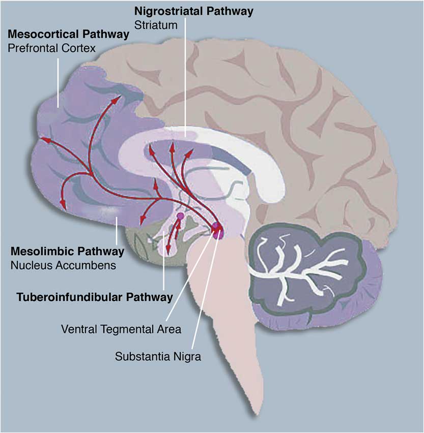
Figure 2 The central nervous system dopamine pathways arising in the ventral tegmental area (VTA) and substantia nigra (SN) of the midbrain. These pathways are implicated in reward processing and are impacted by substance abuse, as described in the text.
More specifically, the VTA projects to the dorsal striatum, prefrontal cortex, thalamus, and parietal regions (the mesocortical DA system), and to core limbic and ventral striatal regions, including the amygdala, anterior cingulate cortex, orbitofrontal cortex, and nucleus accumbens (the mesolimbic DA system).Reference Beaulieu and Gainetdinov 27 , Reference Bjorklund and Dunnett 28
Dysfunctions in these pathways are linked to psychiatric disorders that are associated with compromised emotion regulation and cognitive function, such as schizophrenia, attention-deficit hyperactivity disorder, impulse-control disorders, and affective pathology.Reference Moore, West and Grace 29 , Reference Nemoda, Szekely and Sasvari-Szekely 30 Most relevant to this review, these pathways are also those that are critically involved in drug addiction.Reference Koob and Volkow 19 On the other hand, the SN sends projections to the dorsal striatum, which is implicated more prominently in motor and cognitive processes (nigrostriatal pathway). For example, the degeneration of DA cells in the SN leads to Parkinson’s disease, a prototypical movement disorder.
Dopamine receptors are classified into 2 broad families: D-1-like, consisting of D-1 and D-5 receptors, and D-2-like, consisting of D-2, D-3, and D-4 receptors. D-1-like receptors activate adenylyl cyclase through G-protein coupling; they are relatively prominent in mesocortical terminal regions. Mesolimbic terminal regions contain relatively large numbers of both receptor types, but the mesolimbic D-2 system garners most of the research attention in relation to behaviors facilitated by this circuitry.Reference Joyce 31 , Reference Kohno, Ghahremani and Morales 32 Receptors in the D-2 family are G-protein coupled, inhibit adenylyl cyclase, and activate potassium channels.Reference Missale, Nash, Robinson, Jaber and Caron 33 Mesocortical DA projections and termination sites have been implicated in cognitive control functions, such as behavioral flexibility, working memory, and high level decision-making.Reference Floresco and Magyar 34 Individual differences in functional activation within mesolimbic structures, particularly the nucleus accumbens/ventral striatum, are associated with positive incentive motivation, the motivation-action interface, and heightened responses to substances of abuse.Reference Depue and Collins 35 It has been estimated that 75% of the VTA-to-ventral striatal projection is dopaminergic.Reference Swanson 36
DA models of drug addiction and incentive motivation also posit influences of other chemical systems, given DA’s neuromodulatory regulation of various neuronal populations, its frequent co-release with GABA, and accordingly, the combination of excitatory and inhibitory effects that it exerts within neural pathways.Reference Koob and Volkow 19 , Reference Kaczmarek and Levitan 37 , Reference Trudeau 38
Firing modes
When depolarized, DA cells display 2 firing modes, tonic and phasic. While these exert independent influences over behavior, the difference between the levels of tonic and phasic activity also modulates the intensity of the behavioral output (see referenceReference Luciana and Collins 39 for a discussion). Thus, both tonic and phasic modes are important to understand.
The tonic mode is low in frequency, resulting in low concentrations of extracellular DA. It reflects basal or resting DA neuron firing patterns, regardless of behavioral stimulation.Reference Garris and Rebec 40 , Reference Rice and Cragg 41 Extracellular DA levels are regulated by presynaptic autoreceptors, the action of the DA transporter (via reuptake), and by catabolic enzymes (ie, monoamine oxidase, catechol-o-methyltransferase). Tonic firing is controlled by the neuron’s membrane properties and is regulated by GABAergic inhibition.Reference Floresco, West, Ash, Moore and Grace 42 , Reference Goto, Otani and Grace 43
In contrast, phasic firing is high frequency, characterized by transient bursts of activity.Reference Grace and Bunney 44 – Reference Wanat, Willuhn, Clark and Phillips 46 Relatively high levels of DA are released into neural synapses as a result. Phasic activity is triggered by environmentally salient events, particularly in the context of instrumental learning.Reference Wanat, Willuhn, Clark and Phillips 46 – Reference Schultz 48 Phasic bursts within the striatal region are influenced by glutaminergic excitatory input from brainstem and midbrain sources.
The nature of the relationship between the two firing modes is debated.Reference Goto, Otani and Grace 43 , Reference Niv, Joel and Dayan 49 SchultzReference Schultz 48 demonstrated that phasic DA activity increases in the context of unexpected rewards and in response to reward during initial phases of instrumental reward learning. Furthermore, tonic DA levels transiently decline when anticipated rewards are not delivered or when animals encounter aversive stimuli. For learning to proceed optimally, phasic signals must be detectable. The detection threshold is lowered in the context of decreased background (tonic) firing rates, suggesting that interactions between the 2 firing modes are important in influencing behavioral output. However, increasing attention is directed to the study of tonic dopamine levels, which could contribute to traits that reflect individual differences in incentive motivation.Reference Willuhn, Wanat, Clark and Phillips 47 , Reference Niv, Daw, Joel and Dayan 50 , Reference Ostlund, Wassum, Murphy, Balleine and Maidment 51 Accordingly, each mode of firing independently influences behavior, but interactions are also important. Tonic DA may represent the substrate for basic approach or incentive motivation, while phasic DA reflects learning as it unfolds as a function of experience.
Dopamine system changes with adolescent development
Given the complexities of DA synthesis, release, enzymatic activity in neural synapses, receptor density, and receptor sensitivity, it is beyond the scope of this review to consider developmental changes in all aspects. We refer the reader to recent reviews of this topic.Reference Ernst, Romeo and Andersen 16 , Reference Wahlstrom, White and Luciana 52
We focus here on shifts in DA receptor densities during adolescence as well as changes in tonic and phasic DA activity.
DA receptor changes during adolescence
Dopamine receptors are expressed early in development and change in number through the lifespan. Generally, D-1 receptors are more abundant than D-2 receptors regardless of the lifespan period.Reference Andersen, Dumont and Teicher 53 , Reference Gelbard, Teicher, Faedda and Baldessarini 54 However, their developmental trajectories differ. Regarding D-1 receptors, rat studies have revealed that striatal D-1 receptors increase in number during adolescence and decrease thereafter.Reference Andersen, Dumont and Teicher 53 , Reference Tarazi, Tomasini and Baldessarini 55 , Reference Teicher, Andersen and Hostetter 56 This developmental pattern is most marked in the dorsal striatum, caudate, and putamen.Reference Gelbard, Teicher, Faedda and Baldessarini 54 – Reference Giorgi, De Montis and Porceddu 57 Reports on the temporal pattern of developmental changes in D-1 receptor density in the ventral striatum are more inconsistent, with some studies reporting steady increases,Reference Leslie, Robertson, Cutler and Bennett 58 and others suggesting a periadolescent-limited peakReference Tarazi, Tomasini and Baldessarini 55 followed by a decline and then another peak in late adulthood.Reference Andersen, Dumont and Teicher 53 , Reference Teicher, Andersen and Hostetter 56
Similarly, D-2 receptor density peaks in the caudate and putamen in early puberty.Reference Gelbard, Teicher, Faedda and Baldessarini 54 , Reference Teicher, Andersen and Hostetter 56 , Reference Tarazi, Tomasini and Baldessarini 59 , Reference Rao, Molinoff and Joyce 60 In addition, D-2 density was found to increase linearly in the medial prefrontal cortex (PFC) until adulthood.Reference Tarazi, Tomasini and Baldessarini 59 In the nucleus accumbens, D-2 receptors do not exhibit the same late adulthood increases in density as do D-1 receptors.Reference Andersen, Dumont and Teicher 53 , Reference Teicher, Andersen and Hostetter 56
These changes in receptor profiles are important to describe, because tonic and phasic firing modes may be differentially associated with the activation of distinct receptor subtypes.Reference Luciana, Wahlstrom, Porter and Collins 25 Phasic DA release activates D-1 receptors to facilitate inputs from the VTA to mesolimbic structures, enabling the learning of behavioral strategies during reinforcement learning.Reference Goto, Otani and Grace 43 Accordingly, the increases in D-1 densities observed in the striatum during adolescence might ultimately facilitate aspects of reinforcement learning that are mediated by phasic DA signaling. In contrast, tonic DA influences the VTA-mesocortical projections through D-2 receptor actions. Increases in tonic D-2 stimulation dampen afferent inputs to the PFC, whereas decreases in tonic D-2 stimulation facilitate those inputs.Reference Goto, Otani and Grace 43 These D-2–mediated effects ultimately enable switches to new response strategies when current responses fail to yield anticipated rewards.Reference Goto and Grace 61 There may be increases in tonic D-2 stimulation during adolescence given the observed increase in striatal D-2 receptor density. Ultimately, this increase would serve to enhance behavioral flexibility.
Recently, it was found that there is an age-dependent function of a subtype of D-2 receptors. Activation of these receptors in adolescence, but not other periods of the lifespan, decreases synaptic spine development, providing evidence that these receptors play a role in synaptic pruning, given that a decline in spine density is part of the pruning process.Reference Jia, Zhao, Hu, Lindberg and Li 62 Given the evidence for peak D-2 receptor production during adolescence, this finding suggests an important link between neurochemical changes during development and resultant alterations in regional brain morphology.
The role of DA in the pruning process has also been explained in relation to developmental changes in PFC glutamatergic (GLU) activity. Both DA and GLU systems contribute interactively to neuroplasticity, which is prominent during adolescence.Reference Crews, He and Hodge 63 Particularly, the developmental trajectories of PFC GLU and striatal DA systems may interact across adolescence in ways that increase both the risk for addictive behaviors and the deleterious consequences of exposure to addictive substances on neurodevelopment.Reference Selemon 64 , Reference Selemon 65 Glutamate receptor (GLUR)-mediated synaptic plasticity, via configurations of the N-methyl-D-aspartate (NMDA) and alpha-amino-3-hydroxy-5-methyl-4-isoxazole propionic acid (AMPA) receptor systems, generate long-term depression (LTD), which is thought to underlie synaptic pruning, a characteristic of neural changes in adolescence.Reference Selemon 64 Importantly, D-1 receptor activation promotes LTD through indirect actions on AMPA receptors.Reference Sun, Zhao and Wolf 66 Thus, the proposed increase in striatal dopamine signaling in adolescenceReference Luciana, Wahlstrom, Porter and Collins 25 , Reference Wahlstrom, White and Luciana 52 may, through D-1 and D-2 receptor-mediated mechanisms, contribute to the regulation of synaptic pruning within the striatum, but also in higher cortical regions, through glutamatergic mechanisms. The DA/GLU interactive effects on the progression of the pruning phase may be disrupted by exposure to substances of abuse during the sensitive period of adolescence, and have unique long-term consequences, not seen when drugs are used after this developmental period.Reference Auclair, Otani, Soubrie and Crepel 67 , Reference Pascual, Boix, Felipo and Guerri 68 Although changes in other neuromodulatory systems (ie, GABA) and their maturing interactions with DA function are also important to consider, coverage of these interactions is beyond the scope of this review.
Tonic and phasic dopamine activity
We have previously summarized evidence that tonic DA levels underlie adolescent-specific developmental increases in incentive motivation.Reference Wahlstrom, Collins, White and Luciana 17 , Reference Luciana, Wahlstrom, Porter and Collins 25 , Reference Luciana and Collins 39 , Reference Wahlstrom, White and Luciana 52 Tonic firing rates in the VTA are observed to be higher in adolescents than in adult animals.Reference McCutcheon, Conrad, Carr, Ford, McGehee and Marinelli 69
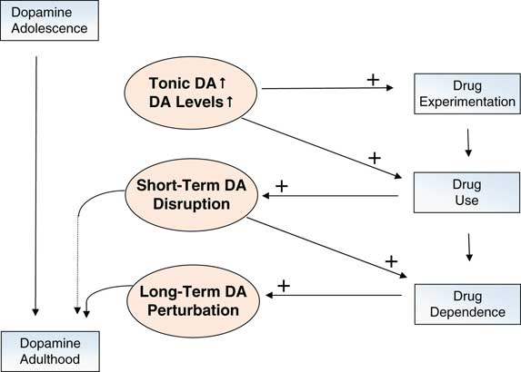
We have suggested that these increases enhance approach behavior, and serve to bring adolescents into contact with reward contexts (even those that confer risks). The tendency to approach contexts that are high in reward salience is supported by adolescents’ social, sexual, and other risk-taking behaviorsReference Eaton, Kann and Kinchen 3 ; by their self-reports of reward-responsivity and sensation-seekingReference Harden and Tucker-Drob 70 – Reference Urosevic, Collins, Muetzel, Lim and Luciana 72 ; and by brain responses as observed in laboratory-based neuroimaging studies of adolescent reward processing. Reference Luciana and Collins 39 , Reference Bjork and Pardini 73 , Reference Richards, Plate and Ernst 74 Reward-relevant structures within the mesolimbic DA system are most strongly activated, relative to children and adults, when adolescents experience or anticipate rewards that are highly salient. In real-world contexts, these reward stimuli may include substances of abuse. Thus, adolescent peaks in tonic dopamine levels encourage experimentation with drugs of abuse as well as engagement with other rewarding stimuli (see Figure 3).

Figure 3 The dopamine system becomes more functionally active during adolescence through increases in tonic DA activity and phasic signaling. These changes are associated with experimentation with drugs of abuse. With increasing drug use, parameters of DA transmission are neuroplastically altered, leading to drug addiction and long-term alterations in reward system function.
Contexts in which rewards are present bring opportunities for learning. When an individual encounters a potentially rewarding object or situation, he or she must make a decision about whether consumption of that stimulus will lead to a positive versus a negative outcome. The decision to consume is partly determined by individual differences in tonic dopamine, since those with higher levels of tonic dopamine (and accordingly, higher levels of incentive motivation) are more responsive to such cues in the environment. For instance, in animal models, increases in incentive motivation are associated with heightened tonic DA activity in the striatum and with increasing response vigor, as demonstrated through behavioral and computational models.Reference Niv, Daw, Joel and Dayan 50 , Reference Joel and Weiner 75 Importantly, though, outcomes of decisions during reinforcement learning may be particularly uncertain in adolescents, who are not necessarily familiar with social contexts, potential romantic partners, illicit substances, or other sources of positive reinforcement. Feedback or outcome-based learning, when outcomes of behavioral choices are uncertain, is accompanied by phasic bursts of DA in striatal regions. Through repeated experiences, the individual must learn how to make decisions despite this uncertainty in order to promote adaptive future behavior. This decision-making process involves regions of the frontal cortex. Accordingly, during the learning process, prefrontal circuits are called upon in a “bottom up” manner, via subcortical-to-cortical relays, to structure behavior. Notably, there is some evidence of relative increases in these learning signals (attributed to phasic dopamine) during adolescence.Reference Cohen, Asarnow and Sabb 76 When individuals use substances of abuse, all of which activate the dopamine system as do natural rewards,Reference Di Chiara 77 – Reference Fiorino, Coury and Phillips 79 there are bursts of phasic dopamine firing.Reference Grace 80
If tonic signals are high in the adolescent as we hypothesize, then these phasic signals must be very strong in magnitude to serve as appropriate learning signals. Accordingly, adolescents appear to be vulnerable to risky behaviors because of a strong drive (related to high dopamine tone) to seek rewards or rewarding contexts, which are extremely compelling. These contexts, in turn, will be particularly potent sources of learning (under the control of the phasic mode). The value of such a system is apparent when behavior is appropriately regulated. However, in adolescence, such regulation is inconsistent, leaving the adolescent vulnerable (behaviorally and neurobiologically) to the effects of illicit substances, especially when they are used in high amounts.
Over time, our assertion is that regular experience with phasic dopamine, in the context of probabilistic learning in novel contexts, “teaches” (through subcortical to cortical afferent signals) the prefrontal cortex to activate in service of behavioral regulation. Ultimately, prefrontally mediated behavioral control can be engaged more readily as the transition into adulthood is reached.Reference Luciana, Wahlstrom, Porter and Collins 25 , Reference Luciana and Collins 39 Concordantly, Mastwal et al Reference Mastwal, Ye and Ren 81 recently compared young adolescent mice with adult mice on phasic and tonic dopamine activity. Using optogenetic stimulation and in vivo 2-photon imaging, they observed in the adolescent animals a potentiation of mesocortical DA circuit activity after a period of induced phasic (but not tonic) DA activation. Moreover, phasic activation led to lasting changes in meso-prefrontal structure and function. Overall, these findings indirectly suggest that the pursuit of motivationally salient events in adolescence is a positive and necessary behavioral change—one that promotes the development of self-regulatory control. This pursuit is facilitated by tonic increases in DA, rendering the adolescent more receptive to, and in fact eager to pursue, positive incentives. This engagement provides critical contexts for learning and enhances the later functional integrity of frontal circuits that are important for behavioral regulation. Changes in tonic and phasic DA levels during reinforcement learning occur very rapidly, at the sub-millisecond level, to promote immediate behavioral responses. Recently, an extended mode of reward-predictive dopamine signaling in the striatum was identified. It enabled animals to approach distant (versus immediate and proximal) goals,Reference Howe, Tierney, Sandberg, Phillips and Graybiel 82 lending support to the notion that adolescent-limited changes in tonic and phasic dopamine signaling have longer-term behavioral relevance.
In sum, the above-described changes in various elements of the dopamine system during adolescence are thought to underlie the typically adolescent behaviors that promote risk for substance use. While the developing neural system is, on the one hand, plastic and open to sources of positive influence,Reference Kolb 83 on the other hand, it is also highly sensitive to adverse experiences, and, accordingly, the introduction of toxic compounds bring risks for life-long alterations in neural structure, function, and behavior.Reference Robinson and Kolb 84 With the advent of imaging techniques we can now study in vivo the dopamine system in humans and its developmental changes. These techniques, their strengths, and their weaknesses are described in the next section and summarized in Table 1.
Table 1 Neuroimaging techniques to measure central dopamine function in humans

Neuroimaging Techniques to Study the Dopamine/Reward System in Humans (Table 1)
Brief review of neuroimaging techniques
The two major types of imaging techniques include radionuclear and electromagnetic techniques. Radionuclear techniques consist of single-photon emission computed tomography (SPECT) and positron emission tomography (PET), which were originally used to assay a nonspecific index of brain activity, ie, regional cerebral blood flow (rCBF) or regional cerebral metabolic rates of glucose (rCMRglc), but are currently utilized to investigate specific neurochemical systems (eg, neurotransmitter systems such as D-2 receptor density with the [11C]-raclopride ligand). Electromagnetic imaging uses magnetic resonance (MR) properties of the body constituents to assess morphology [structural MRI (sMRI)], fiber tracks [diffusion tensor imaging (DTI)], function [functional MRI (fMRI)], and concentrations of chemicals present in large quantities in the brain [magnetic resonance spectroscopy (MRS)].
fMRI has replaced the PET/SPECT nonspecific measurements of brain activity (CMRglc, rCBF) for several reasons. fMRI provides indices of rCBF changes with a temporal resolution down to 1 s (vs. 60 s with PET) and spatial resolution of 1 mm (vs. 4 mm with PET). However, its main advantage is the absence of exposure to ionized radiation, which permits investigators to study pediatric subjects and to conduct multiple repeated studies within the same subject. Using repeated scans, fMRI can also provide measures of functional connectivity, which have raised a huge interest. These studies examine functional connectivity changes in response to a task (connectivity modulated by cognitive processes) or during the resting state.
However, MRI does not permit the study of neurochemical systems in the brain, which is the monopoly of PET/SPECT. Indeed, while MRS provides measures of regional concentrations of brain chemicals, such as amino acids and metabolites [eg, metabolites of glutamate (Glu), N-acetylaspartate (NAA), and gamma-aminobutyric acid (GABA)], it is limited by its high threshold of detection (millimolar range with MRS vs. nanomolar range with PET or SPECT). In addition, the roles of the various biological compounds measured with MRS are still unclear, thus complicating the interpretation of findings.
The scientific yield of these neuroimaging techniques can be potentiated by pharmacological challenges. For example, fMRI studies can be paired with a dopamine-specific drug challenge (eg, methylphenidate) to examine the effects of dopamine modulation on specific regional functions. In PET/SPECT, such a pharmacological challenge can serve to examine the integrity of dopamine receptor binding potential when synaptic dopamine concentration is being manipulated.Reference Volkow, Fowler and Gatley 85 , Reference Mizrahi 86
Specifically, in PET imaging of the dopamine system, a radioactive ligand (eg, raclopride, a D-2 receptor ligand) is injected intravenously into the bloodstream. Using mathematical models, PET/SPECT allows the quantification of the radioactivity being bound to receptors, which is translated into receptor binding potential. Specific ligands have been developed that cross the blood-brain barrier to label dopamine receptors, transporters, precursors, or enzymes that degrade dopamine.Reference Volkow, Fowler and Gatley 85 Other tracers bind to glucose and can be measured to infer more general changes in brain metabolism (CMRglc). When combined with pharmacological manipulations, these tracers/ligands can be used to assess functional changes within the brain following systemic manipulations of DA.
Neuroimaging limitations
It is considered unethical to administer pharmacological probes to pediatric samples, including older adolescents, who are not otherwise being treated for clinical conditions. Furthermore, because PET relies on radioactive ligands, it is not approved for use in pediatric research (however, see referenceReference Jucaite, Fernell, Halldin, Forssberg and Farde 87 ). Accordingly, human pediatric imaging studies have relied on MRI techniques, which offer less direct measures of metabolic activity. Typically, fMRI involves measures of blood-oxygen-level–dependent (BOLD) signal changes, which reflect synaptic activity, but can be affected by rate of blood flow, which may vary as a function of age.
A limitation to the study of the dopamine system with conventional MRI techniques is the difficulty of reliably ascertaining signals in small structures, such as midbrain nuclei (ie, VTA) or brainstem regions, that are the sources of DA synthesis and release (however, see referenceReference D’Ardenne, McClure, Nystrom and Cohen 88 ). This is true regardless of age group studied. The use of more sophisticated MRI scanners, with higher field strengths (7 tesla), might permit us to more reliably capture individual differences in small structures.Reference Eapen, Zald, Gatenby, Ding and Gore 89
Finally, fMRI requires complete stillness of research participants, which is difficult for young participants, and can make certain studies impossible to conduct in youths. Recent studies have also focused on structural attributes of regions that are strongly DA-innervated.
We will show how these various imaging techniques can help in the delineation of dopamine-linked changes associated with drug exposure starting in adolescence. To this aim, we take marijuana studies as exemplars. We focus on sMRI, fMRI, and spectroscopy.
MRI Studies of the Dopamine Reward System in Marijuana Users
Scope of the adolescent marijuana use problem
Marijuana use is becoming a critical issue in view of the spreading legalization of this drug across the U.S. The implicit message sent by the legalization of marijuana is that this compound is not harmful. At the same time, marijuana is the most commonly used “illicit” drug among teenagers in the United States. Whereas 17% of 8th graders have tried marijuana, almost half of teens have used marijuana by 12th grade. 90 , Reference Johnston, O’Malley, Miech, Bachman and Schulenberg 91 Most critically, many youths develop a regular pattern of use after initial exposure, with 20% of 12th graders reporting use in the past month and 5% of 12th graders reporting daily use.Reference Johnston, O’Malley, Miech, Bachman and Schulenberg 91
Together, these data are alarming, particularly since the deleterious effects of the regular use of marijuana in adolescents might have serious, long-lasting consequences. Indeed, the stakes may be higher for adolescents than for adults, because adolescence qualifies as a sensitive period in brain development, and because of the specific dysfunctions associated with marijuana use, including addictive liability, cognitive deficits, and anhedonia.Reference Volkow, Baler, Compton and Weiss 92
First, regarding addiction, albeit still debated, evidence indicates that long-term marijuana use can lead to dependence, but can also predispose users to trying other illicit drugs. In addition, a well-described cannabis withdrawal syndrome (including irritability, sleeping difficulties, dysphoria, craving, and anxiety) has been identifiedReference Gorelick, Levin and Copersino 93 and can impinge cessation attempts and contribute to relapse. The addictive aspect of marijuana is likely to involve changes within the dopamine system—changes that are shared by all addictive drug actions.Reference Oleson and Cheer 94 Second, neurocognitive deficits have been reportedReference Schweinsburg, Brown and Tapert 95 in adult and adolescent marijuana users that affect spatial and visual learning and memory (particularly short-term memory), executive functioning, and psychomotor speed. Although not consistent, studies suggest that these deficits may be long-lasting. Cognitive deficits in adolescence are particularly troublesome because of the academic implications, which can have far-reaching consequences for the optimal future occupational and social success of the adolescent. Here again, the dopaminergic system is thought to play a critical role in “tuning” the prefrontal cortex for optimal cognitive function.Reference Puig, Rose, Schmidt and Freund 96
Akin to tobacco smoking, for which a huge effort has been made to inform the public on tobacco’s adverse effects, marijuana should become a target for informing the population at large of the potential negative short-term and long-term outcomes of regular marijuana use, particularly in adolescence. Neuroimaging findings could help in this mission. Here we focus on studies that address the influence of marijuana exposure on the dopamine reward system in adolescents. Among subcortical regions where dopamine receptors are prominent, the active ingredient in marijuana, delta-9-tetrahydrocannabinol (THC), binds to endogenous cannabinoid receptors in the nucleus accumbens and in the amygdala.Reference Burns, Van Laere and Sanabria-Bohorquez 97 Synaptic transmission in these regions is altered after marijuana use.Reference Gilman, Kuster and Lee 98 We emphasize studies that focus on dopamine-innervated structures in the reward system; among these are the nucleus accumbens of the ventral striatum, medial prefrontal cortex (including orbitofrontal and anterior cingulate regions), and amygdala.
Structural neuroimaging with sMRI
Normative development
Until recently, it had not been possible to reliably measure the volumes or anatomical connectivity of subcortical regions that comprise the dopaminergic reward system. Using automated parcellation techniques, several groups have examined the development of structures such as the nucleus accumbens, amygdala, and medial prefrontal cortex across healthy adolescent development. These findings are important to our understanding of normative developmental trajectories and how substance use might impact those trajectories. For instance, Urosevic et al Reference Urosevic, Collins, Muetzel, Lim and Luciana 72 reported that the nucleus accumbens increased in volume from early to mid-adolescence (a pattern also reported by Dennison et al Reference Dennison, Whittle and Yucel 99 ) and decreased in volume thereafter, demonstrating a quadratic pattern. Self-reported reward responsivity showed a similar pattern, with a peak in mid-adolescence and a decline thereafter, suggesting an association between these processes. This quadratic pattern is important, given that it could reflect an adolescent-specific peak in dopamine-mediated reward sensitivity. Use of substances of abuse during this time has the potential to alter the development of the system. In contrast to these findings for the nucleus accumbens/ventral striatum, Dennison et al Reference Dennison, Whittle and Yucel 99 found that the caudate, thalamus, and putamen declined in volume. Amygdala volume did not change over time. Ostby et al Reference Ostby, Tamnes, Fjell, Westlye, Due-Tonnessen and Walhovd 100 reported heterogeneity in developmental trajectories among basal ganglia regions and between subcortical and cortical regions. Overall, these studies suggest that early-to-mid-adolescence represents a potentially important time period for the development of subcortical regions that are key nodes of the reward processing network.
Individual differences interact with age-related changes. Schneider et al Reference Schneider, Peters and Bromberg 101 used voxel-based morphometry (VBM) and found, in 14 year-olds, that greater risk-taking bias was associated with, and partially mediated by, lower gray matter volumes in the ventral striatum. With increased risk-taking bias, there was a decrease in the functional activation of the ventral striatum during reward anticipation. Given that risk-taking propensity is an important predictor of adolescent substance use, these findings are noteworthy in stressing a role for the ventral striatum in this association. Concordantly, Urosevic et al Reference Urošević, Collins, Muetzel, Lim and Luciana 102 found that substance-naïve adolescents with smaller left nucleus accumbens volumes, as well as those with higher self-reported reward sensitivity, were more likely to initiate substance use in the subsequent 2 years. Accordingly, it is clear that individual differences in baseline measures of brain structures in children are important in predicting who will go on to engage in problematic substance use. Of note, these baseline indicators are linked to the DA system.
Effects of marijuana on brain morphology
With respect to structural brain changes associated with marijuana use, much of the literature is characterized by cross-sectional case-control comparisons through which marijuana-using adolescents or young adults are compared to non-using controls. Baseline assessments of marijuana users, before onset of substance use, are frequently unavailable. Thus, it is unclear whether observed differences from control samples represent baseline differences between groups or consequences of drug use. Nonetheless, alterations in frontal gray matter density have been observed with recreational marijuana use,Reference Gilman, Kuster and Lee 98 and there are many findings of altered white matter structure in frontal regions in users.Reference Arnone, Barrick, Chengappa, Mackay, Clark and Abou-Saleh 103 – Reference Gruber, Silveri, Dahlgren and Yurgelun-Todd 106 The pattern of abnormalities across studies is consistent, with reports of increased mean diffusivity of white matter as well as relative decreases in the directionality of fibers and fiber coherence (measured with DTI) in users.
Cause–effect links are indirectly suggested by associations between these differences and parameters of drug use. Earlier use of marijuana has been found to be associated with more pronounced deviations, as well as with increased behavioral impulsivity.Reference Gruber, Dahlgren, Sagar, Gonenc and Lukas 105 These studies generally have not focused on the reward system per se, and links to the dopaminergic system are tentative at best. However, a recent study addresses this shortcoming.Reference Gilman, Kuster and Lee 98 Young (~age 20) recreational users of marijuana were compared to demographically matched, non-using controls. Analysis of whole-brain gray matter volume densities revealed greater densities in marijuana users in the left nucleus accumbens, extending to subcallosal regions, the hypothalamus, the extended amygdala, as well as in the left amygdala proper.Reference Heimer and Alheid 107 In the left nucleus accumbens, average volume was also higher in users than controls, and its surface topography was altered. Animal studies have indicated that exposure to THC increases dendritic length and branching in the shell region of the nucleus accumbens, but not in other areas of the striatum or cortex,Reference Kolb, Gorny, Limebeer and Parker 108 which could account for increased volumes in marijuana users.
The finding of increased gray matter density for the amygdala coheres with other reports of increased density in relation to severity of marijuana dependence,Reference Cousijn, Wiers, Ridderinkhof, van den Brink, Veltman and Goudriaan 109 but suggests that brain structure is altered even before behavior has reached the level of addiction, given that this study focused on recreational users. Generally, these studies suggest that the structural development of the DA-modulated reward system is important in promoting increases in reward sensitivity during adolescence; these same structures are altered in the context of recreational marijuana use.
Functional neuroimaging
Most of the functional imaging work relevant to marijuana use in adolescents has actually been conducted in adults presenting with variable levels of marijuana use, and most importantly, variable ages of onset of marijuana use. Only a few imaging studies have been conducted in adolescents, and none focused on the dopamine system.Reference Lorenzetti, Lubman, Whittle, Solowij and Yucel 110 – Reference Batalla, Bhattacharyya and Yucel 112 Both types of functional imaging have been used, PET and fMRI. We present selected studies for each methodology.
Using PET and the D-2/D-3 receptor ligand [11C]raclopride, 24 non-user adults were compared with 24 marijuana users on the effects of methylphenidate (MP) on dopamine activity. MP blocks dopamine transporters, and, in turn, increases synaptic dopamine concentration.Reference Volkow, Wang and Telang 113 Findings revealed no group differences at baseline, in contrast to blunted receptor availability when challenged with MP, in marijuana users compared to non-users. The decreased dopamine reactivity to MP in marijuana abusers vs. non-users was associated with higher self-report of negative emotionality and craving.
On the other hand, the absence of group differences at baseline is consistent with other studies of striatal D-2/D-3 receptor availability in 4 studies of chronic cannabis users compared with healthy controls.Reference Albrecht, Skosnik and Vollmer 114 – Reference Urban, Slifstein and Thompson 117 However, in the only study where the chronic cannabis users were not abstinent,Reference Albrecht, Skosnik and Vollmer 114 an inverse correlation between recent cannabis consumption and D-2/D-3 receptor availability was found, suggesting a direct effect of cannabis smoking on the expression of striatal DA receptors in heavy cannabis users.Reference Albrecht, Skosnik and Vollmer 114 Thus, two factors are important to evidence dopamine-related perturbations associated with marijuana exposure: the pharmacologic (eg, MP) activation of DA function and the direct action of the drug (non-abstinent users).
The prominent action of marijuana use that started in adolescence seems to be a blunting effect on dopamine activity. This effect is further substantiated by PET studies of young adult users who present a blunting of striatal dopamine release in long-term cannabis users,Reference Bloomfield, Morgan, Kapur, Curran and Howes 118 as well as reduced dopamine synthesis capacity.Reference Murray, Dilleen and Pelloux 119 These findings may be important to our understanding of the frequently reported observation of anhedonia or lack of motivation in chronic users.
Using fMRI with a decision-making task, Nestor et al Reference Nestor, Hester and Garavan 120 and van Hell et al Reference van Hell, Vink, Ossewaarde, Jager, Kahn and Ramsey 121 recorded neural activity during reward and anticipation of loss with different versions of a monetary reward task. Both studies failed to reveal behavioral differences between the groups. However, Nestor et al Reference Nestor, Hester and Garavan 120 reported a greater right ventral striatal activity in cannabis users than controls during reward anticipation. This measure was significantly correlated with number of years of lifetime cannabis use. In addition, a post-hoc analysis of the contrast loss/win cues vs. no-outcome cues showed greater BOLD response in the right ventral putamen in marijuana users vs. non-users.Reference Nestor, Hester and Garavan 120 Conversely, comparing cannabis users to non tobacco-smoking controls, van Hell et al Reference Nestor, Hester and Garavan 120 demonstrated attenuated activity in the nucleus accumbens and caudate nucleus bilaterally during reward anticipation, as well as left putamen and other prefrontal regions.Reference van Hell, Vink, Ossewaarde, Jager, Kahn and Ramsey 121 These findings could be seen to be at odds with the PET study of MP challenge of DA function. A blunted dopamine response to a stimulant challenge might suggest lower striatal response to a reward-related behavioral challenge. It is difficult to integrate these two findings as of yet, because of the many factors that differ between these studies, eg, the methodology, the significance of the D-2/D-3 receptor measure at the behavioral level, and the nature of the samples.
Finally, a systematic study of the functional connectivity of the dopamine circuitry across development and in response to drug exposure can greatly enhance our understanding of how such exposure impacts the developing brain, as well as the behavioral consequences of drug use.
Spectroscopy
Very few studies have used MRS to investigate the impact of marijuana on brain chemistry.Reference Sneider, Mashhoon and Silveri 122 In addition, only a limited number of these studies have focused on the adolescent period; select studies are described here.
Across studies, individuals aged 16–42 years have been tested using either single or multiple voxel techniques. Some studies have focused only on males, given higher use rates in males versus females.Reference Silveri, Jensen, Rosso, Sneider and Yurgelun-Todd 123 , Reference Hermann, Sartorius and Welzel 124 In most cases, last marijuana use was reported at 20 or more days per month. Lower levels of Glu, N-acetyl-aspartate (NAA), and myo-inositol (mIns) were observed in marijuana users compared to controls in regions known to be associated with substance use, including the basal ganglia (lower Glu, NAA, and cholineReference Chang, Cloak, Yakupov and Ernst 125 ; lower glutamine [Glx] and higher mIns in females),Reference Muetzel, Marjanska and Collins 126 thalamus (higher total creatine [tCr]),Reference Chang, Cloak, Yakupov and Ernst 125 cingulate cortex (lower Glu, NAA, tCr, and mIns),Reference Prescot, Locatelli, Renshaw and Yurgelun-Todd 127 dorsolateral prefrontal cortex (lower NAA),Reference Hermann, Sartorius and Welzel 124 and the striatum, as well as posterior cortical regions (lower mIns).Reference Silveri, Jensen, Rosso, Sneider and Yurgelun-Todd 123
Because of the limitations of the single voxel approach, it is unclear whether these findings would generalize to ventral striatal regions. However, the pattern of findings suggests that marijuana users may be vulnerable to gliosis and white matter injury as a consequence of chronic use. Moreover, because DA interacts with both GABA and glutamate systems in the striatum as well as other regions, and because this network of neurotransmitters is adversely impacted as addiction progresses,Reference Kalivas and Volkow 128 these findings across studies are cumulatively suggestive of a broader range of impairments in the brain’s reward processing and learning networks.
Conclusions
A neurodevelopmental approach to understanding the mechanisms that underlie the transition from drug experimentation to drug abuse is essential to the formulation of primary and secondary treatment strategies. Adolescence is the critical period of vulnerability because of the unique neuroplasticity of the brain, which significantly affects the morphology and functional dynamics of the dopamine system—the key neural substrate of drug addiction.
A large body of work is being devoted to understanding the normative maturation of the dopamine system on the one hand and the transient and long-lasting effects of addictive drugs on dopamine function on the other hand. The integration of these two lines of research is complex and depends on the formulation of models that can be tested in animals and humans, feeding a continual dialogue between both basic and clinical fields.
The complexity of this work is phenomenal because of the nature of the dynamics of drug addiction—a moving target that is defined in, not one, but three distinct time frames: ontogenic trajectory, severity of drug problem (initiation, use, dependence), and addiction cycle (craving, withdrawal). In addition, polysubstance use is the rule rather than the exception, which complicates research in humans. Many other biological and environmental factors interact with the propensity for developing a substance use problem. Therefore, it is only through a wide variety of research approaches that the development and maintenance of drug addiction can be understood.
Neuroimaging techniques, including PET/SPECT and MRI, provide unique tools that can be used in both animals and humans. As illustrated with marijuana use, neuroimaging studies clearly document alterations within the dopamine system that are linked to behavioral changes associated with drug actions. These findings not only provide pieces to the puzzle of addiction, but can serve as persuasive evidence for the profound effects of chronic use of drugs on brain function. These can also be of great benefit to public health programs concerned with preventing drug abuse.
The prevention of drug use starts with reducing drug experimentation. At this stage, three factors can be impacted: drug access, beliefs and attitudes toward drug experimentation, and risk-taking propensity. The issue of drug access is outside the scope of this review. The second factor, beliefs and attitudes, can be influenced by information delivered through the media, school, and home. Clear information on the potential short- and long-term deleterious effects of drugs, particularly marijuana, alcohol, tobacco, the most commonly used first drugs in adolescence, needs to be disseminated. Findings from neuroimaging research can be quite eloquent, as “pictures are worth a thousand words.” The third factor raises the question of how to redirect risk-taking behavior in adolescence to adaptive and constructive experiences. Can we train the reward system to respond more strongly to certain stimuli than others? This type of intervention is already being used in anxiety with the development of attention modification training, which helps anxious individuals to change their attention biases away from negative stimuli.Reference Hakamata, Lissek and Bar-Haim 129 The strategy of targeting attention bias is also receiving interest for the treatment of substance use,Reference Field, Marhe and Franken 130 but has not been considered at the experimentation stage of substance use. Last, we believe that any intervention in adolescents should consider the influence of the adolescent social milieu, particularly peers, given the tremendous importance of social relationships in adolescence.Reference Nelson, Leibenluft, McClure and Pine 10 In sum, more work needs to be done to examine behavioral manipulations that could train and protect the reward system against attraction to dangerous stimuli such as “illicit drugs.”
Regarding the next stage of drug use/dependence, the development of treatment strategies is badly needed for adolescents. One promising approach is the combination of motivational enhancement therapy (MET) and cognitive behavioral therapy (CBT).Reference Sample and Kadden 131 , Reference Dennis, Titus and Diamond 132 MET/CBT has been tested for the treatment of cannabis in youths,Reference Dennis, Godley and Diamond 133 and is particularly relevant to drug-related perturbations of the reward system. This treatment has been found to be efficacious in adolescent marijuana abuse. Identifying how this treatment affects striatal and dopamine function is a critical next step.
In sum, the study of the dopamine/reward system for primary and secondary treatment of substance use problems, combined with a clear understanding of the changes within this system during adolescence, should be a focus of intense scrutiny, and neuroimaging can assist us in the rationale, design, and testing of novel therapeutic approaches.
Disclosures
Monique Ernst does not have anything to disclose. Monica Luciana has the following disclosure: National Institute on Alcohol Abuse and Alcoholism, grant recipient, Grant AA020033.