


Ventricular septal defect is the most common congenital cardiac disorder, characterised by an opening in the septum between the ventricles, which allows blood to shunt between the left and right ventricles, that is, left-to-right shunting.Reference Perloff, Rosove, Child and Wright1, Reference Ammash and Warnes2 This disease accounts for up to 30% of all congenital cardiac defects. Irreversible pulmonary vascular obstructive disease, Eisenmenger syndrome, develops in 10–15% of patients with ventricular septal defects, most commonly in the second and third decades of life.Reference Perloff3–Reference Heath and Edwards6 The Eisenmenger syndrome consists of a congenital communication between the systemic and pulmonary circulation, with resultant pulmonary arterial hypertension and right-to-left reversal of flow through the defect. When pulmonary vascular resistance exceeds systemic vascular resistance, it results in hypoxaemia and cyanosis. Electronic circuit simulation uses mathematical models to replicate the behaviour of an actual electronic device or circuit. This system is available for investigating cardiovascular physiology. Several models for the cardiovascular system have been proposed for understanding the cardiovascular physiology and pathophysiology, such as arrhythmia, myocardial ischaemia, hypertension, and valve diseases.Reference Rupnik, Runovc, Sket and Kordas7–Reference Rideout12 In this study, we have designed an analogue circuit that is able to simulate normal condition and Eisenmenger syndrome with ventricular septal defect.
Methods
In this study, we investigated the cardiovascular system using an electronic circuit. The electrons in an electronic circuit create electrical current by moving from a high voltage point to a lower voltage point similar to fluids, which flow from a high pressure point to a lower pressure point. In the designed circuit, we used resistors, capacitors, inductors, diodes, and variable capacitors. Resistors model the resistance of a vessel against the blood flow because of its length and diameter. Capacitors simulate expansion of blood vessels by the increasing pressure – compliance. Inductances model the physical reactions of the blood to flow changes. Physical reaction is the increase or decrease in pressure, which is proportional to blood density and velocity. Diodes model the heart valves and convey the current in one direction. When the voltage at a diode input is high enough, the diode is ON and current can flow through it. Variable capacitors model the left and right ventricles. Increasing the value of these capacitors causes the current to flow from the circuit through the capacitances – diastole period; decreasing the value of the capacitors causes the current to flow into the circuit – systole period. Therefore, the periodical change of capacitors was designed in accordance with systole and diastole intervals.
Cardiovascular system simulation is done using an electrical circuit model, which is shown in Figure 1. In our model, the left and right ventricle are represented by trapezoid-shaped stiffness, and vessels are represented by modified Westkessel model including Resistor-Inductance-Capacitance pi-sections as used in the Rideout model.Reference Rideout12 Matlab® is used for the model implementation. Electrical units and their physiological equivalent values are given in Table 1. In the design of the cardiovascular circuit simulation, we have taken into account physiological limits. The main pressure and volume curves obtained from the model for normal condition are shown in Figure 2. The results for normal condition obtained from the model are similar to those reported in the literature for the cardiovascular system (Table 2).Reference Levick13 Normal condition is assumed as a healthy male adult whose body weight is 70 kilograms, body surface area is 1.8 square metres, and heart rate is 75 beats per minute – heart period is 0.8 seconds.

Figure 1 The electronic circuit model that is used for the simulation of the cardiovascular system. P = pressure; F = flow; V = volume; R = resistance; C = capacitance; S = stiffness; L = inertance; Aor = aortic; Pul = pulmonary artery; MitV = mitral valve; AorV = aortic valve; TriV = tricuspid valve; PulV = pulmonary valve; LA = left atrium; Mit = mitral; LV = left ventricle; RA = right atrium; sep = septum; R1L = systemic resistance; C1L = systemic capacitance; CvenL = systemic venous capacitance; R1R = pulmonary resistance or pulmonary load resistance; C1R = pulmonary capacitance or pulmonary load capacitance; CvenR = pulmonary venous capacitance.
Table 1 The design of the circuit is based on the principles of equivalent quantities.


Figure 2 The main pressure and volume contours obtained from the model for normal conditions.
Table 2 Cardiovascular model parameters and the model measurement values for normal, large VSD and Eisenmenger syndrome.

VSD = ventricular septal defects
To drive each ventricle, two synchronous pacemakers and two capacitances having similar time-varying stiffness characteristics are connected in the circuit. The pacemakers have trapezoidal stiffness-characteristic waves with adjustable frequency, which is given in Figure 1. The pacemakers operate at 1.25 hertz frequency, 75 beats per minute, with durations of systole and diastole being 0.234 and 0.566 seconds, respectively. The aortic and pulmonary capacitances have exponential characteristics.
Aortic and pulmonary distensibility are given below:
![\[--><$$>\eqalign{ {\rm{Distensibility}}\, = \,{\rm{mean}}\,({\rm{capacitance}})\:\cr \quad = \:\frac{{{ {\Bigg[ \matrix {{\rm{First}} \ {\rm{capacitance}} \ {\rm{volume }}\hfill \cr \quad {{-\ {\rm Second}} \ {\rm{capacitance}} \ {\rm{volume}}}\hfill }}}\Bigg]}} {\Bigg[{\matrix{{ {\rm{Maximum}} \ {\rm{pulmonary}} \ {\rm{capacitance}}}\hfill \cr \quad{{- \ \rm Minimum}} \ {\rm{pulmonary}} \ {\rm{capacitance}}}\Bigg]\eqno<$$><!--\]](https://static.cambridge.org/binary/version/id/urn:cambridge.org:id:binary-alt:20160626130605-81984-mediumThumb-S1047951111001478_eqnU1.jpg?pub-status=live)
Here, Maximum Pulmonary and PC1 are the limit capacitance pressure values that aortic and/or pulmonary capacitances possessed when the model was working, and VC2 and VC1 are the capacitance volume values corresponding to the PC2 and PC1. The parameter values of the model in normal condition are given in Figure 1 and in Table 2.
All cardiac valves are simulated using voltage-controlled switches with a piece-wise linear characteristic plus serial resistances having 0.003 hydraulic resistance unit.
The cardiovascular circuit model is modified for the simulation of normal, large ventricular septal defect and Eisenmenger syndrome. The ventricular septal defect is simulated using a septal resistance connected between the left ventricle and right ventricle points of the model. The Eisenmenger syndrome is simulated mainly using septal resistance and pulmonary resistance. The definitions for the septal defect conditions are given below:
The Q p/Q s (shunt) parameter limit values for different ventricular septal defects and Eisenmenger syndrome are given in Table 3.Reference Therrien, Dore and Gersony14 To simulate large ventricular septal defect and Eisenmenger conditions, mainly septal resistance of the model is changed from 0.15 to 0.01 hydraulic resistance unit values, and pulmonary resistance is changed from 0.15 to 2.5 hydraulic resistance unit, as given in Table 2. Pulmonary capacitance value is 0.02 hydraulic capacitance unit for both large ventricular septal defect and Eisenmenger conditions, different from normal condition, which is 0.04 hydraulic capacitance unit. The model measurement values for normal, large ventricular septal defect, and Eisenmenger conditions are given in Table 2.
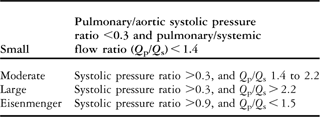
Table 3 The Q p/Q s (shunt) parameter limit values for different ventricular septal defects and Eisenmenger syndrome.
Results
Results for the cardiovascular system under normal, ventricular septal defect and Eisenmenger conditions are presented in Table 2. In this model, although there was a remarkable increase of the pulmonary artery pressure and right ventricle pressure (Fig 3a and b), left ventricle pressure, aortic pressure, aortic flow (Fig 4a–c), and pulmonary compliance (Fig 5a) decreased in the Eisenmenger syndrome. In addition, left-to-right septal flow reversed (Fig 5b) in this disease.

Figure 3 (a and b) An increase in the pulmonary artery pressure and right ventricle pressure.

Figure 4 (a–c) Left ventricle pressure, aortic pressure and aortic flow decreased in the Eisenmenger syndrome.

Figure 5 Pulmonary compliance decreased – the heavy lines show the working areas of the characteristics (a); left-to-right septal flow reversed (b).
Discussion
In this study, we have designed an analogue circuit that is able to simulate normal condition and the Eisenmenger syndrome with ventricular septal defect. Simulation is available for investigating cardiovascular physiology. Several models for the cardiovascular system have been proposed for understanding the cardiovascular physiology and pathophysiology, such as arrhythmia, myocardial ischaemia, hypertension, and valve diseases.Reference Rupnik, Runovc, Sket and Kordas7–Reference Tsalikakis, Fotiadis and Sideris9, Reference Mukkamala and Cohen15–Reference Aguilar, Balsera and Bernal20 Mukkamala et alReference Mukkamala and Cohen15 have designed a forward model-based cardiovascular system identification method that utilises coupling mechanisms related to electrocardiograph-derived heart rate signals, arterial blood pressure, and instantaneous lung volume: circulatory mechanics, heart rate baroreflex, instantaneous lung volume to heart rate, instantaneous lung volume to arterial blood pressure, and sinoatrial node. We did not use these coupling mechanisms in our study. Instead, we applied baroreflex coupling mechanism by changing systemic resistance and pulmonary resistance manually. Resistance values were convenient with physiological output results.Reference Vongpatanasin, Brickner, Hillis and Lange21 In addition, other coupling mechanisms mentioned in Mukkamala's paper can be added to subsequent model studies. However, the principal goal of this study was to focus especially on the Eisenmenger syndrome for educational purposes.
The measurements we obtained for the Eisenmenger syndrome were close to what is reported in the literature. Although the pulmonary artery pressure and right ventricle pressure increased, left ventricle pressure, aortic pressure, aortic flow, and pulmonary compliance decreased.Reference Vongpatanasin, Brickner, Hillis and Lange21 The Eisenmenger syndrome consists of a congenital communication between the systemic and pulmonary circulation, with resultant elevated pulmonary vascular resistance, pulmonary arterial hypertension, and bidirectional or right-to-left shunting. The pathophysiological mechanisms of the Eisenmenger syndrome are not completely known.Reference Vongpatanasin, Brickner, Hillis and Lange21 Chronic exposure of the pulmonary vasculature to increased blood flow produces endothelial cell damage and release, as well as the activation of factors – such as elastase, insulin-like growth factor I, transforming growth factor, thromboxane B2, von Willebrand factor, endothelin – that ultimately lead to vasoconstriction and structural changes, including intimal proliferation and fibrosis, medial hypertrophy, occlusion of the small vessels, plexiform lesions, and necrotising arteritis.Reference Heath and Edwards6, Reference Todorovich-Hunter, Dodo, Ye, McCready, Keeley and Rabinovitch22–Reference Dinh Xuan, Higenbottam, Clelland, Pepke-Zaba, Cremona and Wallwork29 The structural changes lead to increased pulmonary vascular resistance, pulmonary arterial pressure, and reversal of the shunt, which is similar to the result obtained from the model.Reference Vongpatanasin, Brickner, Hillis and Lange21, Reference Bouzas and Gatzoulis30
In our model, we first demonstrated the normal condition of the cardiovascular system and then the ventricular septal defect by using the preceding normal condition model. Finally, we revised our model in order to simulate the Eisenmenger syndrome.
In conclusion, our model is effective and available for simulating normal cardiac conditions and cardiovascular disease. This electrical model proves useful for studying the pathogeneses of the cardiovascular disease, especially the Eisenmenger syndrome. Models can also be built up for teaching purposes; one can easily give information about the system by using its model. We also built up an electrical circuit diagram as a model of the cardiovascular system for training, research, and system classification purposes.
Acknowledgement
We acknowledge a valuable professor of Istanbul Technical University, Tamar Olmez, for helping.


