


Trisomy 7p is a rarely seen genetic abnormality. Very few patients have been reported in the literature. The clinical signs associated with this abnormality have been clearly defined.1 These include psychomotor retardation, a wide and protruding forehead, posterior fontanelle remaining open, hypertelorism, low-set ear, abnormal palmar lines, ocular, genital, and cardiovascular abnormalities. Reference Wilde, Teele and Aftimos1,Reference Schmidt, ten Cate, Weiss and Koehler2 Changes in the number of copes such as chromosomal duplications or deletions may be associated with cardiovascular abnormalities. Reference Schmidt, ten Cate, Weiss and Koehler2 Various cardiovascular abnormalities may be seen in more than one-third of trisomy 7p patients. Reference Ozgun, Batukan, Asbug, Akgun, Caglayan and Dundar3
Tetralogy of Fallot (ToF) is the most frequently observed cyanotic CHD, and its incidence is 0. 34 per 1000 live births. Patients present with a wide perimembranous ventricular septal defect, overriding of the aorta, stenosis in the right ventricular outflow, and right ventricular hypertrophy. Reference van der Ven, van den Bosch, Bogers and Helbing4 The disease is also accompanied by various genetic or chromosomal syndromes in approximately 15% of patients. Reference Athanasiadis, Mylonas and Kasparian5 In this case report, we present an infant patient with a combination of partial trisomy 7p and ToF.
Case report
The female infant delivered through normal spontaneous vaginal birth by a 34-year-old healthy mother at week 36 as G4D2Y2 had a birth weight of 1940 g, which was small for her gestational age, and an APGAR of 6/7. The mother’s antenatal history did not include smoking, alcohol, warfarin, or another drug use or any other chronic conditions. The patient, who had fascial dysmorphism and whose parents were first cousins, underwent abdominal ultrasonography, and the result was normal. ToF was identified during echocardiographic exam (Fig 1). The brain magnetic resonance showed that both occipital lobes were hypoplasic, ectasia was present in the ventricular system, and there was widening of the peripheral cerebrospinal fluid distance in the frontotemporal anterior parts. The patient was identified to have severe sensorineural hearing loss during the hearing test. She was scheduled for chromosome analysis.

Figure 1. In echocardiographic examination; (a) Dextroposition of the aorta and large perimembranous ventricular septal defect (star) are observed.. ( b ) Normal relationship between the aorta and pulmonary artery and RVOT are seen. It is seen that the pulmonary annulus and main pulmonary artery are hypoplasic, and the branches of the pulmonary artery are well developed and confluent. RV: Right ventricle, LV: Left ventricle, Ao: Aorta, LA: Left atrium, RVOT: Right ventricular outflow tract, MPA: Main pulmonary artery, LPA: Left pulmonary artery, RPA: Right pulmonary artery.
The chromosome analysis performed on the short-term lymphocyte culture derived from peripheral blood yielded the result of der(15)t(7;15)(p10;q10). On the meta-phases derived from the patient’s materials, trisomy of the p arm of the chromosome number 7 was identified (Fig 2). The parents underwent chromosome analysis. While the mother had normal karyotype, the father’s karyotype was 46,XY,t(7;15)(p10;q10). The patient received micro-array study, and an increase of 57,963 kbp encompassing the 7p22. 3p11. 1 region was identified.
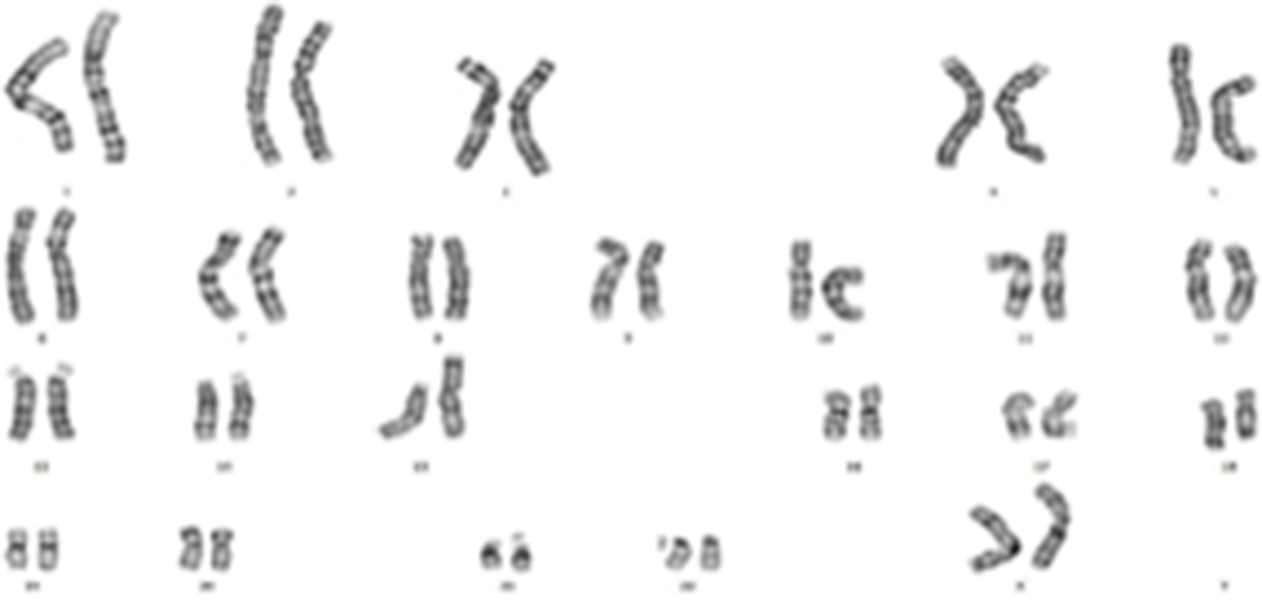
Figure 2. The karyotype image of the case.
When the patient turned became seven months old, her saturation values dropped to 60% and she began to experience spells. The patient, who had developmental retardation, underwent a left Blalock-Taussig aortopulmonary shunt procedure using a 4-mm Goretex graft. The patient’s saturation levels were within the normal ranges. When the patient became 18 months old, she underwent cardiac catheterisation and it was decided to perform total correction. Afterwards, a total correction surgery was performed via sternotomy and transatrial approach using a transannular patch and monocusp. No problems were identified in the follow-up echocardiographic exam.
The patient’s physical exam that was done at the age of 21 months revealed dolichocephaly, a wide and protruding forehead, hypertelorism, a depressed nasal bridge, anteverted nostrils, low-set and dysplastic ears, downturned corners of mouth, a high palate, micrognathia, brachydactyly, bilateral transverse single line on the hands, dimples on the hands, ankles, elbows, and back as well as hypotony. Furthermore, the patient’s fontanelle had not yet closed (8x4 cm). The patient was observed to be behind in terms of neuromotor development steps. She could not hold her head upright or speak. The patient is still under outpatient follow-up. Signed informed consent forms were obtained from the family members to allow the use of clinical findings, laboratory test result and images.
Discussion
Trisomy 7p is a rarely seen genetic disease that develops due to the partial or full duplication of the shorter arm of the chromosome 7, which involves many systems. It was reported for the first time in the year 1968 in the literature. Most cases develop as a result of the malsegregation of parental balanced translocations. Reference Schmidt, ten Cate, Weiss and Koehler2,Reference Arens, Toutain and Engelen6,Reference Kozma, Haddad and Meck7 For our case, it was identified that the father was the carrier of a balanced translocation and that the p arm of the chromosome 7 was triplicated due to the inheritance of an imbalanced product by the child. Parentally inherited balanced-looking changes, regardless of gender, may present with different clinical pictures that may vary according to the chromosomal content after abnormal gamete formation. In our case, der15 was detected after abnormal gamete development in a balanced-looking healthy t(7;15)(p10;q10) father. Chromosome analysis should be done in such cases, and families should be informed about possible situations before pregnancy.
The clinical findings of the trisomy 7p syndrome have been well-defined. Patients may present with abnormalities of the cranio-fascial, musculoskeletal, genital, ocular, and cardiovascular systems. Reference Papadopoulou, Sifakis and Sarri8,Reference AlFardan, Brown, Gessner, Lunt and Scharer9 Half of the cases are lost during the infancy period, and nearly half of them develop mental retardation. Reference AlFardan, Brown, Gessner, Lunt and Scharer9 While the clinical findings may vary per the size of duplication, the most frequent complications develop in the intrauterine period and as a result of the musculoskeletal system involvement associated with the abnormal muscle tone in post-natal life. Hypotony may be seen in one-third of patients. Reference Kozma, Haddad and Meck7 Our patient also had hypotony. The primary cranio-fascial abnormalities seen in cases include micrognathia, hypertelorism, low-set and malformed ears, a large fontanelle and wide sutures. Reference Wilde, Teele and Aftimos1,Reference Kozma, Haddad and Meck7,Reference Lurie, Schwartz, Schwartz and Cohen10 The findings in our case were also consistent with the literature. Severely large front fontanelles that do not close are specifically the most characteristic finding in patients with trisomy 7b syndrome. Reference Wilde, Teele and Aftimos1 The patient’s front fontanelle was still open during her exam performed at 21 months of age, and it was rather large. A large front fontanelle that closes late may also present with different genetic syndromes such as trisomy 13, trisomy 18, achondroplasia, and campomelic dysplasia; however, trisomy 7 should also be considered for differential diagnosis in such a situation.
Various cardiovascular abnormalities may be seen in more than one-third of trisomy 7 patients. Reference Ozgun, Batukan, Asbug, Akgun, Caglayan and Dundar3 A study evaluating the trisomy 7 patients reported the presence of cardiovascular abnormalities in 43% of patients. The most frequently observed abnormalities include atrial septal defect, ventricular septal defect, and patent ductus arteriosus. In the literature, patients with trisomy 7b were also reported to have coarctation of the aorta, ToF, pulmonary and aortic valve stenosis, double inlet left ventricle, and dextrocardia. The spectrum of abnormalities suggests that multiple segments on 7p may be associated with normal development of the heart. Reference Schmidt, ten Cate, Weiss and Koehler2,Reference Lurie, Schwartz, Schwartz and Cohen10 In our case, ToF was present and it was surgically treated with success. A literature review showed that the association of trisomy 7p with ToF is very rare.
Many genetic and chromosomal syndromes may accompany ToF. Approximately 15% of cases are syndromic cases. These may include the DiGeorge syndrome, trisomy 21, 18, and 13 as well as the VACTERL and CHARGE syndromes. Reference Athanasiadis, Mylonas and Kasparian5 A study looking at patients with syndromic ToF reported that hypoplasic pulmonary arteries, aortic dilatation, and major aortopulmonary collateral arteries were more frequent compared to those without syndrome and that the cardiac phenotype was associated with the underlying genetic profile. Similarly, it was reported that patients with ToF had a higher need for staged surgery due to the high frequency of hypoplasic pulmonary artery. Reference Athanasiadis, Mylonas and Kasparian5 In our case, it was also required to perform a systemic pulmonary arterial shunt procedure prior to total repair due to the low saturation in early infancy period. Therefore, staged surgery was performed.
In conclusion, patients diagnosed with trisomy 7p should certainly be scheduled for an echocardiographic exam and be scanned for any CHDs that may accompany it. The CHDs that most frequently accompany this syndrome include atrial septal defect, ventricular septal defect, and patent ductus arteriosis. Yet, it should be known that ToF may also be present, albeit rarely. In patients diagnosed with ToF, one must act with caution with respect to the associated chromosomal and genetic abnormalities, as well. It should be taken into account that ToF, the most frequently encountered cyanotic disease, may be associated with many syndromes and such patients more frequently require palliative intervention.
Acknowledgements
None.
Author contribution
Study conception and design: OG, MS, TAD.
Data collection: OG, TAD.
Analysis and interpretation of results: MS, TAD.
Draft manuscript preparation: OG, MS, TAD.
All authors reviewed the results and approved the final version of the manuscript.
Financial support
This research received no specific grant from any funding agency, commercial or not-for-profit sectors.
Conflict of interest
The author has no conflict of interest.
Ethical standards
This case description does not contain any study with animal performed by any of the authors. All applicable and international, national, and/or institutional guidelines for the case were followed. The procedure performed in the case description was in accordance with the ethical standards of the institutional and/or research committee and with the declaration and with the 1964 Helsinki declaration and its later amendments or comparable ethical standards.
Consent was obtained from the parents of the individual included in the study.


