Although echocardiography has long been used for the assessment of the size of the heart in children, previously, we have applied two-dimensional echocardiography to assess the information on the longitudinal development of the left ventricular end-diastolic dimension and rate of its development by using height as an index in children less than 18 years of age.Reference Nagasawa, Arakaki, Yamada, Nakajima and Kamiya1 We detected a linear relationship between height and the dimensions of the heart in individuals. Height is a simple and accurate index for the assessment of normalcy of cardiac dimensions and there is a sex difference in children. However, in the case of infants less than 1 year of age, the normal dimensions of the heart remain undefined. Our previous study and other similar studies revealed that the cardiac dimensions obtained in these studies were larger than the actual cardiac dimensions. For the purpose of the study, we assumed that the relationship between the height and cardiac dimensions in infants differs from that in children older than 1 year of age. As a result, we assumed that it must be incorrect to apply the regression equations from the above-mentioned studies to the data obtained for infants less than 1 year of age. We were unable to draw an accurate conclusion because of the limited nature of our data.Reference Nagasawa, Arakaki, Yamada, Nakajima and Kamiya1 No previous study has reported differences in cardiac dimensions between these two age groups.
The objectives of our study are to assess normalcy of the left ventricular end-diastolic dimension by using height as an index in infants and premature neonates with a height of less than 75 centimetres; to identify any differences in the relationship between height and cardiac dimensions in infants with a height of less than 75 centimetres and children, above the age of 1 year, with a height of more than 75 centimetres; to show any differences between development in infants born full term and premature neonates; and to show any differences between development in infants on the basis of sex.
Methods
Study subjects
It is difficult to obtain data on cardiac dimensions from a large number of normal infants. We therefore considered the children who met the following requirements to be “normal”: infants without persistent arterial duct, neonatal physiological pulmonary hypertension, and pulmonary disease; those with no history of congenital cardiac disease and no evidence of coronary artery lesions as determined by repeated echocardiograms, electrocardiograms, and other examinations; and those with no significant illnesses or anomalies except for low birth weight. We defined children less than 1 year of age as “infantile” and individuals above the age of 1 year as “children”.
Only those infants who underwent echocardiographic examination at the Gifu Prefectural General Medical Center were included in the study. The study group consisted of 243 Japanese infants – 123 male and 120 female – including 95 premature neonates, whose ages ranged from 10 days to 12 months. Table 1 shows the distribution of the ages of the children on whom echocardiographic examinations were performed. The height of the children ranged from 35 centimetres to 75 centimetres and their weight ranged from 780 grams to 10.5 kilograms. The average body height, body weight, and chest circumference were obtained according to the statistical analysis of children in Japan in 2000.
Table 1 Distribution of ages in 243 echocardiographic examinations performed.

Data acquisition
Two-dimensional echocardiographic studies (Sonos 2500, Hewlett-Packard, New York, United States of America) were performed in the left parasternal short-axis view at the level of the chordae tendinae just below the mitral valve with a 7.5-megahertz transducer at rest. Left ventricular end-diastolic dimension was measured as the distance between the endocardial surface of the ventricular septum and the left ventricular posterior wall at the onset of the QRS complex on the electrocardiogram by M-mode recordings. Each dimension was measured by the “centre convention”, that is, using the middle bright contour excluding the inner and outer areas, with calibrated electronic calipers.Reference Nagasawa, Arakaki, Yamada, Nakajima and Kamiya1 All measurements were made by the same observer (HN). The final values were obtained from an average of more than three successive beats. Intra-observer error was less than 4%, which was calculated on the basis of the data on another 30 normal controls that were measured three times each.
Clinical information on these infants was collected from outpatient and in-patient records. Body height and body weight were measured at the time of echocardiography, and body height was measured in the prone position. Body surface area was calculated according to the equation reported by De Boit and De Boit.Reference Du Boit and Du Boit2
Statistical analyses
The strength of the relationships between the left ventricular end-diastolic dimension and the height or chest circumference was evaluated by using Pearson’s correlation coefficient (r-value). Differences in the slope of the fixed curves were evaluated by the analysis of covariance.Reference Snedecor and Cochran3 A level of p < 0.05 was considered statistically significant.
Results
Physical development of subjects
The mean and 10th and 90th percentiles for the height and weight of these infants were the same across ages, as in the case of the general population of Japanese children.
Relationship between height and left ventricular end-diastolic dimension
The relationship between left ventricular end-diastolic dimension (Y) and body height was best expressed linearly, and the regression equation for the line was Y (millimetre) = 0.41 × [height (centimetre)] + 1.9, excluding premature neonates. The fragment (F) values of this regression equation and those that we had reported previously were 19.5 in male and 18.2 in female infants, which indicates that these equations were different from each other. In addition, we found a discontinuous point. The point was approximately 50 centimetres in height. Next, we divided the data into two categories: height less than 50 centimetres and height more than 50 centimetres. The F-value for these two groups was 5.9 (p < 0.03), and the two groups had different regression equations. Figure 1 shows the relationships between left ventricular end-diastolic dimension and body height for both groups. Table 2 denotes the regression equations. The rate of development of the left ventricle was higher during the first year of life, especially in premature neonates, than at any other age examined.

Figure 1 Relationship between left ventricular end-diastolic diameters and height in 243 subjects. Data for males and females are represented by closed circles and open ones, respectively.
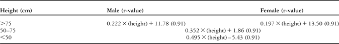
Table 2 Regression equations between left ventricular end-diastolic diameter and height.

The units of height and left ventricular end-diastolic diameter are centimetre, millimetre, respectively
The overall standard deviation was 15% of the average size. Figure 2 shows the lines indicating the mean and the 10th and 90th percentile values of left ventricular end-diastolic dimension with respect to height.

Figure 2 Relationship between left ventricular end-diastolic diameter and height. The mean and the 10th and 90th percentiles of the data are represented by solid and dotted lines, respectively.
Determination of points of division
We determined the height of the border between the regression equations of the lines denoting infants and children. The regression equation of the line was calculated on the basis of the data obtained between the heights of 55 and 70 centimetres. The regression equation was left ventricular end-diastolic dimension (millimetre) = 0.352 × [height (centimetre)] + 1.86. The point of intersection between the formerly reported regression equation for male children and that for infants was calculated to lie at 76 centimetres. The point of intersection between the formerly reported regression equation for female children and that for infants was calculated to lie at 75 centimetres. The height of 75 centimetres was used as a basis for the division of the infants into two categories, namely, infancy and childhood.
Similarly, we determined the height of the border between the regression equations for the lines denoting infants and premature neonates. The regression equation was calculated on the basis of the data obtained at a height of less than 45 centimetres. The regression equation was left ventricular end-diastolic dimension (millimetre) = 0.495 × [Ht (centimetre)] − 5.43. The point of intersection between the lines denoting infants and premature neonates was calculated to lie at 51 centimetres. The height of 50 centimetres was used as a basis to divide the infants into two categories, namely, premature neonates and mature neonates.
Differences in left ventricular end-diastolic dimension by sex
The F-value, which is the index of the probability of a difference in the slopes of the curves for both sexes in terms of height, derived from an analysis of covariance was 1.8 (p > 0.5). Multiple linear regression analysis revealed that the computed t-value for the difference in sex was 1.05. These findings indicate that there was no difference between the sexes in the development of the left ventricle when height was used as an index.
Discussion
Earlier studies have reported regression equations that help identify normal development of left ventricular end-diastolic dimension in childhood by using two-dimensional echocardiography. Body surface area was used as an index in a majority of the regression equations. These equations are not only of no practical use but also provide ambiguous results in cases of infantile ages because logarithms or exponents are used for estimation. In cases of children of infantile ages, the regression equations that use logarithms or exponents show a convex curve on the upper side and a sharp rise, when body weight or body surface area is used as an index. Therefore, in cases of infantile ages, it is difficult to detect the disparity by sight.
Gutgesell et alReference Gutgesell, Paquet, Duff and McNamara4 reported the equations as left ventricular end-diastolic dimension = 9.22 × log (body weight) + 6.94 and left ventricular end-diastolic dimension = 12.88 × log (body surface area) + 37.75.Reference Gutgesell, Paquet, Duff and McNamara4 According to their equations, the estimated left ventricular end-diastolic dimension values were 11.7 and 28.8 millimetres at birth and 15.8 and 32.8 millimetres at the age of 1 year in infants with average height and weight, respectively. In addition, Henry et alReference Henry, Ware, Gardin, Hepner, McKay and Weiner5 reported the equation as left ventricular end-diastolic dimension = 45.2 × [(body surface area) × 0.333] − 6.6. According to their equation, the estimated left ventricular end-diastolic dimension values were 19.8 millimetres at birth and 27.1 millimetres at the age of 1 year. Pearlman et alReference Pearlman, Triulzi, King, Newell and Weyman6 reported the equation as left ventricular end-diastolic dimension = 36.70 × [(body surface area) × 0.514]. According to their equation, the estimated left ventricular end-diastolic dimension values were 16.1 millimetres at birth and 23.4 millimetres at the age of 1 year. We estimated the mean and standard deviation of left ventricular end-diastolic dimension as 19.2 and 0.9 millimetres, respectively, at birth and 27.8 and 0.6 millimetres, same as above, at the age of 1 year. Except for the equation by Henry et alReference Henry, Ware, Gardin, Hepner, McKay and Weiner5, it is apparent that the other equations underestimate or overestimate left ventricular end-diastolic dimension values at birth and at the age of 1 year. However, the equation by Henry et alReference Henry, Ware, Gardin, Hepner, McKay and Weiner5 underestimates the left ventricular end-diastolic dimension values at the 30-week gestational age in neonates by 10%. The disparity at birth was calculated to be approximately 40–50% and approximately 20–40% at the age of 1 year. They might take the data of older ages than infants into confidence or ignore the disparity at these ages. However, the disparity is apparently not small and could lead to wrong conclusions. By using height as an index, disparity can be detected very clearly at sight.Reference Nagasawa, Arakaki, Yamada, Nakajima and Kamiya1 Therefore, we reexamined the problem by using height as an index.
Expectedly, an increase in the rate of development of left ventricular end-diastolic dimension led to differences in the values estimated between infants and children and between infants and premature neonates (Fig 1). The borders were estimated at the height of 50 and 75 centimetres, respectively. It seems that they discontinuously change the increasing rate of left ventricular end-diastolic dimension at those ages.
Statistical analysis revealed that these three equations were distinct from each other. The equations reported in this study are feasible for left ventricular end-diastolic dimension estimation in infants and premature neonates.
Imai et alReference Imai, Satomi and Yasukochi7 reported that the increase in the rate of development of left ventricular end-diastolic dimension correlated closely with body weight in 55 premature and full-term infants, with weight ranging from 543 to 3966 grams. They reported the equation as left ventricular end-diastolic dimension = 9.369 + 2.161 × (body weight in kilogram) and r-value as 0.74. The r-value was relatively small, and the equation may be interpreted in more ways than one, that is, an exponent might assume the relationship between left ventricular end-diastolic dimension and body weight. The average volume reported in their study was lower than that reported in ours. We suspect that the data reported in their study also included data obtained from cases of neonates with physiological pulmonary hypertension.
Hypothesis on the existence of two discontinuous points
The first discontinuous point is located between the lines denoting foetal age and infantile age. This point might be attributed to the end of rapid development of the heart in the foetal period.
The second point is located between the lines denoting infantile age and childhood. This could be attributed to the change in the manner of movement arising due to age, from crawling to walking (bipedalism).
Height as an index of left ventricular end-diastolic dimension
The equations on left ventricular end-diastolic dimension have been reported by using weight or body surface area as an index. Gutgesell et aReference Gutgesell, Paquet, Duff and McNamara4 first reported the linear relationship between height and left ventricular end-diastolic dimension. Nidorf et alReference Nidorf, Picard and Triuzi8 stated that for clinical purposes, body height was an ideal index for the estimation of cardiac dimensions. In addition, our data reveal that height is an appropriate and practical index for evaluating the development of left ventricular end-diastolic dimension in children.Reference Nagasawa, Arakaki, Yamada, Nakajima and Kamiya1, Reference Nagasawa and Arakaki9
The use of two linear equations to evaluate the relationship between height and left ventricular end-diastolic dimension instead of one equation using a logarithm or an exponent might be discouraged. We found that these equations have an intersection and are of no practical use.
In a premature neonate, it is difficult to measure true body weight, because most neonates undergo some medical treatments such as incubation, continuous injection, nasogastric tubing, electrocardiographic monitoring, transcutaneous O2 and CO2 pressure monitoring, etc. Height can be measured more accurately than weight in premature neonates under acute conditions. Therefore, it is an appropriate index for the estimation of cardiac dimensions.Reference Nidorf, Picard and Triuzi8
We reported that there was a sex difference in left ventricular end-diastolic dimension during childhood.Reference Nagasawa, Arakaki, Yamada, Nakajima and Kamiya1 However, in this study, we could not detect a difference. This could be partially because the disparity in left ventricular end-diastolic dimension estimation between the sexes was only 0.4 millimetre, even in infants with a height of 75 centimetres in our previous study. Even if a significant difference could be detected in a larger number of infants, if we measure it in more than 1000 cases, it would still be insignificant and of no practical use. It is expected to be rather small and not worthwhile practically.
Problems of measuring procedure
The difference in the two regression equations for the estimation of left ventricular end-diastolic dimension between infants and children is often attributed to the different methods of measuring height: in the prone and erect positions. In addition, the accuracy of measurements performed in the prone position is doubtful. The difference in height between measurements performed in prone and erect positions, even at the age of 1.5 years was only 1 centimetre – the mean height at the age is 85 centimetres in male and 83 centimetres in female neonates. The difference was small and the accuracy of the measurement of height is not doubtful. The relationship between chest circumference and left ventricular end-diastolic dimension is also well established. Chest circumference can be measured without any error. However, we detected a difference in the two regression equations for the estimation of left ventricular end-diastolic dimension between infants and children in this case as well.
The difference in the two regression equations for the estimation of left ventricular end-diastolic dimension between infants and premature neonates or children and infants is attributed to different body proportions, the change in proportions of the head, trunk, and leg length with age. As proportions of the body change gradually with age, the relationships between the indices and left ventricular end-diastolic dimension cannot be linear.
Is it possible to unify the three equations into one?
The development of left ventricular end-diastolic dimension is in the form of a logarithmic curve; however, we could not find any equation that would satisfy all the conditions in the development of the left ventricular end-diastolic dimension in childhood using a logarithmic index.