Persistent fifth arch is a rare congenital anomaly. It was first presented in 1969.Reference Van Praagh and Van Praagh1 It is defined as a single aortic arch with two separate parts – superior and inferior – with each part having individual adventitial layers, and a location anterior to the trachea.Reference Lambert, Blaysat, Sidi and Lacour-Gayet2 This anomaly is most often associated with other congenital cardiac anomalies. To the best of our knowledge, this is the first report of such an entity incidentally discovered in an adult patient as an isolated anomaly.
Case presentation
A 64-year-old man was referred to our department to undergo multi-slice cardiac computed tomography scan for evaluation of his coronary artery bypass grafts, performed 3 years ago. He had undergone conventional coronary angiography because of a new onset of chest pain, which showed that the two saphenous venous grafts onto the diagonal and right coronary arteries were cut off and that the left internal mammary artery graft onto the left anterior descending artery was not visualised because of severe tortuosity of the left subclavian artery. His past medical history was insignificant except for hypertension and hyperlipidaemia. Echocardiography showed good left ventricular ejection fraction and did not reveal any associated congenital cardiac anomalies. A cardiac computed tomography scan was performed on a 64-slice CT scanner (Somatom Sensation, Siemens, Forchheim, Germany), revealing patent left internal mammary artery graft with no intracardiac structural abnormality. However, a tubular connection between the ascending and descending aorta paralleling the usual aortic arch was detected (Figs 1 and 2).

Figure 1 Sagittal view demonstrating the double-lumen aortic arch anterior to the trachea.

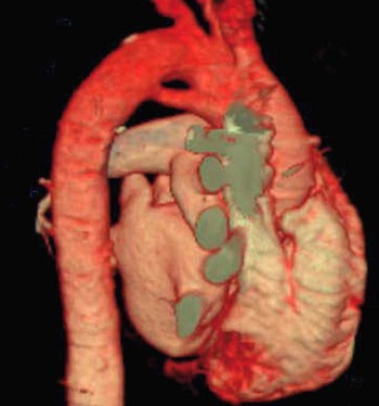
Fig. 2 Volume-rendered image depicting a double-lumen arch.
Discussion
During the embryonic period, the neural cells form the branchial arches. Each of the branchial arches is supplied by an artery called the aortic arch. The fourth aortic arch forms the definitive aortic arch and the right subclavian artery. The fifth aortic arch is located below the fourth arch and is small in 50% of embryos and degenerates. In the remaining embryos, the fifth arch does not exist. A portion of the sixth arch will form the ductus arteriosus and the left pulmonary artery. Persistence of the fifth arch results in a double-lumen aorta; this is due to the connection with the posterior aorta, the left pulmonary artery, or the ductus.Reference Khan and Nihill3 Double-lumen aortic arch has been divided to two main subtypes, systemic-to-systemic and systemic-to-pulmonary connection.Reference Gerlis, Dickinson, Wilson and Gibbs4 In the systemic-to-systemic type, the fifth arch connects the ascending and descending aorta – innominate artery to subclavian artery – and in the systemic-to-pulmonary type the fifth arch connects the ascending aorta to a derivate of the sixth arch, most commonly the left pulmonary artery;Reference Donti, Soavi, Sabbatani and Picchio5 the systemic-to-systemic connection has no functional implication, and if present as an isolated finding the patient is usually asymptomatic. This type is usually underdiagnosed, but it has been reported with other cardiac anomalies, including aortic coarctation, arch interruption, and ventricular septal defects;Reference Donti, Soavi, Sabbatani and Picchio5 more complex anomalies such as tetralogy of Fallot, cor triatriatum, complete d-transposition of the great arteries, truncus arteriosus, pulmonary, and tricuspid stenosis have been associated with the systemic-to-pulmonary subtype.Reference Bernheimer, Friedberg, Chan and Silverman6 In this type, the connection has functional significance and causes a systemic-to-pulmonary shunting. When present with other cardiac anomalies such as pulmonary atresia, coarctation, and arch interruption, this connection serves as an alternative connection and helps in patient survival.Reference Khan and Nihill3 In fact, the clinical presentation depends on which one of these anatomic connections exist and on the associated cardiovascular anomalies. Gerlis et alReference Gerlis, Dickinson, Wilson and Gibbs4 and other authors have stated that this anomaly may not be as rare as thought, as owing to the difficulty in making the diagnosis it is often misdiagnosed as other anomalies, most commonly patent ductus arteriosus and aortopulmonary window in the systemic-to-pulmonary subtype of the disease.Reference Gerlis, Dickinson, Wilson and Gibbs4 In addition, it has been said that persistent fifth aortic arch has to be differentiated from retro-oesophageal right subclavian artery and double aortic arch on conventional angiography.Reference Cabrera, Galdeano and Lekuona7 Interestingly, there are cases in which the abnormality was not clearly characterised and described, for example, as “a single large structure” or “an unusual aortic anatomy” during the surgeries performed for correction of the associated anomalies.Reference Kirsch and Julsrud8
To the best of our knowledge, all previous reported cases of persistent fifth aortic arch are childhood findings, nearly all of them associated with complex cardiac anomalies, and most of them have been detected on conventional angiography or echocardiographic studies. Our case is unique in that it was an isolated finding that was diagnosed incidentally during a cardiac computed tomography scan evaluation of the coronary artery bypass grafts in an adult patient. Up to 20% incidentally discovered extracardiac findings have been reported on multi-detector computed tomography scan evaluation of the coronary artery bypass grafts, where, in comparison with native coronary arteries, more extended parts of the thorax are evaluated.Reference Mueller, Jeudy, Poston and White9, Reference Killeen, Dodd and Cury10 By the use of coronary computed tomography scan before or after coronary artery bypass grafting, the isolated systemic-to-systemic subtype of the double-lumen aortic arch can become a less rare entity; considering that it is mostly reported in children, being familiar with it is becoming important for adult cardiologists and cardiac surgeons who can come across this anomaly incidentally on a cardiac computed tomography scan report.