


Atrioventricular block is defined as congenital if diagnosed in uterine life, at birth, or within neonatal life. Reference Baruteau, Pass and Thambo1 The prevalence is 1 per 15,000–20,000 live births. Reference Hunter and Simpson2 Congenital atrioventricular block can accompany congenital cardiac structural disorders or congenital conduction system pathologies though it can be seen in completely normal hearts without any structural cardiac pathology. Non-structural atrioventricular block generally occurs as a result of immune-mediated injury of conduction system due to transplacental passage of maternal antibodies. Reference Gegieckiene, Vankeviciene, Kinciniene and Liubsys3 There is also a complex genetic background. Reference Syam, Chatel and Ozhathil4
Regardless of the reason, atrioventricular block can be life threatening immediately after birth that can result in sudden death in neonatal period or cardiomyopathy can be seen in childhood. Reference Paiva, Ribeiro and Garcia5
When atrioventricular block is diagnosed prenatally, it must be kept in mind that urgent treatment may be inevitable so all preparations must be made in the operating room. Preferably, in this case, it should be ensured that the delivery is performed in centres with paediatric cardiology and paediatric cardiac surgery.
Transvenous temporary pacemaker implantation in neonates is a very limited and challenging option due to small vessels and may be preferred when urgent surgery is not accessible. Reference Khanna, Arora, Aravindan and Prasad6 In neonates with small vessel sizes, surgical implantation via thoracotomy or limited sternotomy is required. Surgical technique in neonates differs from children and adults. In this study, we aimed to present our method and results of urgent surgical pacemaker implantation technique immediately after birth in neonates due to congenital atrioventricular block.
Material and methods
Local ethics committee approval was obtained in May 2021. Between June 2014 and February 2021, 10 neonates who had congenital atrioventricular block underwent urgent surgical operation to implant permanent epicardial pacemaker by using subxiphoidal mini-sternotomy approach. Six of the patients were female and four of them were male. Mean age was 4.3 days (0–11), while three of them were operated on the day of birth. Mean weight was 2533 g (1200–3300).
Three patients (33%) were diagnosed prenatally, and all of them were operated on the day of birth. Two patients were rescued by using transvenous temporary pacemaker and operated on the second and third day of births after obtaining permanent pacemakers and leads. Five patients were operated in semi-elective conditions.
Two neonates had family history that mothers had diagnosis of rheumatic diseases requiring medical treatment. One patient had sudden sibling death after birth, but the cause of death could not be determined. Two patients had a history of consanguineous marriage.
The mean heart rate before operation was 54 ± 11 per minute.
Results
All operations were achieved by using subxiphoidal mini-sternotomy incision (Fig 1) under general anaesthesia. After the pericardium has been opened limitedly, epicardial 25-mm length dual leads were implanted on right ventricular surface properly and generators were fixed on right (7 patients) or left (three patients) diaphragmatic surfaces by opening the pleura on that sides (Fig 2). There was no complication, morbidity, and mortality related to surgery. Target pacing threshold at 0.5 ms (volt) was below 3 volts and sensing threshold was below 15 mVolt such if that was high, leads were removed and fixed on another surface. Pacemaker effectivity was controlled during and after surgery, and all measurements were made according to the needs of the patients. One patient died on 21th day of operation due to neonatal sepsis.

Figure 1. Limited subxiphoidal mini-sternotomy incision, dotted line shows full sternum.

Figure 2. Intraoperative view of the implanted leads and battery. Arrow shows the battery on the right diaphragmatic surface.
During routine controls, 7 patients had no medical therapy. In 1 patient, junctional ectopic atrial extrasystoles were detected in 6-month Holter ECG and completely disappeared with oral drug therapy. In one patient, rare atrial arrhythmias were detected after infant period and followed without medication and completely disappeared.
Discussion
Congenital heart block is a rare but fatal pathology that can lead to sudden death at birth or in the neonatal period. Cause may not always be known. It is thought to be due to complex maternal and child-related causes or genetic background. Reference Zhou and Hua7 Family history, genetic predisposition, sibling deaths should be well questioned. In our study, 2 patients’ mothers had rheumatic disease requiring treatment, 1 patient had a history of sudden sibling death, and 2 patients had a history of consanguineous marriage, which highlights the importance of questioning this situation during pregnancy.
Congenital heart block can lead to sudden death at birth and immediately in neonatal period. It is also known that cardiomyopathy rarely develops in patients who reach childhood without treatment. Therefore, prenatal diagnosis is very important. Being prepared is lifesaving as immediate postpartum intervention is needed. Delivery should be done in centres with paediatric cardiology and paediatric cardiac surgery. Although the transvenous pacemaker has limited use in newborns, it can save time until the operation. In our study, 2 patients were rescued by using transvenous temporary pacemakers in the period until the obtaining of permanent pacemakers.
The operations performed for permanent pacemaker implantation in the postnatal and neonatal period have differences compared to children and adults; thus, these operations carry more risks. Reference Costa, da Silva, Filho and Carrillo8 The operation is done in a limited area. Epicardial leads are gently implanted in an appropriate area on the right ventricular surface using standard technique. The measurements of sensing and pacing thresholds and lead impedance should be done before connecting the battery; thus, implantation of the leads on a more appropriate place can be possible. Since pacemaker batteries are quite large compared to the baby’s body surface area, it is not possible to place them in a pocket that opens under the skin. For this reason, we prefer to fix the pacemaker battery in the thoracic cavity on the diaphragm in newborns (Fig 3). We did not detect any diaphragmatic dysfunction or lung pathology in these patients so we can say that this method is safe.
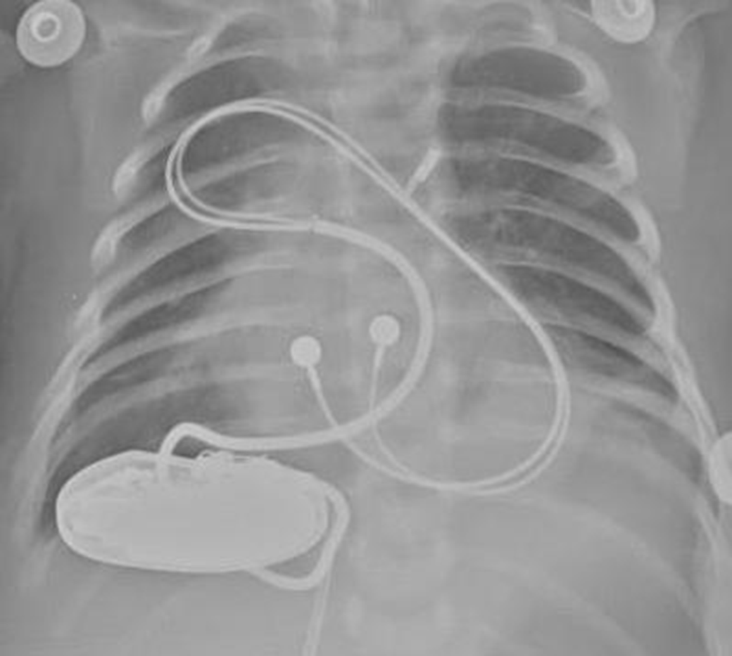
Figure 3. Postoperative view of the pacemaker leads and battery.
Conclusion
Congenital heart block is a rare but important cardiac pathology that requires urgent surgery at birth and immediately in neonatal period. Prenatal diagnosis is lifesaving. Minimally invasive permanent pacemaker implantation technique is a challenging but safe method that differs from children and adults.
Conflict of interest
None.
Ethical standards
This study is designed according to ethical standards.


