


Aetiology of paediatric cardiomyopathy
An aetiological classification of cardiomyopathy was presented by the Pediatric Cardiomyopathy Registry (PCMR) in 2000, in which the following five major categories were identified: familial, metabolic, syndromic, neuromuscular, and idiopathic.Reference Colan, Lipshultz and Lowe 1 – Reference Wilkinson, Landy and Colan 4 In addition, infectious causes are an important cause of dilated cardiomyopathy and heart failure. Data demonstrate that the prognosis varies depending on the aetiological category.Reference Colan, Lipshultz and Lowe 1 , Reference Wilkinson, Landy and Colan 4 – Reference Lipshultz, Orav and Wilkinson 6 The most common causes within each category are shown in Table 1.
Table 1 Genetic causes of paediatric cardiomyopathy.

ARVC=arrhythmogenic right ventricular cardiomyopathy; DCM=dilated cardiomyopathy; HCM=hypertrophic cardiomyopathy; LVNC=left ventricular non-compaction cardiomyopathy; RCM=restrictive cardiomyopathy
Familial cardiomyopathy
The term “Familial” as an aetiological class for cardiomyopathy typically implies an underlying pathogenic sarcomeric or cytoskeletal gene variant. The term is somewhat of a misnomer as, for example, Noonan syndrome is heritable and, therefore, may lead to an autosomal dominant family history of hypertrophic cardiomyopathy. In addition, “familial” cases may result from de novo mutations arising for the first time in the patient. Although these pathogenic variants are heritable, the proband is the first in the family with the mutation, and thus there would not be a family history upon presentation. In the last decade, it has been increasingly recognised that pathogenic variants or mutations in the cardiac sarcomere, cytoskeleton, desmosome, and nuclear envelope give rise to an important subset of paediatric cardiomyopathy cases, including disease in infants.Reference Kaski, Syrris and Burch 7 – Reference Kindel, Miller and Gupta 9 In a single-centre study of consecutive unrelated paediatric cardiomyopathy patients by Kindel et al, 42% of the cases had a familial aetiology based on molecular testing and/or Mendelian inheritance pattern within the pedigree.Reference Kindel, Miller and Gupta 9 The genetic testing for familial cardiomyopathy has been recently reviewed.Reference Tariq and Ware 10
Neuromuscular disease and cardiomyopathy
Neuromuscular disease is commonly associated with cardiomyopathy. Mutations in genes that are important for the functioning of both skeletal and cardiac muscles result in both myopathy and cardiomyopathy. The classic examples of neuromuscular diseases associated with cardiomyopathy are Duchenne and Becker muscular dystrophy. In Duchenne muscular dystrophy, an X-linked condition, boys typically present in childhood with clumsiness, weakness, and progressive difficulty with ambulation. They typically develop evidence of cardiomyopathy in adolescence, but great variability in age of onset exists, and there is much interest in better understanding the genotype–phenotype correlations that might predict the severity of cardiac involvement.Reference Tandon, Jefferies and Villa 11 Cardiac surveillance is indicated beginning with the establishment of the diagnosis of Duchenne muscular dystrophy. Likewise, carrier females of Duchenne muscular dystrophy mutations are at risk for dilated cardiomyopathy in adulthood and require ongoing cardiac screening. Other myopathies that have cardiac involvement include Emery–Dreifuss muscular dystrophy, inherited as an autosomal dominant or X-linked condition and classically characterised by a triad of joint contractures, weakness, and wasting – especially in a humero–peroneal distribution – and cardiac involvement including dilated cardiomyopathy and heart failure. The cardiac features classically present in the second decade. Limb girdle muscular dystrophies are a genetically heterogeneous group of disorders that share weakness of limbs, greater in the proximal than in the distal limbs, and muscle wasting. Many LGMD are associated with cardiomyopathy, and cardiac surveillance is indicated at the time of diagnosis. It is unusual for cardiomyopathy to be the initial presenting feature in these disorders; however, elevations in creatine phosphokinase levels should prompt further evaluation for an underlying myopathy if a diagnosis has not been made. Myotonic dystrophy, myofibrillar myopathies, and congenital myopathies can all have cardiac involvement as well, typically with dilated cardiomyopathy and heart failure.
Friedreich’s ataxia is a neuromuscular disorder that is characterised by hypertrophic cardiomyopathy initially, although dilated cardiomyopathy and heart failure may occur in later stages of disease.Reference Payne and Wagner 12 Caused most commonly by a bi-allelic triplet repeat expansion in intron 1 of the gene encoding Frataxin, the age of onset of Friedreich’s ataxia varies depending on the size of the repeat and residual protein expression. The initial signs are typically clumsiness, falling, and ataxia. Although the symptoms of Friedreich’s ataxia are neuromuscular, Friedreich’s ataxia can also properly be considered a mitochondrial disorder as its pathogenesis is related to defective mitochondrial function resulting from impaired iron handling and abnormal accumulation of intra-mitochondrial iron.
Metabolic disease and cardiomyopathy
The exact incidence of inborn errors of metabolism associated with cardiomyopathy is uncertain. Initial reports quoted 5%,Reference Cox 13 but more recent small studies have demonstrated incidences of 16% of dilated cardiomyopathy and 36% of hypertrophic cardiomyopathy,Reference Payne and Wagner 12 with a second study showing an overall incidence of 13.5%.Reference Badertscher, Bauersfeld, Arbenz, Baumgartner, Schinzel and Balmer 14 The term inborn error of metabolism refers to diseases caused by defects in proteins encoded by genes important for intermediary metabolism or energy production (Table 1). Inborn errors of metabolism are important to recognise causes of cardiomyopathy in children because there are specific treatments for many of them. In addition, they are associated with medical problems in other organ systems that need sub-specialist care. Most inborn errors of metabolism are inherited in an autosomal recessive manner; therefore, recurrence risk estimates within a family differ from “familial” cases of cardiomyopathy. If caused by an autosomal recessive inborn error of metabolism, the recurrence risk would be 25%, whereas autosomal dominant familial cardiomyopathy has a recurrence risk of 50%. Mitochondrial disorders may have an autosomal recessive inheritance pattern if caused by a mutation in the nuclear genome, or exhibit mitochondrial inheritance if caused by a mutation in the mitochondrial genome. Although individual inborn errors of metabolism are quite rare, as an aggregate, they occur in approximately 1 in 4000 individuals and are likely an underappreciated cause of cardiomyopathy in childhood.Reference Byers and Ficicioglu 15
The major categories of inborn errors of metabolism associated with paediatric cardiomyopathy are shown in Table 1 and include disorders of fatty acid oxidation, carnitine transport, storage disorders, organic acidaemias, congential disorders of glycosylation, and mitochondrial disorders. Typical signs and symptoms associated with these inborn errors of metabolism include hypotonia, developmental delay, hypoglycaemia, acidosis or other evidences of metabolic derangement, liver involvement, or evidence of storage such as hepatomegaly or coarse features. Pathognomonic biochemical abnormalities are identifiable in specific disorders, but it must be remembered that metabolic screening represents a snapshot in time, and false negatives can be seen. The metabolic findings in specific disorders have been the subject of a recent review.Reference Byers and Ficicioglu 15
Newborn screening has increased the ascertainment of some inborn errors of metabolism for which there are risks of cardiomyopathy. The American College of Medical Genetics has recommended a core panel of disorders for inclusion in newborn screening, and this includes fatty acid oxidation disorders, propionic acadaemia, and carnitine-uptake deficiency; 16 , Reference Therrell, Lloyd-Puryear, Camp and Mann 17 however, mitochondrial disorders, lysosomal disorders, congenital disorders of glycosylation, and glycogen storage disorders are not on current panels. Similar to the metabolic screening described above, false negatives can also occur on newborn screening. In addition, depending on the timing of each states’ implementation of screening, some children and adolescents with cardiomyopathy have not been screened. As therapy exists for a number of these inborn errors of metabolism – for example, enzyme-replacement therapy for Pompe disease – an early and accurate diagnosis is essential.
Although some children present significant extra-cardiac signs and symptoms associated with their inborn error of metabolism, in others, the diagnosis requires a high degree of suspicion. There is evidence that some inborn errors of metabolism can present later in childhood acutely, with cardiomyopathy as the only symptom of disease, suggesting that ongoing consideration of these disorders in the differential of cardiomyopathy in childhood is warranted.Reference Laemmle, Balmer, Doell, Sass, Haberle and Baumgartner 18 , Reference Lee, Addonizio, Barshop and Chung 19 Cardiomyopathy is a relatively common presenting symptom of mitochondrial disorders, and hypertrophic cardiomyopathy, dilated cardiomyopathy, and left ventricular non-compaction cardiomyopathy have all been described.Reference Scaglia, Towbin and Craigen 20 As an example of the subtlety of these disorders, an 8-year-old child with hypertrophic cardiomyopathy without outflow tract obstruction recently presented after an extensive evaluation at an outside hospital. His medical history was unremarkable, except for attention deficit disorder diagnosed at the relatively young age of 3. His evaluation leading to the diagnosis of hypertrophic cardiomyopathy occurred after the electrocardiogram performed before tonsillectomy demonstrated bradycardia and left ventricular hypertrophy, prompting a diagnostic evaluation. His physical examination was unremarkable except for his cardiac examination, and importantly his neurological and musculoskeletal evaluations were normal. His previous testing included normal genetic panel testing for familial causes of hypertrophic cardiomyopathy, normal urine organic acids, normal acylcarnitine profile, normal urine glycosaminoglycans, and genetic testing for Pompe disease. By report, cardiac biopsy was concerning for storage material, but review of pathology was not immediately available. Tests for lactate, pyruvate, and serum amino acids were ordered. Surprisingly, his lactate level was 12.9 mmol/L and alanine concentrations in the serum were also elevated, consistent with the elevated lactate level. Review of his cardiac biopsy scanning electron micrographs showed evidence of marked mitochondrial proliferation (Fig 1), with some mitochondrial irregularity. The sarcomeres were grossly abnormal. Based on these findings, a mitochondrial disorder was suspected, and sequencing of the mitochondrial genome showed a mutation – m.3303C>T in tRNAleu. The mutation had been previously described in a number of individuals with hypertrophic cardiomyopathy. The diagnosis substantially impacted care, as the patient required surveillance for a number of other medical problems potentially associated with mitochondrial disorders. In addition, specific metabolic precautions were instituted during stressful events such as illness, dehydration, or surgery. Finally, risk assessment for family members and the requirement for familial cardiac surveillance could be precisely determined by testing for the mitochondrial mutation in at risk individuals.

Figure 1 Cardiac biopsy scanning electron micrograph in a patient with a mitochondrial disorder. The patient had a mutation in the mitochondrial genome, tRNALeu. On the scanning electron micrograph, many inter-myofibrillar and sub-sarcolemmal mitochondrial aggregates were observed. Pathological mitochondria included those with paracrystalline inclusions, thumbprint-like cristae, smudged matrix, and cristae dense inclusions.
Genetic syndromes and cardiomyopathy
There are over 100 genetic syndromes in which cardiomyopathy has been described, and the underlying genetic causes of these syndromes are increasingly recognised; one of the most common genetic syndromes associated with hypertrophic cardiomyopathy is the Noonan syndrome. Part of a larger group of RASopathies that include Cardiofaciocutaneous syndrome, Costello syndrome, and Noonan syndrome with multiple lentigenes – previously known as the LEOPARD syndrome – among others, the Noonan syndrome is classically characterised by short stature, dysmorphic features, and cardiac involvement consisting of cardiovascular malformations, hypertrophic cardiomyopathy, or both. Reference Tartaglia, Gelb and Zenker 21 , Reference Wilkinson, Lowe and Salbert 22 Patients with Noonan syndrome are at risk for learning disability. In addition, they are at increased risk for a number of medical problems, and health supervision guidelines exist to guide appropriate management and surveillance.Reference Romano, Allanson and Dahlgren 23 For this reason, it is important to diagnose patients with Noonan syndrome as early as possible. Noonan syndrome is inherited as an autosomal dominant condition, but there is a high de novo rate. As first-degree relatives of patients with de novo mutations are not at risk for cardiomyopathy, cardiac screening would not be required. Genotype–phenotype correlations exist for Noonan syndrome – for example, PTPN11 mutations are more strongly associated with cardiovascular malformations, whereas RAF1 mutations are associated with hypertrophic cardiomyopathy.
Alstrom syndrome is an underrecognised syndrome associated with dilated cardiomyopathy. Infants with Alstrom syndrome may present with dilated cardiomyopathy in infancy but without any other evidence of a syndromic condition.Reference Long, Evans and Olson 24 , Reference Michaud, Heon and Guilbert 25 Often, dilated cardiomyopathy will resolve only to recur during adolescence.Reference Czosek, Goldenberg, Miller, Spicer, Towbin and Ware 26 , Reference Worthley and Zeitz 27 Other medical complications in Alstrom syndrome are age-related in onset and include sensorineural hearing loss and retinal dystrophy leading to blindness. Patients with Alstrom syndrome also have findings similar to metabolic syndromes including obesity, hyperinsulinaemia, early onset type 2 diabetes, and hypertriglyceridaemia. Alstrom syndrome is inherited in an autosomal recessive pattern.
Genetic and metabolic evaluation
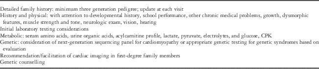
Owing to the significant heterogeneity of causes of paediatric cardiomyopathy, evaluation by a geneticist knowledgeable in cardiac genetics is important. In general, the younger the child, the larger the differential due to limited history and medical information. The parameters for establishing a diagnosis of cardiomyopathy have been well-described. For dilated cardiomyopathy, additional testing should include complete blood count, renal and liver function tests, assessment of levels of creatine phosphokinase, lactate, pyruvate, plasma amino acids, urine organic acids, and an acylcarnitine profile (Table 2). The yield of testing by next-generation sequencing panels for familial dilated cardiomyopathy is ~25%. Additional genetic and enzymatic testing may be useful. Cardiac catheterisation and endomyocardial biopsy are not routine but may be useful in patients with acute dilated cardiomyopathy. Biopsy samples can also be assessed for the presence of mononuclear cell infiltrates, myocardial damage, storage abnormalities, and viral infection or genomes. It is considered standard of care to screen first-degree family members using echocardiography and echocardiography in idiopathic and familial cases.Reference Hershberger, Lindenfeld and Mestroni 28
Table 2 Suggested evaluation of cardiomyopathy in childhood: non-cardiac parameters.
CPK=creatine phosphokinase
In hypertrophic cardiomyopathy, the electrocardiogram typically demonstrates left ventricular hypertrophy with ST segment and T-wave abnormalities. Intra-ventricular conduction delays and signs of ventricular pre-excitation (Wolff–Parkinson–White syndrome) may be present and should raise the possibility of Danon disease – X-linked, caused by LAMP2 mutations – or Pompe disease – autosomal recessive, caused by GAA mutations. Echocardiography is diagnostic in identifying, localising, and quantifying the degree of myocardial hypertrophy. Additional diagnostic studies in hypertrophic cardiomyopathy patients include metabolic testing, genetic testing for specific syndromic conditions, or genetic testing for mutations in genes known to cause isolated hypertrophic cardiomyopathy. The clinical availability of these tests is expanding rapidly and the yield of testing is quite high for hypertrophic cardiomyopathy (50–75%). As with dilated cardiomyopathy, it is considered standard of care to perform cardiac screening and ongoing surveillance in all first-degree family members for idiopathic or familial cases.
Restrictive cardiomyopathy, left ventricular non-compaction cardiomyopathy, and arrhythmogenic right ventricular cardiomyopathy are relatively rare in the paediatric population. Nevertheless, they exhibit the same degree of heterogeneity with regard to cause as hypertrophic cardiomyopathy and dilated cardiomyopathy.Reference Colan, Lipshultz and Lowe 1 , Reference Wilkinson, Landy and Colan 4 , Reference Cox, Sleeper and Lowe 5 Molecular diagnostic rates using currently available genetic testing are not known with certainty.
Cascade screening: family-based care
The present consensus guidelines recommend cardiac screening and known mutation testing for individuals at-risk of developing cardiomyopathy; however, the clinical impact of these recommendations is largely unknown.Reference Hershberger, Lindenfeld and Mestroni 28 – Reference Van Langen, Arens and Baars 33 A recent study of the uptake of cardiac screening and genetic testing amongst first- and second-degree relatives at-risk for hypertrophic cardiomyopathy or dilated cardiomyopathy indicated an uptake rate of 57 and 39%, respectively.Reference Miller, Wang and Ware 34 Not surprisingly, first-degree relatives were more likely to complete cardiac screening and genetic testing than second-degree relatives. When the proband was mutation positive and both cardiac screening and known mutation testing were recommended, relatives were more likely to complete cardiac screening. The number of living affected individuals in a family also impacted the uptake of cardiac screening. In this study, cascade cardiac screening found that 25% of identified at-risk first- and second-degree relatives had cardiomyopathy that was asymptomatic and previously undiagnosed. Genetic testing led to the identification of 22 asymptomatic at-risk relatives for whom ongoing cardiac surveillance was indicated. Known familial mutation testing also identified 33 not-at-risk individuals. Relatives who tested negative for the known familial mutations could be re-assured about the potential risk of disease and ongoing cardiac surveillance could be discontinued. In addition, children of these individuals could be spared genetic testing and cardiac screening.
It should be emphasised that family histories are dynamic, and the indications of testing for affected family members change as new individuals in the family are diagnosed. Therefore, it is important to address family history at each clinic visit and update screening recommendations accordingly. Increasingly, clinicians are being called to incorporate family-based care into medical practice, thus treating the entire family rather than a single individual. This is paradigm-altering in medical practice and has significant implications to the responsibilities and clinical encounters.
Timing of cardiac screening and genetic testing
The timing of genetic testing and cardiac screening needs to be carefully considered for each patient and family. As in other genetic diseases, testing the most severely affected family member before initiating known mutation testing in at-risk relatives is recommended.Reference Hershberger, Lindenfeld and Mestroni 28 , Reference Ackerman, Priori and Willems 29 , Reference Charron, Arad and Arbustini 31 In the case of a symptomatic relative, or a relative participating in potentially high-risk activities such as competitive athletics, cardiac screening before completion of genetic testing in the proband may be indicated to ensure optimal safety.Reference Bos, Towbin and Ackerman 30 If no aetiology is identified, all first-degree relatives should undergo routine cardiac screening. If relatives are diagnosed with disease, subsequent relatives should undergo screening based on the cascade approach. If a disease-causing mutation is identified, all the affected relatives and first-degree unaffected relatives should be offered genetic counselling and genetic testing. Recommendations for genetic testing and cardiac screening are unique to each family and depend upon accurate interpretation of results by professionals with expertise in molecular genetics. An important benefit of establishing a cause of cardiomyopathy in a family is risk stratification for potentially affected family members. A cascade approach to genetic testing in family members is likely to lead to significant cost savings but future studies are warranted to further define the benefit.
Implications for clinical practice
The genetic basis of cardiomyopathy in childhood is complex. An accurate and precise diagnosis is important to better direct patient management, including extra-cardiac management, and to assess risk to family members. Genetic testing for cardiomyopathy is increasingly available. The yield of testing continues to increase, but the interpretation of results is also becoming more complex. Accurate interpretation of genetic test results is necessary to make appropriate recommendations for cardiac screening, and genetic testing and should be carried out in the context of the family history. It is not uncommon to identify more than one genetic variant in a proband and/or a variant of uncertain significance making interpretation more complicated. Frequently, family-based cardiac screening recommendations may not routinely be discussed and/or genetic testing may not be offered in a standardised manner. In addition, this increasingly brings new scenarios for which most physicians have little training, such as the disclosure of family-based information. It is important that paediatric cardiologists receive training in clinical genetics to facilitate appropriate referral and testing. Further, there is an urgent need for more genetics professionals, including genetic counsellors and geneticists, with cardiac disease-specific knowledge. Developing the appropriate infrastructure to increasingly incorporate genetics in the care of patients and families is an important goal.
Acknowledgement
None.
Financial Support
This review was made possible by the Indiana University Health-Indiana University School of Medicine Strategic Research Initiative.
Conflicts of Interest
None.


