


A double aortic arch, as described in 1964 by Edwards,Reference Edwards 1 represents a persistence of both right and left fourth aortic arches that form a complete vascular ring around the trachea and oesophagus, which rarely, by encirclement and compression of mediastinal structures, may produce symptoms of airway obstruction and dysphagia, requiring surgery.Reference Yoo, Min, Lee, Roman, Jaeggi and Smallhorn 2 Arterial duct or ligament is often involved in vascular rings, but probably is also involved in their pathogenesis. We recently encountered a patient with a double aortic arch in whom the closure of the left duct might have led to obliteration of the left aortic arch.
Case report
In a 28-week gestation foetus, a prenatal echocardiogram revealed a double aortic arch with a predominant right and a double arterial duct. Both arches were perfused (Fig 1a). The 1-month postnatal echocardiogram showed the closure of both arterial ducts and the partial obliteration of the left aortic arch between the left subclavian artery and the dorsal aorta (Fig 1b). The infant became progressively symptomatic for dysphagia and stridor on inspiration. Physical examination was normal. The patient underwent a barium esophagogram, which demonstrated a deep persistent extrinsic indentation in the posterior aspect of the oesophagus, consistent with the diagnosis of double aortic arch. Computed tomography scan confirmed the diagnosis, and it was no longer possible to detect flow through the left aortic arch (Fig 2a). The patient was operated at 4 months of age, and the surgical procedure, through a left 4th intercostal space thoracotomy, consisted of the resection of both the left aortic arch remnant, ligament, close to the dorsal aorta, and the left arterial ligament (Fig 2b). The post-operative course was uneventful and the patient was discharged on post-operative day 4.
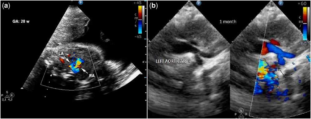
Figure 1 ( a ) Prenatal echocardiography at gestational age of 28 weeks revealing a double aortic arch with a predominant right and a double arterial duct, both perfused. ( b ) Postnatal echocardiography performed at 1 month showing the closure of both arterial ducts and the partial obliteration of the left aortic arch between the left subclavian artery and the dorsal aorta (black arrow). LA = left arch; RA = right arch.

Figure 2 ( a ) CT scan. Asterisk: left aortic arch between left carotid artery and left subclavian artery. White arrow: remnant of left aortic arch post left subclavian artery. Tip of white arrow: left subclavian artery. ( b ) Intraoperative aspect. Asterisk: left aortic arch between left carotid artery and left subclavian artery. White arrow: remnant of left aortic arch post left subclavian artery. Tip of white arrow: left subclavian artery. Tip of black arrow: left arterial ligament. ( c ) Schematic representation of the vascular ring. Tip of black arrow, right arterial ligament. Black arrow, left arterial ligament. Asterisk, obliterated left arch after left subclavian artery. DA = descending aorta; E = esophagus; LA = left arch; LCA = left carotid artery; LSA = left subclavian artery; PA = pulmonary artery; RA = right arch; RSA = right subclavian artery; RCA = right carotid artery; T = trachea.
Discussion
Double aortic arch and the right arch with an aberrant subclavian artery are the two most common forms of vascular ring, a class of congenital anomalies of the aortic arch system in which the trachea and oesophagus are completely encircled by connected segments of the aortic arch and its branches.Reference Edwards 1 Usually, this anomaly is associated with right arterial ductus patency.Reference Kanne and Godwin 3
We describe a postnatal obliteration of the left arch in a double aortic arch probably due to the closure of the left-sided arterial duct. This combination of events created a symptomatic vascular ring: predominant right aortic arch, a left obliterated aortic arch, ligament, and a double-sided arterial ligament.Reference Craatz, Künzel and Spanel-Borowski 4 In this patient, we had patency of both left and right arterial ducts during the prenatal period; the postnatal closure of both of them had different effects on the anatomy of the two arches.Reference Thankavel, Brown and Lemler 5 The underlying mechanisms responsible for these changes probably are the same described for the aetiology of the aortic coarctation. The two theories are: the ductal tissue theory and reduced-flow theory. In the former, tissue from the arterial duct invades the aorta, and when it obliterates it results in an aortic constriction. In the latter, the defect can develop secondary to haemodynamic changes that reduce flow to the involved vessel.Reference Liberman, Gersony, Flynn, Lamberti, Cooper and Stare 6 Embryogenesis of vascular rings is interpreted as the consequence of the reabsorption or obliteration of different segments of the paired original pattern of aortic arches.Reference Lee, Chen and Tsao 7 The ring caused by double ducts/ligaments, left aortic arch fibrous continuity in a right aortic arch in the present case is considered of interest because the obliteration of the mentioned segment occurred in infancy, starting from a classic double aortic arch pattern.
Acknowledgment
The authors would like to thank Professor Pietro Gallo for his precious advise.
Financial Support
This research received no specific grant from any funding agency, commercial or not-for-profit sectors.


