


Introduction
Wildlife infectious diseases are gaining more attention due to their increase in the last few decades and because of the major threats they pose to conservation, animal and human health, and food security. The emergence of novel or introduced diseases in wildlife is often mediated through complex and large-scale processes that are not susceptible to traditional reductionist approaches of causal inference (Plowright et al., Reference Plowright, Sokolow, Gorman, Daszak and Foley2008). Thus, there is a need to facilitate multidisciplinary research linking ecology, animal and human health studies by building bridges between the different disciplines, to better understand emerging infectious disease events.
Chamois border disease (BD)
Pestiviruses in wild ruminants are widespread around the world, but evidence for border disease virus (BDV) infection in these species is scarce (Van Campen et al., Reference Van Campen, Frölich, Hofmann, Williams and Baker2001; Vilcek and Nettleton, Reference Vilcek and Nettleton2006). In 2001, BDV was identified as the cause of a previously unreported disease in Pyrenean chamois (Rupicapra pyrenaica) in Spain. For the first time, a BDV was associated with disease in a free-ranging wild ruminant species. Since then, the disease has expanded to nearly the entire distribution area in the Pyrenees and has caused a dramatic decrease, and in some cases collapse, of Pyrenean chamois populations (Marco, Reference Marco, Gavier-Widén, Duff and Meredith2012). However, there are populations that have not been affected by the disease despite the fact that BDV circulation has been demonstrated, suggesting the existence of different viral strategies for persisting in the host population.
Study area and population
The Pyrenean chamois is a small-sized ungulate found along the border between Spain and France, including in the Principality of Andorra, in the Pyrenean Mountains. It is widely distributed along the axial Pyrenees and Pre-Pyrenean massifs and is mostly managed through National Hunting Reserves and National Parks (Fig. 1). For the past few decades, the positive trend in their populations has allowed their use as game, even in private hunting areas. The total population was estimated to be about 53,000 individuals in 2004, but many Pyrenean chamois populations have since declined locally following severe mortality episodes associated with BDV infection (Herrero et al., Reference Herrero, Lovari and Berducou2008).

Fig. 1. Map of the Central and Eastern Pyrenees showing the main protected areas: 1a. Alt Pallars National Hunting Reserve (NHR), Northern Sector; 1b. Alt Pallars NHR, Southern Sector, namely Boí; 2. Cerdanya-Alt Urgell NHR; 3. Cadí NHR; 4. Val d'Aran; 5. Principality of Andorra; 6. Freser-Setcases NHR; 7. Orlu Wildlife Reserve; 8. Benasque NHR; 9. Los Circos NHR.
Etiology
The genus Pestivirus of the family Flaviviridae consists of four species: bovine viral diarrhea virus 1 (BVDV-1), bovine viral diarrhea virus 2 (BVDV-2), classical swine fever virus and BDV, and a new putative species tentatively named ‘HoBi-like pestivirus’ (or BVDV-3) (Haider et al., Reference Haider, Rahman, Khan, Mikolon, Gurley, Osmani, Shanta, Paul, Macfarlane-Berry, Islam, Desmond, Epstein, Daszak, Azim, Luby, Zeidner and Rahman2014). Traditionally, pestiviruses have been classified according to their mammalian host species but this may not always be accurate, as they are not strictly host-specific and can infect a variety of artiodactyl species. In particular, with the relatively small number of BDV isolates described in the literature, there is no strict correlation between the pestivirus genotypes identified and their hosts. These include varied species of artiodactyla such as sheep, pig, cattle, reindeer, bison and Pyrenean chamois. Thus, molecular techniques are being used for classification of pestivirus species according to genotypic diversity rather than animal hosts (Vilcek et al., Reference Vilcek, Willoughby, Nettleton and Becher2010).
Chamois BDV has been characterized as BDV-4 genotype (Arnal et al., Reference Arnal, Fernández-de-Luco, Riba, Maley, Gilray, Willoughby, Vilcek and Nettleton2004; Frölich et al., Reference Frölich, Jung, Ludwig, Lieckfeldt, Gibert, Gauthier and Hars2005; Marco et al., Reference Marco, Rosell, Cabezón, Mentaberre, Casas, Velarde, López-Olvera, Hurtado and Lavín2008). Comparison of the full chamois BDV sequence with that of other BDV strains revealed that it is a typical BDV, but with considerable differences in the lengths of the 5′ UTR and 3′ UTR and in the insertion of four amino acids at the N-terminus of NS2 (Vilcek et al., Reference Vilcek, Willoughby, Nettleton and Becher2010). Experimental studies confirmed that this virus was the primary agent of the disease (Cabezón et al., Reference Cabezón, Velarde, Mentaberre, Fernandez-Sirera, Casas-Díaz, Lopez-Olvera, Serrano, Rosell, Riquelme, Lavin, Segales and Marco2011; Martin et al., Reference Martin, Duquesne, Guibert, Pulido, Gilot-Fromont, Gibert, Velarde, Thiéry, Marco and Dubois2013).
Epidemiology
Susceptible species
Pyrenean chamois is the only wild artiodactyl species that to date has been affected by the disease. Mouflon, red deer, fallow deer, roe deer and wild boar share habitat with Pyrenean chamois in the Pyrenees, but have never been reported with clinical disease similar to that seen in Pyrenean chamois. However, positive ELISA antibody results have been obtained in mouflon. A comparative virus neutralization test (VNT), using chamois and ovine strains, confirmed the ELISA results and showed higher titers to ovine strains, indicating that it was likely that these animals were infected with strains of ovine origin (Marco et al., Reference Marco, Rosell, Cabezón, Beneria, Mentaberre, Casas, Hurtado, López-Olvera and Lavín2009a). Seropositive results have also been obtained in wild boar, but the VNT, although confirming BDV infection, did not clearly indicate the origin of the strain as either ovine or Pyrenean chamois (Rosell et al., Reference Rosell, Cabezón, Mentaberre, Casas, Lavín and Marco2008).
Domestic ruminant species that share their habitat with Pyrenean chamois are mainly sheep, goat and cattle. Sheep and cattle showed a high seroprevalence of antibodies against pestiviruses, and with the comparative VNT, higher titers were observed to ovine and bovine strains in sheep and cattle (BDV Spain 97 and BVDV-1 NADL), respectively, when compared to titers to Pyrenean chamois strain. Thus, it was unlikely that Pyrenean chamois infected domestic ruminants (Marco et al., Reference Marco, Rosell, Cabezón, Beneria, Mentaberre, Casas, Hurtado, López-Olvera and Lavín2009a). However, a study of clinical cases of BD in sheep on the French side of the Pyrenees confirmed that the etiological agent was a BDV strain of chamois origin (Bertin-Cavarait, Reference Bertin-Cavarait2006). Therefore, the possibility that chamois strains could infect domestic species sharing their habitat cannot be ruled out.
Spatiotemporal distribution
The Pyrenean chamois is found in the Pyrenean Mountains and its distribution is continuous across the range with no important ecological or physical barriers that would limit animal movements. Chamois BDV was detected for the first time in 2001 in the Northern Sector of Alt Pallars National Hunting Reserve, in the Central Pyrenees of Catalonia (Fig. 1). This first outbreak lasted 2 years and had a marked seasonal pattern, with affected animals concentrated at the end of winter and beginning of spring (February and June). Piroplasms were detected in blood smears, by PCR, and spleen and kidney lesions consistent with hemolytic anemia were reported, indicating that pestivirus infection may have triggered the development of the infection (Hurtado et al., Reference Hurtado, Aduriz, Gómez, Oporto, Juste, Lavín, López-Olvera and Marco2004; Marco et al., Reference Marco, López-Olvera, Rosell, Vidal, Hurtado, Juste, Pumarola and Lavín2007). Morbidity and mortality are difficult to evaluate in wildlife in remote areas, such as high mountain terrain with limited accessibility where diseased and dead animals are quickly removed by scavengers. Mortality can be estimated indirectly, through the census of the population that is conducted yearly. Thus, the cumulative decrease in this area for the 2 years of the outbreak was 42% (Marco et al., Reference Marco, López-Olvera, Rosell, Vidal, Hurtado, Juste, Pumarola and Lavín2007).
Unexpectedly, in winter 2005, after 2 years of epidemiological silence with just one isolated case of disease on the Spanish side of the Pyrenees and several others cases on the French side, a new severe outbreak occurred in Cerdanya-Alt Urgell National Hunting Reserve. It yielded the highest estimated mortality recorded until now for chamois BD (86%) in just a couple of months, between the end of January and March, with pneumonia as the most prevalent secondary process. Shortly afterwards (June), the first cases were detected in the neighboring Hunting Reserve of Cadí. The disease spread slowly for about two and a half years to cover the entire reserve and nearby private hunting areas. Bacterial pneumonia was also the most common finding at necropsy and the estimated mortality was 63% (Marco et al., Reference Marco, Rosell, Cabezón, Mentaberre, Casas, Velarde and Lavín2009b).
These BD outbreaks in Pyrenean chamois followed different epidemiological patterns and had different mortality outcomes, but with outstanding high mortality in common. Nevertheless, during the same period, isolated cases and mild outbreaks of BD were recorded in some other areas, such as Val d'Aran and the French side of the Pyrenees. At the same time, there was an absence of disease in some areas located between affected zones, such as in the Principality of Andorra (Fernández-Sirera, Reference Fernández-Sirera, Riba, Cabezón, Rosell, Serrano, Lavín and Marco2012a). Unexpectedly, in the easternmost National Hunting Reserve, namely Freser-Setcases, chamois BDV was detected in healthy Pyrenean chamois but no clinical signs or mortality were observed in the population (Marco et al., Reference Marco, Rosell, Cabezón, Mentaberre, Casas, Velarde and Lavín2009b).
In 2009, additional mild outbreaks of disease occurred in other previously unaffected areas (southern sector of the Alt Pallars National Hunting Reserve, namely Boí) and in 2010, a severe outbreak took place in the Principality of Andorra (Fernández-Sirera et al., Reference Fernández-Sirera, Riba, Cabezón, Rosell, Serrano, Lavín and Marco2012a). In 2011, the disease was detected for the first time in Benasque National Hunting Reserve, in the Aragón Region, and gradually expanded westward with a regular epidemiological pattern and mortality ranging between 30 and 45%. As of 2014, more than two-thirds of the Pyrenean chamois population was infected and it is expected that in a short time the entire Pyrenean chamois distribution area will be affected by the infection, with the exception of some apparently isolated, small populations that may remain free of infection.
The Freser-Setcases National Hunting Reserve, with an area of 20,000 ha, is an extraordinary case. It has the highest Pyrenean chamois density in the region and is the only important chamois population that to date has not suffered an epizootic outbreak, despite the fact that chamois BDV has been isolated from healthy hunted animals. Only one isolated case of clinical disease was reported in 2007. Antibody prevalence in the population is high, indicating that there is active virus circulation (Marco et al., Reference Marco, Rosell, Cabezón, Mentaberre, Casas, Velarde and Lavín2009b). Population dynamics are not affected by the disease, as total numbers of chamois increase each year and must be regulated by increasing the number of hunting permits. Therefore, BDV is silently persisting in the chamois population of this area, without adverse population effects, in contrast to most of the other affected populations, suggesting the existence of two different simultaneous viral strategies for persisting in the host population.
Following the severe outbreaks of disease, the infection is now endemic in the Pyrenees region. However, different epidemiological scenarios have appeared: on the one hand, frequent BDV circulation with possible negative impacts on population dynamics in some areas (Alt Pallars National Hunting Reserve) with no recovery of total numbers; and on the other hand, lack of virus circulation and rapid recovery of the chamois population (Cadí and Cerdanya-Alt Urgell National Hunting Reserves) (Fernández-Sirera et al., Reference Fernández-Sirera, Cabezón, Allepuz, Rosell, Riquelme, Serrano, Lavín and Marco2012b). In the latter case, the extremely high mortality may have jeopardized the long-term maintenance of BDV in the chamois population leading to an increasing proportion of naïve animals. Recently, this epidemiological scenario has led to a second BD outbreak in Cadí, milder than the first, with lesser impact on population dynamics. The same is expected to occur in Cerdanya-Alt Urgell in the near future.
In France, at the Orlu Wildlife Reserve in the Eastern Pyrenees, although no mass mortality has ever been observed, a decrease in population numbers has been reported for the last two decades. The high prevalence of viremic chamois in this area suggested that BDV infection might have had a clinical effect on reproduction and on juvenile mortality (Pioz et al., Reference Pioz, Loison, Gibert, Dubray, Menaut, Le, Artois and Gilot-Fromont2007).
Transmission
Routes of transmission of the disease in Pyrenean chamois are not fully known. However, the epidemiological pattern of the outbreaks observed, with short course epizootics and high mortality, indicates a high horizontal transmission rate. This is supported by field studies that confirmed the excretion of the virus via the oro-nasal route and by experimental studies on BDV infection, which used these routes of inoculation (Cabezón et al., Reference Cabezón, Rosell, Velarde, Mentaberre, Casas-Díaz, Lavín and Marco2010b, Reference Cabezón, Velarde, Mentaberre, Fernandez-Sirera, Casas-Díaz, Lopez-Olvera, Serrano, Rosell, Riquelme, Lavin, Segales and Marco2011; Martin et al., Reference Martin, Duquesne, Guibert, Pulido, Gilot-Fromont, Gibert, Velarde, Thiéry, Marco and Dubois2013). Thus, the oro-nasal route may be the most likely and frequent route of BDV transmission during severe outbreaks.
Pestiviruses can cause acute or persistent infections (Simmonds et al., Reference Simmonds, Becher, Collet, Gould, Heinz, Meyers, Monath, Pletnev, Rice, Stiansny, Thiel, Weiner, Bukhet, King, Adams, Carstens and Lefkowitz2011). Acute infections of immunologically naïve animals are generally subclinical, transient and of variable duration but may be lethal with highly virulent viruses. But pestiviruses also can cross the placenta and infect non-immunocompetent fetuses, leading to reproductive problems or apparently healthy offspring, generating persistently infected (PI) animals that may excrete high amounts of virus and spread the infection. PI animals in Pyrenean chamois have never been observed and were unlikely during the outbreaks of disease because infected females may die prior to giving birth due to the severity of the disease. In addition, this is supported by the age of diseased chamois, including adult and older animals unlikely to be PI, and the low seroprevalence reported before the outbreaks in some populations (Marco, Reference Marco, Gavier-Widén, Duff and Meredith2012). In fact, the confirmation of PI animals in domestic ruminants by identification of the virus in two separate samples taken within a minimum of 21 days of each other, without seroconversion, is not valid for Pyrenean chamois (Nettleton et al., Reference Nettleton, Gilray, Russo and Dlissi1998). Diseased Pyrenean chamois of all ages that were captured and studied, but died shortly after capture and could only be analyzed at a single sampling point, were always viremic and most of them antibody negative. In addition, an affected 4-year-old female chamois captured and hospitalized in captivity in 2002 that survived for more than 1 month was viropositive and did not seroconvert during this period (Hurtado et al., Reference Hurtado, Aduriz, Gómez, Oporto, Juste, Lavín, López-Olvera and Marco2004; Marco, Reference Marco, Gavier-Widén, Duff and Meredith2012). This long-lasting viremia in naturally infected chamois agrees with experimental studies in which infected animals shed virus constantly for the 34 days of the experiment, but differs in that all challenged chamois seroconverted. However, these neutralizing antibodies were not sufficient to clear the virus. The mechanisms for this long-lasting viremia co-existing with high titers of neutralizing antibodies in infected viremic chamois remain unclear (Cabezón et al., Reference Cabezón, Velarde, Mentaberre, Fernandez-Sirera, Casas-Díaz, Lopez-Olvera, Serrano, Rosell, Riquelme, Lavin, Segales and Marco2011).
In the Freser-Setcases National Hunting Reserve, where BDV is persisting silently, viral transmission may be different. With a moderate to high seroprevalence and a very low viroprevalence in the chamois population, we presume that PI animals may be the key to viral persistence in this particular epidemiological scenario. However, it has not yet been possible to confirm this.
Pathogenesis, clinical signs and lesions
The pathogenesis of ruminant pestiviruses is complex, as indicated by a wide variety of clinical signs and the variable results following intrauterine infections (Nettleton and Entrican, Reference Nettleton and Entrican1995). However, severe pestivirus infections are much less frequent in domestic ruminants than mild or even unapparent infections. The severe disease associated with chamois BDV is unprecedented in pestivirus infections in wild ruminants. Field and experimental studies in Pyrenean chamois and experimental infection in non-pregnant sheep, using the same chamois BDV strain, showed that the pathogenesis of the disease in chamois is completely different from that of sheep. Pyrenean chamois develop a long-lasting viremia and severe disease, with the virus able to be isolated from all tissues and via all excretion routes. In contrast, viremia was not detected in sheep and disease was absent, consistent with reports of acute infections in sheep with other BDV strains of ovine origin (Marco et al., Reference Marco, López-Olvera, Rosell, Vidal, Hurtado, Juste, Pumarola and Lavín2007; Cabezón et al., Reference Cabezón, Velarde, Rosell, Lavín, Segalés and Marco2010c, Reference Cabezón, Velarde, Mentaberre, Fernandez-Sirera, Casas-Díaz, Lopez-Olvera, Serrano, Rosell, Riquelme, Lavin, Segales and Marco2011; Martin et al., Reference Martin, Duquesne, Guibert, Pulido, Gilot-Fromont, Gibert, Velarde, Thiéry, Marco and Dubois2013). Similar results were obtained in an experimental infection in pigs with the same chamois BDV. Despite a longer viremia, clearance was observed at 2 weeks post-infection and agrees with previous studies involving transmission and infection of BDV from ruminants to swine (Cabezón et al., Reference Cabezón, Rosell, Sibila, Lavín, Marco and Segalés2010a).
To investigate the possibility of PI animals in Pyrenean chamois, three females at two-thirds of pregnancy were experimentally inoculated with the same virus as the experiment performed in non-pregnant chamois. However, pregnancy was unsuccessful in all inoculated animals because they either died or aborted and later died. Viral RNA was detected in all organs of the fetuses, confirming intrauterine infection and suggesting that PI offspring may be possible (Martin et al., Reference Martin, Duquesne, Guibert, Pulido, Gilot-Fromont, Gibert, Velarde, Thiéry, Marco and Dubois2013). Vertical transmission was also demonstrated in the fetus of a naturally infected female chamois that died before parturition (Cabezón et al., Reference Cabezón, Rosell, Velarde, Mentaberre, Casas-Díaz, Lavín and Marco2010b).
Clinical signs in naturally infected Pyrenean chamois include non-specific signs, such as emaciation, weakness and difficulty moving, but also neurological signs such as depression and abnormal behavior, including the absence of flight reaction and loss of fear of humans (Fig. 2). Skin lesions are commonly found, consisting of different degrees of alopecia and skin hyperpigmentation (Marco et al., Reference Marco, López-Olvera, Rosell, Vidal, Hurtado, Juste, Pumarola and Lavín2007, Marco, Reference Marco, Gavier-Widén, Duff and Meredith2012). Other clinical signs observed in diseased chamois are dyspnea, skin abscesses and keratitis, but these may be associated with secondary infections. At necropsy, emaciation is the most common finding, together with bacterial bronchopneumonia. The most significant microscopic lesions are found in the brain. They consist of edema, diffuse moderate spongiosis and gliosis with astrocyte hypertrophy, and neuronal degeneration and necrosis throughout the brain. Inflammatory infiltrates of mononuclear cells surrounding blood vessels are observed occasionally. In the skin, follicular atrophy and telogenization of the remaining hair follicles are observed, with epidermal hyperplasia and melanosis with evident orthokeratotic hyperkeratosis. In the dermis, mild mononuclear interstitial inflammatory infiltrates are occasionally seen (Marco, Reference Marco, Gavier-Widén, Duff and Meredith2012).
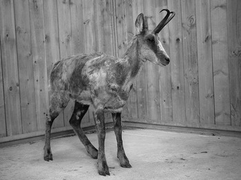
Fig. 2. Pyrenean chamois with BDV infection showing extensive alopecia, skin hyperpigmentation and depression.
Diagnosis
The differential diagnosis list for BD in chamois can be extensive depending on the clinical signs and secondary infections of affected animals. Alopecia, depression and abnormal behavior are usually indicative of the process. The most important entity in differential diagnosis is sarcoptic mange. Initially, intense pruritus, alopecia and erythematous eruptions are observed, but lesions may pass unnoticed due to the thick fur. In chronic stages, crusting, hyperkeratosis, lichenification, and thickening of the skin are seen (Pence and Ueckermann, Reference Pence and Ueckermann2002). However, this process has not been detected in the Pyrenees to date. Pneumonia, dermatophytosis, toxoplasmosis and cerebral coenurosis are diseases described in Pyrenean chamois that should be considered in the differential diagnosis. Pneumonia, very common due to a secondary infection in chamois BD, is also a common primary cause of death in chamois populations and severe epizootics have been reported (Ferroglio, Reference Ferroglio, Gavier-Widén, Duff and Meredith2012).
Laboratory confirmation of BDV infection is mainly performed by RT–PCR and virus isolation, and final viral characterization by the immunoperoxidase monolayer assay (IPMA) test using monoclonal antibodies C-16 (pestivirus-specific), CA-3 and CT-6 (BVDV1-specific) and WS363 (BDV-specific) (Marco et al., Reference Marco, López-Olvera, Rosell, Vidal, Hurtado, Juste, Pumarola and Lavín2007). Immunohistochemistry on formalin-fixed, paraffin-embedded organ samples has also been conducted using the monoclonal anti-pestivirus antibody 15C5 as an ERNS-specific capture antibody (Hurtado et al., Reference Hurtado, Aduriz, Gómez, Oporto, Juste, Lavín, López-Olvera and Marco2004). Commercial competitive ELISA for detection of seropositive reactors has been shown to be useful for Pyrenean chamois and is recommended, as there is often a lack of specific anti-species antibodies for wildlife (Frölich, Reference Frölich, Gavier-Widén, Duff and Meredith2012). VNT is the ‘gold standard’ for the detection of specific antibodies and has frequently been used to confirm the ELISA results in Pyrenean chamois (Marco, Reference Marco, Gavier-Widén, Duff and Meredith2012).
Management
Since the first BD outbreak, Pyrenean chamois management decisions have been made that reduce or stop hunting. After mortality episodes and depending on the evolution of the populations, evaluated through annual census, hunting is reestablished with limited numbers of permits. Control and eradication measures for BDV infection in Pyrenean chamois are unlikely in the near future. Detection and removal of PI animals and vaccination, which are the main strategies to control the disease in domestic ruminants, may not be feasible in large populations of wild ungulates, such as the Pyrenean chamois.
Pathogen, host and environmental factors
Changes in the interplay of pathogen, host and environmental factors may have contributed to the pathogenesis and the different epidemiological patterns of BD in Pyrenean chamois. Novel disease patterns are related to changes in these factors and are keys to creating novel transmission patterns and selection of novel pathogens. This is especially complex in wildlife and there is a critical need to better understand these new scenarios to manage emerging diseases. In many of these disease events in wildlife, human actions play a role and can moderate these factors (Engering et al., Reference Engering, Hogerwerf and Slingenbergh2013).
Pathogen factors
Pathogen evolution has been described as an important driver in the spread of diseases to novel host ranges or in changes to virulence (Engering et al., Reference Engering, Hogerwerf and Slingenbergh2013). A key factor influencing BD emergence in 2001 in Pyrenean chamois may have been pathogen invasiveness through viral mutation. Among major pathogens, RNA viruses have the highest rate of mutation and in large animal populations may adapt easily to new selective conditions (Lauring et al., Reference Lauring, Frydman and Andino2013).
A retrospective study was performed in archived sera and organ samples of Pyrenean chamois from the years 1990 to 2000, before the appearance of the first case of the disease. Antibodies against BDV were detected by ELISA and VNT in 36 of the 74 chamois sera tested from the Catalan Pyrenees, indicating that BDV infection had been present in the chamois population since at least 1990, 11 years before the first outbreak of disease. In addition, two BDV genotype-4 infections were detected by RT–PCR and isolated, suggesting that the emergence of the disease may have been due to factors other than the introduction of a new virus in the chamois population (Marco et al., Reference Marco, Cabezón, Rosell, Fernández-Sirera, Allepuz and Lavín2011).
Host factors
The dynamics of infectious diseases are also shaped by individual and population host level responses to infection. Thus, host factors such as Pyrenean chamois immune status and genetic variability may have also driven different epidemiological scenarios and can play an important role in buffering populations against widespread epizootics (Altizer et al., Reference Altizer, Harvell and Friedle2003).
Immunological status of the different chamois populations before BD epizootics may have determined the final outcome of mortality in Pyrenean chamois. In two of the study areas, Cadí National Hunting Reserve and the Principality of Andorra, the prevalence of antibodies in the population before disease appearance was 5.1 and 5%, respectively (Marco et al., Reference Marco, Rosell, Cabezón, Mentaberre, Casas, Velarde and Lavín2009b; Fernández-Sirera et al., Reference Fernández-Sirera, Riba, Cabezón, Rosell, Serrano, Lavín and Marco2012a, Reference Fernández-Sirera, Cabezón, Allepuz, Rosell, Riquelme, Serrano, Lavín and Marcob). In the southern sector of the Alt Pallars National Hunting Reserve (Boí), moderate seroprevalence, estimated to be 38%, was observed before the first case of the disease in 2009. Mortality could not be evaluated because annual censuses of the population could not be completed due to weather conditions, but few diseased animals were found and observations by rangers and hunters suggested a low impact on population dynamics. Consequently, hunting was not banned but reduced, and continued in subsequent years. To date, the population is stable and since 2010 no more clinical cases have been reported. Freser-Setcases National Hunting Reserve remains the only important chamois population that has not been affected by BD epizootics, despite the fact that BDV is circulating in apparently healthy animals and an isolated diseased chamois was found in 2007 (Marco et al., Reference Marco, Rosell, Cabezón, Mentaberre, Casas, Velarde and Lavín2009b). Seroprevalence in this protected area is high, ranging between 40 and 70% for the last few years. Thus, there may be high virus circulation in this area with no negative effects on population dynamics. Population numbers are growing each year according to official censuses and hunting permits are on the increase to maintain the population at stable numbers, as it has one of the highest densities in the Pyrenees and is considered to have reached its carrying capacity.
The high seroprevalence in some areas, compared to others, may be related to contact with other pestivirus strains, such as BDV of ovine origin. In the comparative VNT of ELISA-positive healthy chamois, high titers of neutralizing antibodies for ovine BDV strains were observed, even more than 8–10-fold higher than those for the chamois strain (Marco et al., Reference Marco, Rosell, Cabezón, Mentaberre, Casas, Velarde, López-Olvera, Hurtado and Lavín2008). This suggests the existence of an additional infection process due to ovine BDV strains in the chamois populations, with apparently low pathogenic viruses that may not have negative effects at the population level. In mountain areas, sheep and cattle share grazing areas and water sources with wild ungulates and it is feasible that they play a key role in the spread of BDV among chamois (Olde Riekerink et al., Reference Olde Riekerink, Dominici, Barkema and de Smit2005). Infection of chamois with BDV of ovine origin could even be beneficial and protective against virulent BDV strains by cross-immunity. In an experimental infection study, BDV-seropositive chamois with higher VNT titers against ovine BDV strains, were inoculated with a chamois BDV isolate. They did not become viremic or diseased, compared to inoculated seronegative chamois, and confirmed that previously acquired humoral immunity protected them against chamois BDV infection (Cabezón et al., Reference Cabezón, Velarde, Mentaberre, Fernandez-Sirera, Casas-Díaz, Lopez-Olvera, Serrano, Rosell, Riquelme, Lavin, Segales and Marco2011). However, during the last few years, sheep numbers have dropped drastically in the Catalan Pyrenees and it is not known whether it may have influenced the epidemiology of BD in the Pyrenean ecosystem.
Genetic variability of the major histocompatibility complex (MHC) is considered to be important for conferring resistance to a variety of pathogens. An investigation into the genetic variability at the MHC class II DRB1 exon 2 locus in the different Pyrenean chamois populations concluded that the only non-affected chamois population (Freser-Setcases Reserve) had a higher genetic diversity than previously reported for other populations of chamois (Cavallero et al., Reference Cavallero, Marco, Lavín, D'Amelio and López-Olvera2012). However, further studies would be necessary to clarify if genetic variability had a role in disease epidemiology or if the disease has led to selection for a narrower MHC gene range in severely affected populations.
The apparently erratic occurrence of BD epizootics in the Catalan Pyrenees may be partly explained by Pyrenean chamois ecology and behavior. Different space-use patterns among groups of this species have been reported within the same population (Crampe et al., Reference Crampe, Bon, Gerard, Serrano, Caens, Florence and Gonzalez2007). Coexistence of sedentary groups, which use the same areas throughout the year, and migratory groups, which use different summer and winter ranges, could explain the spatial epidemiology of diseases such as infectious keratoconjunctivitis (Crampe et al., Reference Crampe, Caens, Empain, Florence, Kieser, Laffeuillade, Llanes and Morceau2008).
Environmental factors
There is ongoing evidence that climate change increases the occurrence of diseases in natural systems (Altizer et al., Reference Altizer, Ostfeld, Johnson, Kutz and Drew Harvell2013). Environmental factors such as climate and its influence on chamois winter survival, availability of food resources, pathogen survival and transmission, and the role of possible vectors and their seasonal abundance have not been evaluated in chamois BD, but deserve more attention.
In mountain ungulates, pneumonia is one of the main health concerns because outbreaks are relatively frequent and can play a significant role in population dynamics (Ferroglio, Reference Ferroglio, Gavier-Widén, Duff and Meredith2012). Climatic and other ecological factors may trigger outbreaks of pneumonia in wild animals, including the Pyrenean chamois. However, in the case of pneumonia in BDV-infected chamois, the high frequency of this and other secondary infections suggests their magnification by immunosuppressive effects of coincident BDV infection. The most severe outbreak of BD in Pyrenean chamois occurred in the Cerdanya-Alt Urgell National Hunting Reserve and resulted in a population collapse, with the highest mortality recorded (Marco et al., Reference Marco, Rosell, Cabezón, Mentaberre, Casas, Velarde and Lavín2009b). Pneumonia was, in this case, a major contributing factor in the high mortality rate observed.
Conclusions
Three categories have been proposed for infectious emergence disease events (Engering et al., Reference Engering, Hogerwerf and Slingenbergh2013): (i) pathogens appearing in a novel host; (ii) mutant pathogens displaying novel traits in the same host; and (iii) disease complexes emerging in a new geographic area. BD in Pyrenean chamois would fall into the second category, as BDV may have become hypervirulent for this wild ungulate species, with a combination of drivers as the underlying causal factors of emergence. However, all existing pathogens at some time may have undergone changes, switched host species and increased their geographic distribution. A preliminary study into the origin and dispersion of the Pyrenean chamois BDV genetic variants suggests that this BDV phylogenetic group originated from sheep BDV genotype-4, generating a founder effect due to intra-species spread and spatial dispersion (Luzzago et al., Reference Luzzago, Ebranati, Lanfranchi, Cabezón, Lavín, Rosell, Rossi, Zehender and Marco2014).
The transmission of infectious disease agents between wild and domestic animals has become a global issue of growing interest. A more integrated health approach and multidisciplinary research will be required to make further gains in managing pestiviral diseases, especially those at the wildlife and domestic animal interface in complex ecosystems. Pyrenean chamois mortality related to BD may be a primary factor limiting population recovery and management in some areas, with important economic and social consequences. In addition, if control and eradication programs are implemented in domestic ruminants, as they have been in many countries, wildlife populations should be monitored for pestiviruses to ensure success in the long-term.
Acknowledgments
The authors thank the staff of the National Hunting Reserves and Hunters Associations of Catalonia. This research was funded by Generalitat de Catalunya and by Ministerio de Economía y Competitividad of the Spanish Government (grant numbers CGL2006-11518/BOS, CGL2009-09071/BOS and CGL2012-40057-C02-01).


