Significant outcomes
• A high-fat diet (HFD) for 8 weeks significantly increases hypothalamus–pituitary–adrenal (HPA) axis response to acute restraint stress.
• Prenatal dexamethasone (DEX) exposure (a model of intra-uterine growth restriction) and HFD in combination exacerbate depressive-like behaviour.
• In the current model, intra-uterine growth restriction does not predispose to a greater susceptibility to the detrimental effects of a HFD.
Limitations
• Only a crude metabolic assessment was made, that is, caloric intake, fat pad weights, fasting glucose and insulin levels.
• Our studies were performed in late adolescent male rats only.
Introduction
An adverse foetal environment is a well-known cause of poor health outcome in later life (Reference McMillen and Robinson1–Reference Godfrey, Inskip and Hanson3). Specifically, intrauterine growth restriction (IUGR) is known to increase the risk of metabolic disease including obesity, insulin resistance (IR) and diabetes in humans (Reference McMillen and Robinson1,Reference Hales and Barker4,Reference Desai and Ross5). Furthermore, IUGR has been linked to neurodevelopmental and neuroendocrine disturbances in animal studies and may predispose to psychiatric illnesses such as schizophrenia and depression in humans (Reference Cannon, Jones and Murray6–Reference Rees, Harding and Walker9). Dysregulation of the HPA axis has been proposed to orchestrate some of these abnormalities (Reference Phillips10), but the exact mechanisms and whether they are associated are still unclear. Additionally, recent meta analyses have shown that depression is an independent risk factor for developing metabolic syndrome as well as diabetes and vice versa, suggesting that some pathophysiological mechanisms could be shared in these disorders (Reference Mezuk, Eaton, Albrecht and Golden11–Reference Pan, Keum and Okereke13).
Experimentally, IUGR may be modelled in rodents by supraphysiological prenatal glucocorticoid exposure (Reference Reinisch, Simon, Karow and Gandelman14). Pups of female rats that are treated with DEX in the last part of pregnancy are consistently characterised by low birth weight (Reference Nyirenda, Lindsay, Kenyon, Burchell and Seckl15–Reference Kjaer, Wegener, Rosenberg and Hougaard23). Other abnormal findings in these animals have been described as well, although with some inconsistency. Some authors reported increased activity of the HPA axis at baseline or following acute stress in males only (Reference Buhl, Neschen and Yonemitsu16,Reference Hauser, Feldon and Pryce17,Reference Shoener, Baig and Page20,Reference O'Regan, Kenyon, Seckl and Holmes21), another study found impaired DEX suppression test in both sexes (Reference Oliveira, Bessa and Mesquita22), and yet another study concluded that a healthy rearing dam protects against HPA axis hyperactivity in both sexes (Reference Brabham, Phelka, Zimmer, Nash, Lopez and Vazquez19). With regard to behaviour, increased depressive-like behaviour in the Forced Swim Test (FST) has been described in females (Reference Kjaer, Wegener, Rosenberg and Hougaard23), but not in males (Reference Welberg, Seckl and Holmes18,Reference Nagano, Ozawa and Suzuki24). Pre-diabetic metabolic disturbances such as decreased glucose tolerance, hepatic IR and increased fasting glucose levels have been described in males only by some authors (Reference Nyirenda, Lindsay, Kenyon, Burchell and Seckl15,Reference Buhl, Neschen and Yonemitsu16,Reference O'Regan, Kenyon, Seckl and Holmes21), whereas others found no abnormalities in glucose metabolism in males (Reference Drake, Raubenheimer, Kerrigan, McInnes, Seckl and Walker25,Reference Shahkhalili, Moulin, Zbinden, Aprikian and Mace26).
Thus, it can be hypothesised that prenatal DEX treatment per se may give rise to subtle adverse effects that are not always conspicuous. However, aberrant traits may be provoked by or exacerbated in a later life stressful environment. This hypothesis is further strengthened by certain findings. The acoustic startle response (ASR), a characteristic sequential contraction of the skeletal musculature evoked by a sudden and intense acoustic stimulus (Reference Koch27), was found to be increased only in prenatally DEX-treated animals when previously exposed to a blood sampling procedure under restraint (Reference Hougaard, Andersen, Kjaer, Hansen, Werge and Lund28). Another study showed that a chronic mild stress paradigm induced depressive-like behaviour in rats only when exposed to DEX prenatally (Reference Oliveira, Bessa and Mesquita22). Furthermore, while being slightly hypotensive at baseline, blood pressure rose to a higher level in maternally DEX-treated rats in stressful surroundings compared with rats from healthy mothers (Reference O'Regan, Kenyon, Seckl and Holmes29). Similarly, liver steatosis as a consequence of an HFD was seen only in prenatally DEX-treated rats (Reference Drake, Raubenheimer, Kerrigan, McInnes, Seckl and Walker25).
HFDs are often used to induce obesity and metabolic disorders in rodents in order to model the human metabolic syndrome (Reference Buettner, Scholmerich and Bollheimer30). In addition, HFD has been shown to impair cognitive function in rodents (Reference Winocur and Greenwood31,Reference Pathan, Gaikwad, Viswanad and Ramarao32) as well as exacerbate depressive-like behaviour in a genetic animal model of depression (Reference Abildgaard, Solskov, Volke, Harvey, Lund and Wegener33).
Therefore, the aim of the current study was to test whether behavioural, metabolic or neuroendocrine disturbances could be provoked or exacerbated by a HFD in a rat model of IUGR induced by maternal gestational DEX treatment.
Materials and methods
Animals
Forty time-mated young adult rats (Wistar, HanTaC:WH, SPF) arrived at gestational day (GD) 3 and were randomly distributed in pairs to white plastic cages (27 × 43 × 18 cm, EUROSTANDARD III, Scanbur, Denmark) with pine-bedding (Lignocel® S8, Brogaarden ApS, Lynge, Denmark) and nesting material (Enviro-Dri, Brogaarden®, Denmark) in a light-controlled room (12/12 h light/dark cycle, lights off at 6 a.m.). Standard chow (Altromin standard diet 1324) and tap water were provided ad libitum. Clean cages and new bedding were provided twice weekly. Body weights were recorded at GD 4, 7, 10 and 13–21. The animals were randomly assigned to two groups, vehicle (VEH, n = 20) or dexamethasone (DEX, n = 20).
The animal welfare committee, appointed by the Danish Ministry of Justice, granted ethical permission for the studies. All procedures complied with the EC Directive 86/609/EEC and with the Danish law regulating experiments on animals (permission ID 2007/561-1378).
DEX exposure
DEX (Sigma-Aldrich Denmark ApS, Brøndby, Denmark) used for animal experimentation was dissolved in 4% ethanol/isotonic saline to 100 μg DEX/ml. From GD 14 to 21, the rats were injected daily s.c. in the nape of the neck with 150 μg/kg DEX or vehicle solution as assigned, between 10.45 and 11.45 a.m. The daily dose was calculated based on body weight from the previous day +5 g.
Pregnancy and lactation data
At gestation day 21, the dams were housed individually. The expected day of delivery, GD 22, was designated postnatal day (PND) 0. Litter size and gender were registered on PND 3, as were body weights. Pup weights were also recorded on PND 10 and at weaning, on PND 21. Litters were not culled, but were left with their dam throughout rearing to minimise stress on the dam as well as the offspring.
Post-weaning conditions
At 4 weeks of age, the animals were transferred by coach to Centre for Psychiatric Research, Aarhus University. Two to three males per litter were randomly selected from eight dams belonging to each exposure group (n = 40 in total) and housed in non-sibling pairs of similar prenatal exposure in clear Macrolon Type IIIH cages with shelter and nesting material in a temperature (20°C) and light-controlled room (12/12 h light/dark cycle, lights on at 7 a.m.). Each cage was then randomised to standard chow or HFD, comprising four study groups in total (n = 10). To lessen stress from the novel environment during assessments, the animals were moved to the experimental locations 1 day before each separate test. Study design is depicted in Fig. 1.

Fig. 1 Timeline and study design VEH = prenatal vehicle solution; DEX = prenatal dexamethasone; CHOW = standard chow diet; HFD = high-fat diet; FST = Forced Swim Test; OF = open field; ASR = acoustic startle response; blood = fasting blood samples; stress = acute restraint stress.
Diets, weights and caloric intake
From PND 44 (study initiation), the rats were fed their respective diets ad libitum. The HFD (Research Diets Inc., D12492) was composed of 60 kcal% fat (mainly lard) and refined carbohydrates (sucrose and maltodextrin), total kcal/g 5.24. Standard chow (Altromin 1324) was composed of 11 kcal% fat, total 2.84 kcal/kg. All rats and diet remnants were weighed twice a week. Caloric intake was estimated as (rat weight gain/cage weight gain × cage caloric intake).
FST
The animals were tested in the FST, a commonly used screening procedure for depressive-like behaviour in rodents (Reference Porsolt, Le and Jalfre34). Specifically, increased immobility (floating) during forced swim has been suggested as an index of depressive-like behaviour in rodents. Immobility behaviour is defined as the rat making no movements beyond those needed to keep its head above the water. Briefly, each rat was tested twice, 24 h apart. The animal was placed in a transparent, water-filled (25°C), plastic cylindrical tank (H: 54 cm, ∅: 24 cm, water depth: 40 cm) and allowed to swim for 15 and 7 min in the pre-test (day 1) and test trial (day 2), respectively. The water was changed between each trial. After each session, the animals were dried in a towel. All swim sessions were video-recorded and scoring consisted of determining the dominant behaviour (struggling, swimming, immobility) within 5 s intervals as described elsewhere (Reference Cryan, Markou and Lucki35,Reference Detke, Rickels and Lucki36). The first 5 min of the pre-test trial as well as the entire test trial were scored. In the test trial, a separate analysis of the last 4 min was performed to assess the plateau phase, in which behaviours reach a stable level over time. All scoring was performed blinded to study groups.
Open field
Locomotor activity was tested immediately before the FST test trial (day 2). The rats were individually placed in a video-recorded square box (100 cm × 100 cm, wall height: 20 cm) for 7 min, and their movements were subsequently tracked by Noldus EthoVision XT7 software. Total distance travelled (cm) was used as indicator of locomotor activity.
ASR and pre-pulse inhibition (PPI)
In animals and humans, stimuli that induce fear and anxiety or administration of anxiogenic agents increase the ASR (Reference Grillon37). PPI denotes the ability of a weak lead stimulus to attenuate the ASR to another immediate and more powerful stimulus. Impairments of PPI may reflect neural gating deficits as seen in, for example, schizophrenia (Reference Swerdlow, Weber, Qu, Light and Braff38).
AST was tested in all animals at the age of 3 months (cf. Fig. 1) using SR-Lab™ SDI startle response system (San Diego Instruments (Europe) Ltd., Birmingham, UK). Testing was conducted as previously described (Reference Hougaard, Andersen, Hansen, Hass, Werge and Lund39). White background noise (70 dB(A)) was delivered continuously inside the chambers from a 3.5″ tweeter (model BT2, MG electronics, Hauppauge, NY, USA) 14 cm above the animal holder (a plexiglass tube, ∅: 8.8 cm). The internal chamber light was on during testing. A 5 min acclimatisation period commenced test sessions that lasted ∼20 min and consisted of 45 trials. The startle-eliciting stimulus consisted of a 40 ms broadband 120 dB(A) noise-burst. Each session started and ended with five 120 dB(A) startle trials, and 35 test trials in between were delivered in semi-randomised order. The 35 test trials were of six different kinds: 10 startle trials (120 dB(A)) without pre-pulse (denoted S120); 20 similar trials preceded by a pre-pulse (i.e. five trials with each of four pre-pulse intensities, 72, 74, 78 or 86 dB(A), denoted PPI72, PPI74, PPI78 and PPI86, respectively); and five trials with no stimulus except background noise (denoted NOISE). Movement of the tube was registered for 100 ms after onset of the startle stimulus (sampling frequency 1 kHz), amplified, and the average response over 100 ms (AVG) was calculated. For each rat, mean AVG in each of the six kinds of test trials (S120, PPI72, PPI74, PPI78, PPI86 and NOISE) was calculated. PPI was calculated as percent reduction in mean of AVG responses: %PPI = 100%−(mean AVG preceded by pre-pulse)/(mean AVG of S120) × 100%.
Neuroendocrine stress response to acute restraint
To assess HPA axis activity, the corticosterone (CORT) response during acute restraint stress was measured. Blood was sampled from a tail snip immediately before (0 min) the rat was placed in a plexiglass restraint tube in bright spotlight. Additional blood samples were collected after 20, 40, 60 and 80 min. All samples were collected in heparinised capillary tubes, and plasma was immediately frozen on dry ice. Corticosterone EIA kit (Immunodiagnostic Systems Nordic A/S, Copenhagen, Denmark) was used for CORT measurements.
Fasting glucose and insulin levels
On study day 50 (PND94), following a 12-h fast, blood was sampled from a tail snip. Blood glucose level was determined on a Precision Xceed glucose monitor (Abbott Diabetes Care, Copenhagen, Denmark) as a mean of two measurements. Additional blood was collected in heparinised capillary tubes, and plasma was immediately frozen on dry ice. An ultrasensitive rat insulin ELISA kit (DRG Diagnostics GmbH, Marburg, Germany) was used for the insulin measurements. As an indicator of IR, the updated HOMA2 IR index (Reference Wallace, Levy and Matthews40) was calculated.
Organ weights
All animals were euthanised on study day 61 (PND 105) by decapitation. The weights of liver, heart and epididymal fat pads were measured. Relative fat pad weight was calculated as percentage of body weight.
Statistical analysis
All values are expressed as mean ±SEM, and an α level of 0.05 was used as marker of statistical significance. In analysis of pregnancy and lactation data, litter means were used in order to eliminate a litter effect, and litter size was included as a covariate. A two-way analysis of variance (ANOVA; prenatal treatment × diet) was performed. With regard to CORT response and PPI, a combined repeated measures analysis and two-way ANOVA was applied with time and pre-pulse intensity, respectively, as the varying condition within subjects. Tukey's correction for multiple comparisons was used in post-hoc analysis. All analyses were repeated with litter size as a covariate, but data are presented unadjusted unless specified otherwise. All analyses were performed by Systat 12. Area under the curve (AUC) for CORT levels was calculated in GraphPad Prism 5. Immobility in FST (day 2), CORT levels, basal startle response, insulin andHOMA2 index were logistically transformed in order to obtain equal variances and/or normal distributions.
Results
Pregnancy and lactation data
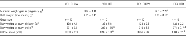
As expected, pups of DEX-treated dams had significantly lower birth weights (F (1,31) = 213; p < 0.001) (Table 1) with litter size included as a significant covariate (F (1,31) = 37.4; p < 0.001). DEX-treated mothers also gained significantly less weight in pregnancy (GD4 to GD21) (F (1,31) = 145; p < 0.001) with significant litter size effect (F (1,31) = 43.9; p < 0.001) (Table 1). No differences were seen in litter size, still-births or gestational length between maternal treatment groups (data not presented).
Table 1 Study groups, body weight and caloric intake

CHOW, standard chow; DEX, prenatal dexamethasone; HFD, high-fat diet; VEH, prenatal vehicle solution.
aSignificantly different from VEH (p < 0.001).
bSignificant covariate effect of litter size (p < 0.03; unadjusted data shown).
cSignificantly different from VEH–CHOW group (p < 0.04).
dSignificantly different from DEX–CHOW group (p < 0.02).
Weights and caloric intake
Body weights and caloric intake are presented in Table 1. At study initiation (PND 44), animals from DEX-treated mothers had caught up on control body weights (F (1,36) = 2.4; p = 0.13). At study end (PND 105), animals on HFD weighed the most (F (1,35) = 40.0; p < 0.001) and had consumed more calories, whereas DEX groups tended to be lighter (F (1,35) = 3.29; p = 0.08), all compared with their respective controls. Interestingly, when included as covariate, a big litter size was still associated with a lower body weight at study initiation (F (1,35) = 10.6; p < 0.01) as well as at study end (F (1,34) = 5.86; p = 0.02).
Organ weights
Relative weight of the epididymal fat pad was significantly increased among HFD animals (F (1,34) = 135.0; p < 0.001) (Table 2). Interestingly, prenatal DEX was also associated with increased fat pad percentage (F (1,34) = 11.5; p < 0.01). Liver weights were increased by HFD (F (1,34) = 7.55; p = 0.01) (Table 2). No differences in heart weights were seen.
Table 2 Fasting glucose and insulin levels
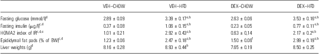
BW, body weight; CHOW, standard chow; DEX, prenatal dexamethasone; HFD, high-fat diet; IR, insulin resistance; VEH, prenatal vehicle solution.
Three insulin samples were below detection limit. Fat pad and liver weights from two animals were missing due to technical difficulties.
aSignificantly different from VEH–CHOW (p < 0.05).
bSignificantly different from DEX–CHOW (p < 0.01).
cSignificant main effect of DEX (p < 0.05).
dSignificant main effect of DIET (p < 0.01).
eSignificant covariate effect of relative epididymal fat weight (p = 0.001).
fTrend towards difference to VEH–CHOW (p = 0.08).
FST
In the FST test trial (day 2), prenatally DEX-treated animals overall spent more time being immobile compared with VEH (F (1,36) = 4.27; p < 0.05). This corresponded with decreased swimming behaviour among DEX-treated animals (F (1,36) = 6.2; p = 0.02). Diet did not affect depressive-like behaviour (F (1,36) = 0.73; p = 0.40). No post-hoc differences between individual groups were manifest. In analysis with litter size as covariate, the depressive-like behaviour was surprisingly exacerbated with increasing litter size (F (1,35) = 5.7; p = 0.02).
When looking at the plateau phase only (the last 4 min) (Fig. 2a), the effect of DEX was enhanced (F (1,36) = 8.5; p < 0.01). Specifically, only the DEX–HFD group presented with a higher level of depressive-like behaviour compared with VEH–CHOW animals (p = 0.01), although no prenatal treatment × diet interaction was present (F (1,36) = 0.14; p = 0.7). This was corresponded by less swimming in DEX–HFD animals compared with VEH–CHOW (p = 0.04). Diet (F (1,36) = 3.0; p = 0.09) or litter size (F (1,35) = 2.58; p = 0.12) did not affect depressive-like behaviour in the plateau phase.
Fig. 2 Forced Swim Test depressive-like behaviour (immobility) in the Forced Swim Test during (a) the first 5 min of the pre-test trial (day 1) and (b) the plateau phase of the test trial (last 4 min of day 2). Only prenatally DEX-treated animals on high-fat diet showed a higher level of depressive-like behaviour compared with chow-fed animals from vehicle-treated mothers. *Significantly different from VEH–CHOW. VEH, prenatal vehicle solution; CHOW, standard chow diet.
A similar pattern was seen in the pre-test trial (day 1; Fig. 2b). DEX increased immobility (F (1,36) = 4.47; p = 0.04) while diet only tended to do so (F (1,36) = 3.19; p = 0.08). The two variables did not interact (F (1,36) = 0.13; p = 0.72). Again, only the DEX–HFD group was significantly more immobile compared with VEH–CHOW in post-hoc analysis (p = 0.04), and litter size significantly affected the level of depressive-like behaviour (F (1,35) = 4.58; p = 0.04) in the same way as in the test trial.
Open field
There was no effect of diet (F (1,36) = 0.0; p = 0.95) or prenatal DEX (F (1,36) = 0.1; p = 0.77) on distance travelled.
ASR and PPI
ASR tended to increase as a consequence of prenatal DEX treatment in the middle 10 startle trials to the 120 dB(A) pulse (Fig. 3), only narrowly missing significance (F (1,36) = 4.0; p = 0.053). No effect of diet was seen (F (1,36) = 0.06; p = 0.8). ASR in the five opening trials was lower compared with the five final trials (610 ± 56 vs. 316 ± 41 mV; F (1,36) = 39.5; p < 0.001), suggesting that a tolerance developed during the test. This tolerance, however, was independent of prenatal treatment (F (1,36) = 0.2; p = 0.7) as well as diet (F (1,36) = 0.3; p = 0.6).
Fig. 3 Acoustic startle response basal startle response in the middle 10 test trials, mean of AVG (120 dB). Almost significant effect of prenatal DEX treatment in two-way ANOVA. DEX = dexamethasone.
Considering PPI, some differences were observed (Fig. 4). PPI was enhanced with increasing pre-pulse intensity (dB) in the repeated measurements analysis (F (3,108) = 28.5; p < 0.001). Interestingly, an overall intensity × diet × prenatal treatment interaction was seen (F (3,108) = 3.3; p = 0.02). Specifically, the interaction was present in first order (linear) polynomial test (F (1,36) = 6.8; p = 0.01). Looking at the two lower intensities, 72 and 74 dB, this interaction was confirmed in multiple comparisons. In PPI72 trials, DEX–HFD animals had a lower startle response compared with DEX–CHOW animals (p = 0.02), but this difference was absent in PPI74 trials (p = 0.99). In PPI74 trials, nevertheless, prenatal DEX treatment overall attenuated the startle response (F (1,36) = 6.7; p = 0.01) without differences between individual groups. No differences were seen for PPI78 and PPI86 trials.
Fig. 4 Pre-pulse inhibition of startle response Inhibition in % of the basal startle response by an immediately preceding pre-pulse of four different intensities. At 72 dB (a) there was a significant interaction between prenatal treatment and diet. At 74 dB (b) a significant effect of prenatal treatment was seen in ANOVA. No differences were seen at 78 (c) or 86 dB (d). *p < 0.05.
Neuroendocrine stress response to acute restraint
Baseline levels (0 min) of CORT were neither affected by prenatal conditions (F (1,36) = 0.16; p = 0.7) nor diet (F (1,36) = 1.0; p = 0.3). A significant effect of time in repeated measurements analysis was seen, reflecting an overall increase in CORT during restraint stress (F (4,140) = 102.2; p < 0.001) (Fig. 5a). Of greater interest, a significant time × diet interaction was found (F (1,140) = 3.11; p = 0.02). Thus, AUC was calculated and revealed that both HFD groups had higher CORT responses in the stress test compared with both CHOW groups (p < 0.03; Fig. 5b). Prenatal DEX did not affect CORT response to acute stress (F (1,35) = 2.68; p = 0.1).
Fig. 5 Acute restraint stress corticosterone response following acute restraint stress. (a) A significant time × diet interaction was seen in repeated measurements analysis (p = 0.02). (b) Area under the curve. *p < 0.03 compared with both CHOW groups. p < 0.001 compared with both CHOW groups. One sample (80 min) was missing due to haemolysis (excluded from AUC). AUC = area under the curve; CHOW, standard chow diet.
Fasting glucose and insulin levels
In animals on HFD we found increased fasting glucose (F (1,36) = 29.2; p < 0.001) and insulin levels (F (1,36) = 34.8; p < 0.001) on study day 50 (PND94). In fact, both HFD groups had higher levels compared to CHOW groups (Table 2). Interestingly, prenatal DEX treatment just narrowly resulted in lower overall insulin levels (F (1,33) = 4.3; p < 0.05), whereas glucose levels were unaffected by DEX (F (1,36) = 0.2; p = 0.6). The HOMA2 index of IR showed the same pattern, as it was increased by HFD (F (1,33) = 36.4; p < 0.001) and decreased by DEX (F (1,33) = 4.3; p < 0.05) (Table 2). Interestingly, a high relative epididymal fat pad weight had a significantly increasing effect on HOMA2 index when included as a covariate (F (1,30) = 14.0; p = 0.001).
Discussion
Our main hypothesis that 8 weeks of HFD would provoke or exacerbate aberrant behavioural, metabolic or neuroendocrine parameters in a rat model of IUGR was not convincingly proved. However, we found that chronic HFD dramatically increased the CORT response to acute restraint stress independently of prenatal DEX treatment. Similarly, others have found increased or prolonged HPA axis response to acute restraint stress following chronic HFD (Reference Tannenbaum, Brindley, Tannenbaum, Dallman, McArthur and Meaney41–Reference Sharma and Fulton43). Paradoxically, a dampening of HPA axis activity by HFD is seen in rats exposed to an early life adverse experience (maternal separation) (Reference Maniam and Morris44). Also, when rats are given the choice to eat high-fat ‘comfort food’ or standard chow, a decreased restraint stress response is described (Reference la Fleur, Houshyar, Roy and Dallman45,Reference Pecoraro, Reyes, Gomez, Bhargava and Dallman46). This stress relief by limited palatable food is probably mediated by its rewarding properties (Reference Ulrich-Lai, Ostrander and Herman47). Nevertheless, functional hypercortisolism is often seen in human obesity as in our model (Reference Pasquali, Vicennati, Cacciari and Pagotto48,Reference Champaneri, Xu and Carnethon49).
As opposed to other studies in male rats (Reference Buhl, Neschen and Yonemitsu16,Reference Hauser, Feldon and Pryce17,Reference Shoener, Baig and Page20,Reference O'Regan, Kenyon, Seckl and Holmes21), we did not observe an increase in HPA axis activity with prenatal DEX treatment. One potential explanation could be pre-weaning conditions. Brabham et al. (Reference Brabham, Phelka, Zimmer, Nash, Lopez and Vazquez19) have shown that the CORT response is markedly influenced by the rearing dam, as the increased CORT response in DEX-exposed pups was fully reversed by cross-fostering to a vehicle-treated mother. However, like us, Shoener et al. and O'regan et al. (Reference Shoener, Baig and Page20,Reference O'Regan, Kenyon, Seckl and Holmes21) did not use a cross-fostering protocol but still observed an increased CORT response. Furthermore, the tested offspring were approximately of the same age as ours (3 months), and both studies actually used a slightly lower dose of DEX (125 and 100 μg/kg, respectively) than we did (150 μg/kg). Thus, other factors than the intensity of prenatal manipulation must be crucial. In fact, we have previously neither found increased CORT response in prenatally DEX-exposed females nor in offspring of dams exposed to a chronic mild stress paradigm in pregnancy (Reference Hougaard, Andersen, Kjaer, Hansen, Werge and Lund28,Reference Hougaard, Mandrup, Kjaer, Bogh, Rosenberg and Wegener50,Reference Kjaer, Wegener, Rosenberg, Lund and Hougaard51). We therefore speculate that the strain of rat, that is, the Wistar rats involved in our studies, may somehow be resistant to HPA axis modulation by prenatal manipulations. As a parallel, the degree of susceptibility to early postnatal manipulations has been found to vary between Fischer and Lewis rats in behavioural tests (Reference Skripuletz, Kruschinski, Pabst, von Horsten and Stephan52).
Our results from FST suggest that a combination of prenatal DEX and HFD was associated with a higher level of depressive-like behaviour, as only DEX–HFD animals were found to be more immobile compared with VEH–CHOW animals both in the pre-test and the test trial. The fact that prenatal DEX per se is not sufficient to induce depressive-like behaviour in the offspring is in agreement with similar studies in male rats (Reference Hauser, Feldon and Pryce17,Reference Welberg, Seckl and Holmes18,Reference Nagano, Ozawa and Suzuki24). No interaction was seen between diet and prenatal conditions, and this makes an additive effect of the two factors most likely. In contrast, we recently found that HFD exacerbated depressive-like behaviour only in a genetically predisposed rat, the Flinders Sensitive Line rat, an inbred genetic model of depression (Reference Abildgaard, Solskov, Volke, Harvey, Lund and Wegener33). Still, our findings indicate that diet may play a role in affecting depressive symptomatology, and clinical findings support this notion (Reference Akbaraly, Brunner, Ferrie, Marmot, Kivimaki and Singh-Manoux53–Reference Jacka, Pasco and Mykletun55). In any case, HFD does not seem to ameliorate aberrant behaviours related to depression as opposed to what has been reported in a study of HPA axis hyperactivity because of early maternal separation (Reference Maniam and Morris44). Further studies are needed to elucidate the possible pathophysiological interplay.
The ASR markedly tended to be stronger in our prenatally DEX-treated animals at 3 months of age, only narrowly missing significance in ANOVA analysis (p = 0.053). Increased ASR has been associated with fear and anxiety (Reference Grillon37). We have previously observed exaggerated startle in prenatally DEX-treated female rats at 6 months of age, but only in animals having undergone blood sample under restraint before the ASR test (Reference Hougaard, Andersen, Kjaer, Hansen, Werge and Lund28). Our rats were born in Copenhagen and moved to Aarhus by coach (300 km) at 4 weeks of age. Similarly to the blood sampling procedure, this stressful experience could potentially have triggered an increase in the ASR. However, we have also found that prior exposure to the FST could not substitute restraint stress in increasing ASR levels in prenatally DEX-treated female rats (Reference Kjaer, Wegener, Rosenberg and Hougaard23). Hauser et al. (Reference Hauser, Feldon and Pryce56) also observed no changes in basal startle among 3-month-old rats using a lower dose of DEX (100 μg/kg) than we did (150 μg/kg). These findings may suggest a complex interplay between prior stressful experiences, DEX dose and age. Interestingly, ASR in male rats also tested at 3 months of age, but exposed to DEX during PNDs 1–3, resulted in lower ASR (Reference Claessens, Daskalakis, Oitzl and de Kloet57). These animals also presented with a lower body weight during lactation, but nevertheless, it seems to be crucial whether DEX exposure takes place in utero or in the early PNDs.
Considering PPI of ASR, some changes related to treatments were seen at the lower pre-pulse intensities. Prenatal DEX increased PPI at 74 dB, and an interaction between diet and prenatal conditions was seen at 72 dB. Previously, we also found increased PPI at 74 dB in prenatally DEX-treated females (Reference Kjaer, Wegener, Rosenberg and Hougaard23). This makes the possibility of chance findings unlikely. Prenatal DEX-exposure is consequently capable of altering neural circuits related to gating mechanisms, and this effect may be enhanced by HFD as seen at 72 dB.
Metabolic parameters were mainly affected by diet. HFD elevated body weight, caloric intake and fasting glucose as well as insulin levels, but prenatal DEX exposure did not seem to predispose to metabolic derangement. Correspondingly, Drake et al. (Reference Drake, Raubenheimer, Kerrigan, McInnes, Seckl and Walker25) did not find altered glucose tolerance as a consequence of prenatal DEX treatment in 6-month-old rats, but did, however, demonstrate exacerbated liver steatosis when the animals were fed a HFD. Buhl et al. (Reference Buhl, Neschen and Yonemitsu16) did not find significantly altered fasting levels either, but still prenatally DEX-treated animals had liver-specific IR when investigated in a hyperinsulinemic euglycemic clamp. Thus, it is still possible that our animals suffered from other abnormalities in glucose homeostasis that was not revealed in crude fasting levels. Nevertheless, our prenatally DEX-treated animals were actually associated with lower fasting insulin levels. In this context, experimental and clinical studies have demonstrated that poor growth in utero results in an initial increase in insulin sensitivity that is associated with accelerated postnatal growth and that these events precede the emergence of IR (Reference Morrison, Duffield, Muhlhausler, Gentili and McMillen58). Correspondingly, the lower insulin levels in our animals could potentially be due to a catch-up growth, as pups of DEX-treated mothers had a markedly lower birth weight compared with pups of VEH-treated mothers. However, a catch-up growth levelled this difference in body weights at study initiation (PND 44). Further studies are needed to conclude on this topic.
Interestingly, litter size exerted a significant influence on body weight at birth, and this was sustained during the entire study. Surprisingly, with increasing litter size, the depressive-like behaviour in the FST during both swimming sessions was reinforced, and this effect was not associated with body weight at the time of swimming. We therefore speculate that a big litter size may result in placental insufficiency and IUGR or reduced maternal care during rearing that could potentially increase the tendency towards later life depressive-like behaviour. Further studies on this topic are urgent to exclude the possibility of chance findings.
A certain limitation of this study is that we are only examining late adolescent rats of male sex. As human obesity prevalence is increasing in all age groups, including children (59,60), we found it important to perform this study comprising not yet adult rats. Furthermore, we have previously shown that HFD may exacerbate depressive-like behaviour in genetically susceptible rats of same age (Reference Abildgaard, Solskov, Volke, Harvey, Lund and Wegener33).
We conclude that chronic HFD, in addition to inducing aberrant metabolic disturbances, is capable of exacerbating the stress responsiveness of the HPA axis independently of prenatal DEX exposure and IUGR. This potentially implies an increased risk for developing psychiatric and metabolic disease. Furthermore, our findings suggest that IUGR and HFD may cumulatively result in depression-related behaviour as well as alterations in central nervous system gating mechanisms.
Acknowledgements
The authors thank Michael Guldbrandsen and Lisbeth Petersen for their indispensable laboratory assistance and care taking of the animals. Furthermore, they thank Heidi Jungland, Christina Weide Fischer, Nadia Knudsen, Pia H. P. Poulsen and Elsebeth Hornemann for their skilful assistance and Kirsten Mønsted Christensen for linguistic comments.
Financial support
AA was supported by The Danish Council for Independent Research (Medical Sciences) and the Health Research Fund of Central Denmark Region. S.L. was supported by ‘Augustinus Fonden’ and ‘Aase og Ejnar Danielsens Fond’. K.S.H. was supported by the National Research Centre for the Working Environment.
Statement of Interest
None.
Ethical Standards
The authors assert that all procedures contributing to this work comply with the ethical standards of the relevant national and institutional guides on the care and use of laboratory animals.